Determination of Heat Acclimatization by Capacitance Hygrometer-Sweat Capture Capsule Method*
Yu-Jen FAN
Department of Environmental Physiology, Institute of Tropical Medicine, Nagasaki University, 12-4 Sakamoto-machi,
Nagasaki 852, Japan
Abstract: Measurements of sweat rate in tropical and Japanese subjects were perform- ed in an environmental control chamber (room temperature 28•Ž, humidity 60%). Local
heat load (43℃ water bath) was applied on lower legs (30min, and 20min) to induce
thermal sweating responses. The indicators of thermoregulatory heat loss responses such as sweat onset time, threshold oral temperature for sweating and sweat volumes (for 20 min during heat load, for 5min after heat load) etc. were simultaneously measured by using thermography and capacitance hygrometer-sweat capture capsule method. By analyzing the data of tropical inhabitants and Japanese (sportsmen and non-sportsmen), the central and peripheral mechanism of heat acclimatization were investigated. In this study, a new quantitative calibration by using capacitance hygrometer-sweat capture capsule method was devised, i.e., on the top of the capsule fixed to the skin of sub- ject, a small hole was made, through which, subject's sweat or 30•Ž 0.45% NaCl solu- tions (0.01, 0.02, 0.03 and 0.04 ml) were precisely dropped into the capsule with a micropipette and hole sealed. Relative humidity changes (%RH) of the capsule were con- tinuously recorded by capacitance hygrometer. By calculating the absolute numidity from relative humidity, sweat rate (mg/cm2•Emin) could be obtained and sweat volume was quantitatively decided by the sweat rate. (Fan-Kosaka method). Further, it was found that the sweat onset time detected by thermography (i.e., change of skin temperature) and by sweat capture capsule method were consistent. This new method made the measurement of sweat volume simple and accurate, and that these experimen- tal modalities may be utilized in further determination of physiological mechanisms of heat acclimatization.
Key words: Capacitance hygrometry, Sweat capture capsule method, Calibration of sweat volume, Heat acclimatization, Temperature regulation
Received for publication, May 15, 1987
Contribution No. 1982 from the Institute of Tropical Medicine, Nagasaki University
*A portion of this paper was presented at the 64th Annual Meeting of the Physiological
Society of Japan, Chiba, 1987.
INTRODUCTION
The onset of thermal sweating in heat-unacclimatized subject during application of heat load does not occur untill after core temperature has started to rise and the max- imum sweating rate (about 0.2ml/sec) is not reached until core temperature has risen by about 1°C (Benzinger, 1969; Bligh, 1973). In the process of heat acclimatization, the onset of thermal sweating occurs after a progressively shorter delay and at a lower core temperature, in response to repeated exposures to the same heat load on succeeding days (Ladell, 1964; Hori, 1976; Fan et al., 1984). Recently, there appeared to be many in- formation about an importance for measurement of fine changes of sweat rate in the pro- cess of heat acclimatization, and pulsatile nature of thermal sweating was observed by us- ing various recording techniques, such as an infrared gas analyzer (Albert and Palmes,
1951), thermography (Ohwatari et al., 1983) resistance hygrometry (Nakayama and Takagi, 1959; Custance, 1962; Ogawa an Bullard, 1971, 1972) and capacitance hygrometry (Ogawa and Bullard, 1971, 1972; Sugenoya and Ogawa, 1985; Ogawa, 1987). Regarding capacitance hygrometry, the response during the calibration and much finer fluctuation of sweat rate curve during measurement by using capacitance hygrometer were quicker and more ac- curate compared to those of resistance hygrometer (Sugenoya and Ogawa, 1985). In both resistance and capacitance hygrometry, however, there seems to be something devised on the calibration for measureing sweat volume. Therefore, the purpose of the present study was to devise a new method for accurate calibration of sweat volume in the actual state of sweating skin. And analysis of thermal sweating responses induced by heat load was porformed by using the newly devised sweat capture capsule method from the view point of determination of heat acclimatization.
METHODS
Subjects: 25 healthy tropical male residents and 38 Japanese males were the subjects of this study. 26 out of 38 Japanese subjects, were the athletic students of Nagasaki University and the rest were sedentary Japanese. Tropical subjects were those who had just arrived in Japan within (the past) three months, while the experiments were being performed. Their native countries were Southeasten Asia (Thailand, Phillipine, Taiwan, In- donesia, Burma, Bangadesh) and Africa (Tanzania, Zaire, Sudan). Two kinds of method for detecting the thermal sweating-thermography and capture capsule method-were ap- plied, and 40 out of 76 experiments were carried out with capture capsule method in the present investigation.
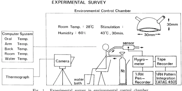
Experimental procedures: an environmental control chamber (28"C, 60%) as shown in Fig. 1 was used for this study. All subjects were submitted to sit on the chair for at least 60 minutes before the experiment began. Attachments were made as shown in Fig.
1. Ten minutes after the beginning of experiment, heat load (43°C hot water, 30min) was
applied on bilateral lower extremities to induce thermal sweating. Thermoregulatory
EXPERIMENTAL SURVEY
Environmental Control Chamber
C omputer System Oral Temp.
Arm Temp.
Back Temp.
Room Temp.
Water Temp.
rhermograph
Room Temp. : 28C Stimulation =
Humidity : 60% 43°C,SOmin.
sensor
Camera
N2
SOmnr
/ IHygro- (JTape
4
30mm f
meter I I Recorder
water bath
%RH Pen- Recorder
%RH Pattern Integration (ATAC 450)
Fig. 1. Experimental survey in environmental control chamber.
(For details see text)
responses such as temperature changes of oral cavity, rectum and skin (chest, abdomen, back, forearm) were simultaneously measured with thermistor connected to computer (PC 8801). Mean skin temperatures of a certain area of chest and abdomen were measured every minute and analyzed by using thermography and computer system (NIHON KODEN, K. K.) in order to detect the sweat onset time of each subject. In the present experi- ment, a continuous quantitative measurement of thermal sweating was performed by us- ing plastic capsules (30mm diameter, 30mm height) attached onto the chest and abdominal skin with a liquid adhesive (10% celloidin). Dry N2 gas was flowed into the capsule with a constant flow rate of 1 £/min, and the change of the humidity of effluent gas were detected by Hygrometers (H211 TAKARA Instruments. Co.) connected to DC-pen-recorder, as shown in Fig. 1. For the calibration of sweat volume, a new method was developed.
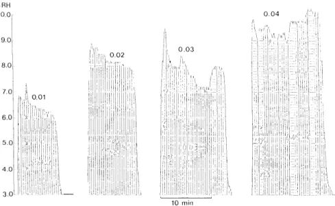
On the top of the capsule, a small hole to fit to the tip of micropipette (Finnpipette 2, LABSYSTENS OY Finland) was made. At the end of experiment the subject was submit- ted to supine position on a bed in order to calibrate the sweat volume. Through the hole, sweat or 30°C 0.45% NaCl solution was dropped into the capsule with the micropipette precisely. Fig. 2 shows the time courses of humidity change when 0.01, 0.02, 0.03 and 0.04 ml of 0.45% NaCl solution dropped in the capsule attached to forearm skin. The area of each tracing of hygrometer recording were quantitatively measured in mg and plotted against the volumes of NaCl solutions added for calibration. A regression line [Y=17.09x+0.0494 (correlation coefficient r=0.993)] for volumes 0.01, 0.02, 0.03 and 0.04 ml was obtained. (Fig. 3)
For calculating the sweat rate (mg/cm2-min), absolute humidity (x) is induced first by following equation from relative humidity (%RH).
x=0.622x-^ ' P^
Po-<pPs
E nvironmental control chamber: 28°C, 60%
Diameter of capsule attached to human forearm skin: 30mm Test solution: 30°C, 0.45% Nacl solution
0 .02
%RH
10.0-
9.0
8 .0
7 .0
6 .0
5.0
4 .0
3.0
0 .01
1
II
II
ii Ij
i I
0 .03
\
II
ii 6
1 ,
I'l
\
I\I
;l
I
""10 .04
VI
ii
'I
10 min
Fig. 2. The time courses of humidity change of 0.01, 0.02, 0.03 and 0.04 ml of 0.45% NaCl
solution dropped in the capsule attached to forearm skin. (For details see text)
mg
0.8
0.7
0.6
0.5
0.4
0.3
0.2
0.1
0 à" à"
Y =17.09X +0.0494
r=0.993
0.01 0.02 0.03 0.04 ml 0.05
Fig. 3. Correlation between sweat (0. 45% NaCl solution) volume (ml) and integrated
area of relative humidity. (For details see text)
Where x:Absolute Humidity (kg/kg Dry air)
<p:Relative Humidity (%RH) Ps :Saturated Pressure (mmHg) Po:Whole Pressure (mmHg)
For example when Po=760mmHg t=30°C (Ps=31.82mmHg)
^=10% diameter of capsule:30mm
N2 à"gas flow:1 fi/min
Therefore (%RH) 10% > 0.416mg/cm2-min
RESULTS
The sweating responses of a tropical inhabitant and Japanese evoked by a local heat load were monitored in an environmental control chamber. The changes of chest and abdominal skin temperature during sweating was recorded and analyzed by ther- mography system. The duration between starting heat load application and sweating onset was found to be longer in tropical subjects (n=20 mean onset time and SE: ll.9+
0.6 (chest) ll.4±0.7 (abdomen) compared with those of Japanese sportsmen (n=20 mean onset time and SE: 9.1±0.7 (chest) 9.1±0.7 (abdomen) and Japanese non-sportsmen (n=
10 mean sweat onset time and SE: 6.5±0.6 (chest) 6.5±0.6 (abdomen)) as shown in
mm
15r
10
50
Comparison of sweat-onset time between tropical residents and Japanese
** fTVN chest (ch)
**
T- T-
H
I abdomen
* p<0.05
** p<0.01
**-* p<0.05
MeaniSE (ab)
(di) (ill)) (ch) (ab) (cli) (ah)
Tropical Japanese Sedentary
residents sportsmen Japanese
(n=20) (n=20) (n=10)
Fig. 4. Comparison of sweat-onset time between tropical inhabitants and Japanese
in 43°C hot water stimulation. Asterisks show the significant difference of
Comparisons to the sedentary Japanese (For details see text)
Fig. 4. Oral temperature rise rate during heat load of 12 subjects of each group were analyzed. It was concluded that tropical residents have a higher rate (0.045°C/min) than Japanese sportsmen (0.035°C/min) and also tropical residents showed a bigger ATo (ATo=0.
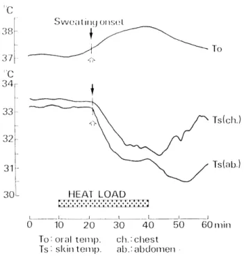
24-0.291C) than that of Japanese sportsmen (ATo=0.18-0.19°C). As shown in Fig. 5, in- dividually, the mean skin temperature on abdomen during sweating was always lower than that on chest, and the abdomen tended to sweat faster compared to chest. After terminating the heat load, the mean chest skin temperature instantly rised whereas the mean abdominal skin temperature showed a little but definite dip of temperature dissocia- tion of which origin and mechanism are not clear yet. Fig. 6 shows the experimental pro-
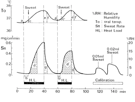
cedure of sweating analysis by using the sweat capture capsule method with calibration curve obtained by Fan-Kosaka method. Sixty minutes after the entry of an environmen- tal control chamber (28°C, 60%), the experimental recording was begun and 10 minutes later the subjects was applied the first 30 minutes heat load (43°C water bath) on bilateral lower extremities. After the end of heat load, it took 20 minutes before the humidity of capsule returned to initial level. Then, the second 20 minutes heat load was applied, 30 minutes after the end of second heat load, calibration of sweat volume was executed by dropping 0.01, 0.02 ml of subject's sweat into the capsule. Further in this
"C
Swoiilincj ousel
37 ^
"'C
*
A\ /^ Ts(ch.)
31 - ""^V / Ts(ab-)
30 ,,y^Lk9á",,,.
0 10 20 30 40 50 iJOmin
To'oraltemp. ch.:chest
Tsà"skintemp. ab.'-abdomen
Fig. 5. Sweating onset time detected by thermography and the skin temperature
dissociation after heat load, white arrow: the sweating onset time of ab-
domen, black arrow: the sweating onset time of chest. (For details see text)
°c
38
37
To Sweat
ai 36^
mg/cm-min
0.6
SR
0.4
0.2
d 0L Pb~
H.L.
Sweat
fG' ef
a\ 2i Vj
20 40
g
t-
d '
liy H.L.
%RH : Relative Humidity To : oraltemp.
SR ; Sweat Rate HL: Heat Load
0 .02ml
Sweat
0 .01ml
Sweat
Calibration
60 80 100 120 140 min
Fig. 6. The experimental procedure of sweating analysis with calibration.
(For details see text)
15 10 5 0
figure (Fig. 6), the latent time of sweat onset (b, b'), the sweat volume during 20 minutes heat load (d, d'), the change of oral temperature (ATo) at sweating onset (c, c') and the sweat volume for 5 minutes after end of heat load (g, g') were schematically demonstrated.
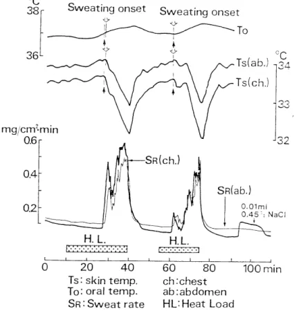
Recording of thermal sweating of a Japanese sportsman induced by the first and the second heat load (Fig. 6) is shown in Fig. 7. Sweat rate (mg/cm2-min) and calibra- tion of sweat volume were determined by sweat capture capsule method (Fan-Kosaka method). Mean chest and abdominal skin temperature were measured by thermography.
Sweating onset oral temperatures during the first and the second heat load were 37.01°C and 37.06°C, respectively. These oral temperatures seem to be the threshold temperature for the initiation of sweat response. Therefore, the threshold temperature of 37.01 °C-37.
06°C is the set point temperature (Hammel et al., 1963) for sweating of this subject. The
sweat onset times dectected by the thermograghy and the sweat capture capsule method
were similar both at the chest and the abdomen. This finding indicates the universal out-
break of thermal sweating (Kuno, 1953), which may be achieved by the central
sudomotor drive, i.e., via the central mechanism for sweating located in the pre-optic
area and anterior hypothalamus (PO/AH). However, the latent time of second sweat onset
evoked by the second heat load was shorter than that of the first heat load, this being
due to the fact that the oral temperature at the beginning of second heat load was still
Sweating onset
à"o
36
*
à"o
mg/cm2.min
0 .6
0.4
0.2
0
S à"o weating onset To
Tsd'n ab. cap.)
JC i34
Ts(ii Ts(a
nb
ch.cap)
33 32
31
I
tf Sfi(ab.)
=/ Sfilch.) E vTvrr^:^ H.L.
/
H.L.
30 Tsich.)
0.02ml
Sweat
0.01ml
Sweat
0 20 40 60 80 100 120 140min
ch:chest To: oral temp. ab:abdomen SR:Sweat rate HL:Heat Load
7. Sweating pattern induced by heat load (hot water: 43C; 30min, and 20min) on lower legs of a Japanese sportsman. A regression line of calibration Y=19.66x-0.0656 (correlation coefficient r=0.9997) (For details see text)
high, though the sweat rate had returned to the initial level. The sweat volume during
20 minutes of the second heat load was larger than that of the first heat load. The in-
creased sweating capacity observed during the second heat load may depend not only on
increased activity of the central sudomotor drive but also on the increased activity of the
peripheral sweat gland (Kuno, 1956, Nadel et al, 1974). The sweat volume for 5 minutes
after the end of the second heat load was less than that of the first heat load. The
decreased sweating capacity is possibly due to the phenomenon of the so-called
hidromeiosis (Brown, et al., 1965). During sweating, the chest and abdominal skin
temperature decreased by evaporative heat loss which was able to detect with ther-
mography system. At the same time, on the contrary, the sweat rate detected by the
capsule method increased. So the decreased and increased curves composed a mirror im-
mage as shown in Fig. 7. The sharp spike of sweat rate curve appeared at the beginn-
ing of the second heat load is considered to be mental sweating.
Fig. 8 demonstrates a similar recording of thermal sweating of a Chinese sport- sman. Both sweating onset oral temperatures during the first and the second heat load were 37.01°C and 37.04°C respectively. These threshold temperatures for the initiation of sweating are similar to those of Japanese sportsman in Fig. 7. The other ther- moregulatory responses such as sweat volume and sweat onset time observed during sweating induced by the first and the second heat load were considerably similar to those of Japanese sportsman as summarized in Table 1. However, after terminating both the first and the second heat load, definite dips of temperature dissociation were observ-
°c
38r S\A/eatingonset o --x~N -5 -7 \ }f 37 - yff 36
mg/cnr.miin
0 .6
0.4
0.2
0
^>
f I
<y
I
S weating -O ^-x onset
*
^>
o
To
<r /Ts in ch. cap.)
/
!T
H.L.
JV "|-Sfifch.)
SR(ab.) IP SL
H.L.
[à":-:-:-:-:-:-^m
Ts(ab.) Ts(ch.)
0 .02ml
0.45% Nacl
20 40 60 80 100
To-"oral temp.
SR:Sweat rate
°c 34
33 32 31
30
29
120 140min
ch-'chest ab:abdomen HL:Heat Load
Fig. 8. Sweating pattern induced by heat load (hot water: 43°C; 30min, and 20min)
on lower legs of a Chinese sportsman. A regression line of calibration was
Y=15.33x-0.0288 (correlation coefficient r=0.9973) (For details see text)
T able 1. Analysis of oral temperature changes and sweat volumes correspond to Fig.
A: Japanese sportsman B: Chinese sportsman C & D: Tropical residents (For details see text)
b c d e f g
(min) CO (mg) CO CO (mg)
subject initial To of 1st
H. L.
l atent time ATo of sweat vol. ATo of ATo of sweat vol.
of 1st 1st sweat of1st20min 1st SOmin 5min after of5minafter
sweatonset onset H.L.duration H. L. H. L. off 1st H. L.
12
0 .31 0.46 0.10 0.20
15.0 10.6 0.4 0.8
0 .82 1.04 0.46 0.55
0 .53 0.83 0.39 0.36
13.0 13.6 0.4 32.5
b'
subject initial To of 2nd
H. L.
A | 36.83
B 36.88
C 36.98
D 36.40
latent time ATo of
of 2nd 2nd sweat
sweat onset onset
6767
d' e' f' g'
sweat vol. ATo of ATo of sweat vol.
of2nd20min 2nd 30min 5min after of5minafter
H.L.duration H. L. H. L. off 1st H. L.
0 .23 0.16 0.03 0.26
33.0 20.0 20.0 74.0
0 .69 0.56 0.37 0.47
0 .41 0.44 0.34 0.29
0 .3 0.9 0.3 30.6
ed between chest and abdominal skin temperatures as shown in Fig. 5. The change of the chest skin temperature in the capsule (in ch. cap.) measured with thermistor during sweating showed the same tendency as those detected by thermography.
Fig. 9 shows a recording of thermal sweating of a tropical inhabitant just arrived in Japan one month before the experiment was performed. The threshold oral temperatures of the first sweat onset and the second sweat onset were 36.92°C and 37.01
°C, respectively. These threshold temperatures were almost similar to those of Japanese sportsman and Chinese sportsman as shown in Fig. 7 and Fig. 8, respectively. However, the first latent time of the first heat load (17 minutes) is markedly longer compared to those of Japanese sportsman (9 minutes) and Chinese sportsman (12 minutes). This long latent time caused the small amount of sweat volume in first 20 minutes heat load. This result clearly indicates the phenomenon of tropical heat acclimatization.
Analytical results of sweating responses of 4 typical subjects: Japanese sportsman Chinese sportsman and 2 tropical residents were summarized in Table 1. Subjects A, B, and C correspond to subjects of Fig. 7, 8 and 9, respectively. The figure of subject D is exceptionally omitted in this paper. The alphabets of this table (a-g, a'-g') were also cor- responded to those of Fig. 6, respectively. The analytical data on heat loss responses of thermoregulation-the present indicators of thermal sweating-were not complete, never- theless, the tendency in the present results demonstrated in this table was expectable.
Therefore, the results of the present experiment may be utilized in further determination
of physiological mechanisms of heat acclimatization.
°c
38
36
mg/cm-min
0.6 r
0 .4
0 .2
o
S weating onset Sweating onset
*
^>
^
j
4
H .L.
vI oi
SR(ch.)
H.L.
To
c
à"Ts(ab.) -,34
Ts(ch.) 33
32
SR(ab.)
0 .01ml
0.45:: Nad
_\
0 20 40
Ts: skin temp.
To: oral temp.
SR:Sweat rate
60 80 100 min
ch:chest ab:abdomen HUHeat Load
Fig. 9. Sweating pattern induced by heat load (hot water: 43°C; SOmin, and 20min) on lower legs of a tropical inhabitant. A regression line of calibration was Y=16.96x-0.0011 (correlation coefficient r=0.9971) (For details see text)
DISCUSSION
Observation and measurement of sweating responses have been determined by
Minor method, Ohara's filter paper method, thermography system available for various
skin areas (Ohwatari et al, 1983), and Galvano-Skin-Reflex (GSR) technique of electric
resistance change of the skin. Pulsatile nature of mental and thermal sweating was
observed under various experimental conditions using newly developed techniques, such
as an infrared gas analyzer (Albert et al., 1951), resistance hygrometry (Nakayama and
Takagi, 1959; Custance, 1962) and capacitance hygrometry technique for measuring local
sweat rate (Ogawa and Bullard, 1971, 1972; Sugenoya and Ogawa, 1985). The comparison
of two recording curves taken by the use of a capacitance hygrometer and a resistance
hygrometer indicated a quickened response during the calibration and much finer fluctua-
tions of sweat rate curve during measurement in the record by capacitance hygrometry
(Sugenoya and Ogawa, 1985). Therefore, in the present investigation recording of sweat
rate was achieved by the capacitance hygrometry by using a newly developed hygrometer
(H211, TAKARA Instruments, Co.) which is highly suitable for picking up fluctuations of sweat expulsion. Here, a new trial was performed to develop a new accurate method for calibration of sweat volume in the actual experimental state of the subject's skin under the sweat capture capsule. To attain the aim, a small hole fit to the tip of micropipette was made on the top of capsule. In addition to Fig. 2 and Fig. 3, calibration of humidity
change of 0.01, 0.02 and 0.03 ml 0.45% NaCl solution was carried out, and a regression line Y=18.6x-0.0109 (correlation coefficient r=0.998) was obtained. The results of these two regression lines with two correlation coefficients indicate a high reliability of the pre- sent method for the determination of sweat volume.
The principle and developmental process of thermography and its application to the measurement of sweating were theoretically and experimentally reported in the previous paper (Ohwatari et al, 1983). And the sweat onset time detected by thermography were consistent with those of the present sweat capture capsule method which confirmed the reliability and accuracy of these two different methods for measurement of sweating.
Therefore, we called this measuring method as sweat capture capsule method (Fan- Kosaka method). The results of the sweat onset times demonstrated in Fig. 4 agreed with other investigators. According to Hori et al., (1976) in similar environment condition (30"C, 70%), inhabitants of Naha, Okinawa and Honshu showed the sweat onset time of 8.
9 and 3.5 minutes by 42C water bath heat load on the legs, respectively.
Regarding the longer sweating onset time of tropical inhabitants (see Fig. 4, Fig. 9 and Table 1) compared to Japanese inhabitant, though there exists some differences bet- ween Japanese sportsman and sedentary Japaneses the following explanations are possi- ble: a) a shift of threshold core temperature of sweating onset time, b) a set of core temperature to lower level, and c) a decline of rising curve of core temperature during heat load. The factor which determine the set-point of body temperature are lower basal metabolism concerning to heat production and the physical constitution relating to heat
dissipation of the tropical inhabitants (Hammel et al., 1963).
The universal outbreak of sweating (Kuno, 1953) was confirmed not only at the chest and abdominal skin but also the face, neck, back and extremities surface areas of present subjects. However, the phenomenon of temperature dissociation between the chest and abdominal skin was observed after terminating the heat load, (see Fig. 5 and 8)
Although the origin and thermoregulatory mechanism of the temperature dissocia-
tion is not clear, but the following factors: a) regional differentiation of sympathetic ef-
ferents on the chest and abdominal skin blood flow during thermal stimulation (Iriki, 1976,
1983), b) active vasoconstriction of abdominal skin vessel, c) difference of subcutaneous
fatty layer between the chest and abdomen, d) the effect of skin pressure reflex caused
by sitting position (Takagi and Sakurai, 1950), e) difference of concentration and distribu-
tion of active sweat glands between the chest and abdominal skin, f) air flow of en-
vironmental control chamber, g) the phenomenon of hidromeiosis due to exhaustion of
sweat gland activity (Brown and Sargent, 1965; Ogawa 1987), h) after heat load off, an in-
creasing of evaporative heat loss capacity due to the decrease of sweat rate on the ab-
dominal skin etc, are considered.
Hemihidrotic phenomenon i.e., the inhibition of thermal sweating in human beings as a consequence of postural changes (Kuno, 1953; Ogata and Ichihashi, 1935) was prov- ed to be the effect of mechanical pressure applied on the skin (Takagi and Sakurai, 1950;
Kawase, 1952). The mechanism of afferent pathway of the skin pressure reflex was par- tially clarified by the findings of reflex inhibition of shivering by pressure stimulation on the skin surface of the rabbit (Takagi, 1960; Kosaka, 1967, 1969). Therefore, effect of this skin pressure reflex during sitting on a chair may be considered as an important fac- tor of the present temperature dissociation between the chest and abdominal skin.
The origin and physiological mechanism of hidromeiosis (Brown.and Sargent, 1965;
Ogawa, 1987) is recently proved to be the decreased sweat rate due to organic displace- ment of sweat gland caused by excess sweating during severe heat load. This phenomenon may be available to explain the physiological mechanism concerned with temperature dissociation and the decreased sweating capacity for 5 minutes after ter- minating the second heat load as shown in Figs 7, 8, 9 and Table 1. Concerning sweat volume in the present experiment, sweat volume collected from sweat skin during the first and the second heat load applied on bilateral under extremities was not enough to analyze the concentrations of electrolytes such as sodium, chloride and potassium, the result of which could explain the mechanism of Hidromeiosis and of increased sweat volume during 20 minutes of the second heat load (Fujishima and Kosaka, 1971).
In fact, natives in the tropical zones have the high capacity of sweating but they have acquired the ability to avoid excess sweating by heat acclimatization (Kuno, 1956).
The long term heat acclimatization in contrast to short term heat acclimatization is enigmatic except to the teleologic thinking which can't be ruled out as incorrect in the present investigation.
The present results of sweating responses in Japanese and tropical sportsmen sub- mitted to heat load and physical training agreed with those of the similar experiments designed from the view-point of cross adaptation (Hori, 1977), and which is marked by in- creased sweating capacity observed in a Japanese sportsman (Fig. 7) and of a tropical sportsman (subject D in Table 1) This may be due to both increased activity of the cen- tral sudomotor mechanism in the pre-optic area and anterior hypothalamus (PO/AH) through heat load and of the peripheral vaso-motor activity on the sweat gland through mechanical stimulation of physical training (Kuno, 1956; Nadel et al, 1974).
ACKNOWLEDGEMENTS
The author is deeply grateful to Prof. Mitsuo Kosaka for his review of the
manuscript and discussion, and to all the staffs in the Department of Environmental
Physiology, Institute of Tropical Medicine, Nagasaki University, especially to Miss. Junko
Kawashima & Miss. Junko Hayashima for their typing and drawing. The author also
wishes to thank Dr. Nobu Ohwatari for his technical suggestions and advices.
REFERENCES
1) Alvert, R. E. & Palmes, E. D. (1951): Evaporative rate patterns from small skin areas as measured by infrared gas analyzer. J. Appl. Physiol., 4, 208-214.
2 ) Benzinger, T. H. (1969): Heat regulation; Homeostasis of central temperature in man. Physiol.
Rev., 49, 671-759.
3 ) Bligh, J. (1973): Temperature regulation in mammals and other vertebrates, pp 273-274. North- Holland/American Elsvier.
4) Brown, W. K. & Sargent, F. (1965): Hidromeiosis. Arch. Environ. Health., ll, 442-453.
5) Custance, A. C. (1962): Cycling of sweat gland activity recorded by a new technique. J. Appl.
Physiol., 17, 741-742.
6 ) Fan, Yu-Jen, Ohwatari, N., Kosaka, M. & Ye-Win (1984): Studies on heat adaptation (Report H) -Sweating responses of tropical inhabitant-. Trop. Med., 26(4), 191-195.
7) Fujishima, K., & Kosaka, M. (1971): Sweating on the immersed human skin in hot bath.
Nagoya Med. J., 17(1 -2), 25-31.
8) Hammel, H. T., Jackson, D. C., Stolwijk, J. A. J., Hardy, J. D. & Stromme, S. B. (1963):
Temperature regulation by hypothalamic proportional control with adjustable set temperature. J.
Appl. Physiol., 18, 1146-1154.
9 ) Hori, s., Ihzuka, H. & Nakamura, M. (1976): Studies on physiological responses of residents in Okinawa to hot environmet. Jap. J. Physiol., 26, 235-244.
10) Hori, S., et al. (1977): Comparison of physical characteristics, body temperature and basal
metabolism between Thai and Japanese in a neutral temperature zone. Jap. J. Physiol., 27, 525
-538.
ll) Iriki, M. (1976): Regional differentiation of sympathetic efferents. (in Japanese). IGAKU NO
AYUMI. 98(5). 255-261
12) Iriki, M. (1983): Regional differentiation of sympathetic efferents during thermal stimulation. J. Therm. Biol., 8, 225-228.
13) Kawase, T. (1952): Further studies on "pressure sweat reflex" Jap. J. Physiol., 3, 1.
14) Kosaka, M. (1969): Reflex inhibition of cold shivering by pressure on the eye-ball and the ear root of the rabbit, and its afferent pathway. Jap. J. Physiol., 19. 149-
159.
15) Kosaka, M., Takagi, K. & Koyama, Y. (1967): Reflex inhibition of shivering by
pressure to the skin and the histological investigation of its afferent spinal pathway.
Experientia, 23, 453.
16) Kuno, Y. (1956): Human Perspiration. Charles C Thomas, Springfield. Illinois.
17) Ladell, W. S. S. (1964): Terrestrial animals in humid heat: man. In : Handbook of Physiology, sect. 4, Adaptation to the environment, D. B. Dill, ed. (Washington D. C.: Amer. Physiol. Soc.) pp. 625-659.
18) Nadel, E. R., Pandolf, K. B., Roberts, M. F. & Stolwijk, J. A. J.: (1974): Mechanisms of ther- mal acclimation to exercise and heat. J. Appl. Physiol., 37, 515-520.
19) Nakayama, T. & Takagi, K. (1959): Minute pattern of human perspiration observed by a con- tinuously recording method. Jpn. J. Physiol., 9, 359-364.
20) Ogawa, T. (1987): Basic knowledge on mechanisms of sweating of clinical tests. J. Clin. Exp.
Med., 140(6), 413-417.
21) Ogawa, T, & Bullard, R. W. (1971): Sudomotor activity with and without generalized sweating.
J. Physiol (Paris)., 63, 371-373.
22) Ogawa, T. & Bullard, R. W. (1972): Characteristics of subthreshold sudomotor neural impulses.
J. Appl. Physiol., 33, 300-305.
23) Ogata, K. & Ichihashi, T. (1963): J. Orient. Med. (in Japanese), 23, 1127.
24) Ohwatari, N., Fujiwara M. Iwamoto J. Fan Yu-Jen, Tsuchiya K., & Kosaka M. (1983): Studies on heat adaptation. -Measurement of sweating reaction of a tropical inhabitant., Trop. Med., 25,
235-241.
25) Sugenoya J. & Ogawa T. (1985): Characteristics of central sudomotor mechanism estimated by frequency of sweat expusions. Jap. J. Physiol., 35, 783-794.
26) Takagi, K. (1960): Influences of skin pressure on temperature regulation. In: Essential problems in Climatic Physiology, ed. by Yoshimura, H., Ogata, K., & Itoh, S., Nankodo, Kyoto, pp. 212-
249.
27) Takagi, K. & Sakurai, T. (1950): A sweat reflex due to pressure on the body surface. Jap. J.
Physiol., 1, 22.
容量式湿度計‑発汗カプセル法による暑熱順化の解析 范育仁(長崎大学熱帯医学研究所環境生理)
気温28℃,湿度60%に調節された環境制御実験装置で暑熱地住民や日本人スポーツマンの両 下肢に温熱刺激(43℃温水,30分)を負荷して体温調節の熱放散反応(特に温熱発汗)を誘発 し,サーモグラフィ装置や容量式湿度計‑発汗カプセル法によって発汗開始時間,発汗閾値口 腔温,刺激中(20分間),刺激後(5分間)の発汗量などを各種熱放散指標と同時記録・解析し て、暑熱地住民・日本人スポーツマン・日本人非スポーツマンの3群についてデーターを比較
し暑熱順化の中枢性・末梢性機序解明に資した.
本研究では特に(1)容量式湿度計‑発汗カプセル法による発汗定量の校正曲線(calibra‑
tion)の描記に新考案を試み,被験者に装着した発汗カプセルの頂部にもうけた小孔からカプセ ル下の皮膚の上に汗や30℃・0.45% NaCl溶液を0.01,0.02,0.03,0.04mlの容量ずつマイ
クロピペットを用いて段階注入,高感度容量湿度計を介して相対湿度(%RH)曲線を連続記録 し,これを絶対湿度に変換,最終的には発汗量をmg/cm2 minの単位で測定し,実記録中の 発汗量の計測を可能とした.
(2)上記,発汗カプセル法(Fan‑Kosaka method)による発汗潜時,発汗開始閾値口腔温 はサーモグラフィ装置による胸部・腹部平均皮膚温の変化時点のそれらの一致,さらに容量式 湿度計‑発汗カプセル法は発汗量の正確な測定が可能である点,発汗解析に極めて有効な手段 であることが明らかになった.
熱帯医学 第29巻 第2号107‑121頁, 1987年6月