Contribution of anti-CCP antibodies, proximal interphalangeal joint involvement, HLA-DRB1 shared epitope and PADI4 as risk factors for the development of rheumatoid arthritis in palindromic rheumatism
Mami Tamai*, Atsushi Kawakami*, Naoki Iwamoto*, Kazuhiko Arima*, Kiyoshi Aoyagi
¶& Katsumi Eguchi*
*Unit of Translational Medicine, Department of Immunology and Rheumatology
and
¶Department of Public Health, Graduate School of Biomedical Sciences, Nagasaki University, 1-7-1 Sakamoto,
†Nagasaki 852-8501, Japan, Graduate School of Health Sciences, Nagasaki University, 1-7-1 Sakamoto, Nagasaki 852-8520, Japan
Short running title: Prediction of the development of RA from PR
Key Indexing Terms: palindromic rheumatism, rheumatoid arthritis, anti-CCP antibodies, proximal interphalangeal joints, HLA-DRB1*shared epitope, PADI4
Grant Support: This study was supported by a grant from The Ministry of Health, Labour and Welfare, Japan
Address for correspondence and reprint requests:
Mami Tamai, M.D., Ph. D., Unit of Translational Medicine,
Department of Immunology and Rheumatology, Graduate School of Biomedical Sciences, Nagasaki University, 1-7-1 Sakamoto, Nagasaki 852-8501, Japan
Phone: + 81-95-819-7266 Fax: + 81-95-849-7270
E-mail: [email protected]
Abstract
Objectives. To determine which variables at baseline are predictive for development of rheumatoid arthritis (RA) from palindromic rheumatism (PR) in a Japanese population.
Methods. Anti-cyclic citrullinated peptide (anti-CCP) antibodies, joint involvement pattern, genotypes of HLA-DRB1, peptidylarginine deiminase (PADI4) and protein tyrosine phosphatase (PTPN22) were examined in 28 patients with PR at baseline, and their clinical outcome was prospectively evaluated. The same variables were also investigated in 38 healthy controls.
Results. Eleven out of 28 patients with PR developed RA. The prevalence of anti-CCP antibodies in the PR patients who developed RA was significantly higher compared to the patients who did not. Proximal interphalangeal (PIP) joint involvement at baseline was also predictive towards the development of RA. Compared with the controls, differences in the frequency of single-nucleotide polymorphism (SNP) on padi4_104
[T(RA susceptible)→C(RA non-susceptible)]and the presence of an RA susceptible homozygote of the PADI4 haplotype were detected in patients with PR whereas we could not find any further difference in PR patients who developed RA compared to PR patients who do not develop RA in PADI4. None of the subjects possessed the PTPN22 SNP (1858 C→T). Cox regression analysis revealed that anti-CCP antibodies as well as PIP involvement are the most relevant variables for the development of RA from PR.
None of the PR patients with either HLA-DRB1*SE alleles (or HLA-DRB1*0405 allele) or anti-CCP antibodies developed RA.
Conclusions. Anti-CCP antibodies, in relation to HLA-DRB1*SE carriership, and PIP
involvement are predictive for the development of RA from PR in the Japanese
population.
Abbreviations
ACR: American College of Rheumatology
anti-CCP antibodies; anti-cyclic citrullinated peptide antibodies IgM-RF: IgM-rheumatoid factor
PADI4: peptidylarginine deiminase 4 PCR: polymerase chain reaction
PIP joint: proximal interphalangeal joint PR: palindromic rheumatism
PTPN22: protein tyrosine phosphatase 22 RA: rheumatoid arthritis
SE: shared epitope
SNPs: single nucleotide polymorphisms
Introduction
Palindromic rheumatism (PR) is a disorder defined by the presence of sudden, recurrent attacks of joint pain, swelling and redness (1). Initially, there is complete remission between episodes and physical findings, but about one-third of PR patients later develop rheumatoid arthritis (RA) (1). Because significant joint damage in RA occurs within the first few years of the disease (2, 3), the early prediction of the development from PR to RA is an important issue, providing practical clinical value in helping physicians to decide which patients with PR are at high risk and should be considered for aggressive therapeutic intervention.
Serologic variables, including anti-cyclic citrullinated peptide (anti-CCP) antibodies, are clinically valuable for the classification and early definition of RA (4-7). The immunological similarity of RA to PR, especially in persons who have developed RA, has been described in Caucasian patients with positive anti-CCP antibodies at entry, which is the best serologic predictor of subsequent development of RA (4).
Gonzalez-Lopez, et al reported that the involvement of hand joints, especially
proximal interphalangeal (PIP) joint affected at baseline is predictive for further
development of RA (1). In addition, HLA-DRB1*SE is associated with the
development of RA (8). Establishing evidence from Japanese PR patients is crucial
because there is a distinct difference in the *SE allele genotype, peptidylarginine
deiminase (PADI) 4 and protein tyrosine phosphatase (PTPN22) requirement in RA
between Caucasians and Asians (9-11). Anti-CCP antibodies at first visit may also be
important in further development of RA in Japanese patients with PR (12), whereas, in
Chinese patients with RA, no association of HLA-DRB1*SE with anti-CCP antibodies
is reported (13). Therefore, we have examined the above variables in PR patients, and
this is the first prospective clinical study of PR by autoantibodies, joint involvement
pattern, and gene polymorphisms in a Japanese population to assess future progression
into RA.
Patients and Methods
Patients. Patients with PR were enrolled from the Early Arthritis Clinic at the Unit of Translational Medicine, Department of Immunology and Rheumatology, Graduate School of Biomedical Sciences, Nagasaki University. The present study consisted of 28 PR patients at entry, and 38 healthy controls, who gave their informed consent to the protocol, which was approved by the Institutional Review Board of Nagasaki University.
Twenty-eight PR patients were prospectively examined by our department and their clinical status (PR or its progression to RA), was determined. The diagnosis of PR was based on the criteria described by Gonzalez-Lopez et al. (1), and that of RA was based on the 1987 criteria of the American College of Rheumatology (ACR) for RA.
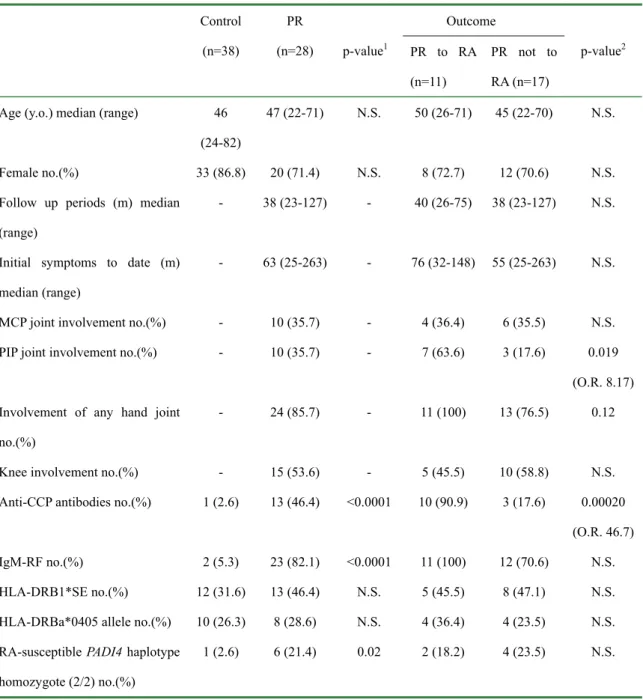
Demographic data at baseline of patients with PR, and also those of the control subjects are presented in Table 1.
Serological tests, HLA-DRB1 typing and polymorphism of PADI4 and PTPN22
Serological tests included testing for anti-CCP antibodies (DIASTAT Anti-CCP, Axis-Shield, Dundee, UK; cut-off value 4.5 U/ml) and the Immunoglobulin (Ig) M rheumatoid factor (IgM-RF) (latex-enhanced immunoelectometric assay; Dade Behring, Marburg, Germany; cut-off value 14 IU/ml) (14-16). High resolution analysis of HLA-DRB1 genotyping was performed, as described previously (16, 17), by polymerase chain reaction (PCR) according to the protocol recommended by the manufacturer, from genomic DNA in peripheral blood samples. Single nucleotide polymorphisms (SNPs) of PADI4 were genotyped (GenBank accession no. NT004610) according to the previous reports (9, 10). Three lesions of PADI4 SNPs, padi4_94 T(RA susceptible)→C(RA non-susceptible) rs 2240340, padi4_104 T(RA susceptible)→C(RA non-susceptible) rs 1748033 and padi4_102 T(RA susceptible)→C(RA non-susceptible) rs 2240337 were examined in a GeneAmp PCR System9700 (Applied Biosystems, Foster City, CA, USA) using the following PCR cycling conditions: incubation at 95℃
for 10min, followed by 45 cycles of 94℃ for 30s, 60℃ for 30s, 72℃ for 3min and
final extension at 72℃ for 7min, which is essential for the classification of the
haplotype of PADI4 (9, 10). The Invader assay was performed as described previously
(10). The primer and Invader probes for each SNP were designed using Invader Creator
software (Third Wave Technologies, Madison, WI, USA) to have theoretical annealing
temperatures of 63°C and 77°C, respectively (Table 2). In addition, analysis of the
protein tyrosine phosphatase (PTPN22) SNP (1858 C→T, rs2476601) was performed based on the previous report (18).
Statistical analysis. Differences between groups were examined for statistical significance using the Mann-Whitney U test, Chi-Square test or Fisher’s exact probability test. We compared the progression from PR to RA in the presence or absence of anti-CCP antibodies by using the Kaplan-Meier method and the log-rank test.
A Cox regression analysis was used (using the SAS system) to examine which variables
at baseline affected the further development of RA. A P value < 0.05 denoted the
presence of a statistically significant difference.
Results
Variance of anti-CCP antibodies, IgM-RF and SNPs of PADI4 and PTPN22 in PR and controls
Table 1 summarizes the demographic data for patients and controls. The prevalence of
anti-CCP antibodies and IgM-RF was significantly high in patients with PR compared
to the controls (see p-value Table1). Of the 28 patients with PR, 11 developed RA. The
median period of initial symptoms to the development of RA in the 11 patients was 29.5
months. No statistical difference was found in the follow up periods as well as in the
initial symptoms to date between the 17 PR patients who had not progressed to RA and
the 11 PR patients who had progressed to RA. The prevalence of anti-CCP antibodies in
the PR patients who developed RA (n = 11) was significantly high compared to the
patients who did not (n = 17) [10 (90.9%) versus 3 (17.6%), p = 0.0037], whereas the
distribution of IgM-RF was not. The prevalence of PIP involvement in the PR patients
who developed RA (n = 11) was significantly high compared to the patients who did not
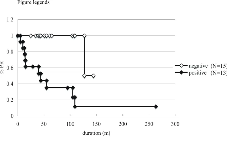
(n = 17) [7 (63.6%) versus 3 (17.6%), p = 0.019]. We compared the progression to RA
from PR in the presence or absence of anti-CCP antibodies by the Kaplan-Meier
method and the log-rank test. These analyses revealed that PR patients who are
anti-CCP antibodies-positive at the first visit develop to RA further at high frequency
compared to anti-CCP antibodies-negative PR patients (Figure 1). The genotype of
PADI4 in padi4_94, padi_104 and padi4_102 was screened. The difference of padi4_94
and padi4_102 was not found between PR patients and controls. However, the
distribution of the minor allele frequency of padi4_104 in PR was 0.50 whereas that of
the controls was 0.263, demonstrating a difference of padi_104 in PR in comparison
with the controls (p = 0.048). The frequency of the RA susceptible PADI4 haplotype
(haplotype 2; padi4_94 T, padi_104 T and padi4_102 C) tended to be high, because the
haplotype frequency of PR in the present study (0.375) was high compared with that
found in a Japanese established RA study (0.32 and 0.314 in Refs 9 and 10,
respectively), although it did not reach statistical significance (p = 0.086). However, the
frequency of the RA susceptible homozygote of the PADI4 haplotype was high in
patients with PR (Table 1, see p-value Table 1). Although we could not examine all of
the DNA samples for PTPN22, PTPN22 SNP (1858 C→T, rs2476601) was not found in
the 37 controls or 17 PR patients (data not shown).
Characteristics of the PR, who developed RA, by anti-CCP antibodies and HLA-DRB1 SE
As shown in Table 1, anti-CCP antibodies were a predictive serological marker for the further development of RA, although IgM-RF was not (see p-value Table 2). PADI4 is thought to be a susceptibility gene for PR, but the distribution of padi_104 and also the RA susceptible PADI4 haplotype were not different between the PR patients who developed RA and those who did not (data not shown). We investigated the contribution of HLA-DRB1*SE in the development of RA from PR. A low percentage of anti-CCP antibodies-negative PR patients at entry progressed to RA (two out of 15, Table 1).
Furthermore, none of the 7 patients with PR who possessed neither HLA-DRB1*SE
alleles nor anti-CCP antibodies progressed to RA (p-value; 0.0021 vs 5 patients positive
with HLA-DRB1*SE+ and anti-CCP antibodies+ population who progressed to RA at
80%, by Chi-Square test). Similar results were obtained by the combination of the
HLA-DRB1*0405 allele and anti-CCP antibodies, demonstrating that none of the 11
patients with PR who possessed neither HLA-DRB1*0405 alleles nor anti-CCP
antibodies progressed to RA (p-value; 0.0017 vs four patients positive with
HLA-DRB1*0405+ and anti-CCP antibodies+ population who progressed to RA at 75%,
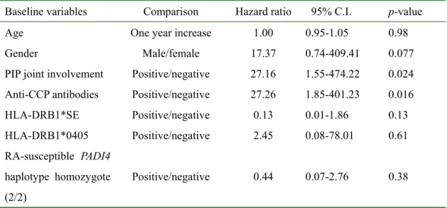
by Fisher’s exact probability test). Finally, Cox regression analysis was used to examine
which variables at first visit affect the further development of RA. We included the age
at entry, gender, anti-CCP antibodies at entry, HLA-DRB1*SE, HLA-DRB1 *0405
allele, RA-susceptible PADI4 haplotype homozygote and PIP involvement as
parameters. Accordingly, anti-CCP antibodies and PIP involvement were extracted as
significant parameters for the prediction of development to RA from PR (Table 3) .
Discussion
We found that the relative value of anti-CCP antibodies in predicting the progression of PR to RA was superior to IgM-RF. Cox regression analysis also revealed that among the variables examined in the present study, anti-CCP antibodies provide a significant predictor for further development to RA. PIP involvement at baseline is also a good predictor. Homozygosity for the HLA-DRB1*SE alleles in Caucasians with PR is reported to be a risk factor for disease progression to chronicity in PR (8); however, none of the present 28 PR cases expressed the diplotype of HLA-DRB1*SE alleles (data not shown). The presence of one allele of HLA-DRB1*SE, including the *0405 allele, which is the most frequent HLA-DRB1*SE allele in Japanese RA patients (16), did not differentiate the progression to RA. However, of note, none of the PR patients who had neither HLA-DRB1*SE nor anti-CCP antibodies progressed to RA. The use of HLA-DRB1*0405 allele showed the same result. These data demonstrate that anti-CCP antibodies constitute a principal predictor of RA from PR, and the combination of anti-CCP antibodies with HLA-DRB1*SE analysis may more precisely determine the outcome of Japanese PR patients.
A genome-wide association study of Japanese RA patients has identified other RA-associated genes besides HLA-DRB1, one of which was PADI4 (9-11). Our present study suggests that, despite the small number of patients examined, PADI4 is associated with PR. This is a novel finding, and strengthens the idea that PR, especially when it later develops into RA, could be a prototype of RA. Probably because of the small number of patients examined, we did not find any additional association of PADI4 with clinical outcome of PR. Among the SNPs of PADI4, the strongest association was observed not in padi4_104 but padi4_94 in RA (9-11), which is different from the present result. A particular polymorphism of PADI4 responsible for the association might differ between RA and PR, and this needs to be clarified in the future work.
In conclusion, anti-CCP antibodies and PIP involvement are significant predictor in
PR for further development of RA. To a lesser extent, the presence of HLA-DRB1*SE
is a predictor of the progression to RA from PR in Japanese population, being similar to
immunologic risk profile in RA. PADI4 may be related to the autoimmune-prone
phenotype of PR. Genetic associations of common diseases, including RA, are often of
modest magnitude (11). Accordingly, Cox regression analysis of the present study
revealed that anti-CCP antibodies and not genetic polymorphism is a significant
predictor for development of RA from PR. Our data indicated that a genetic polymorphism is one of the surrogate markers; however, the clinical features and anti-CCP antibodies are more promising variables at present for consideration in the development of RA from PR. In addition, other variants of these genes which have been suggested to alter the signal transduction in RA, including Fc receptor-like 3 (FCRL3) (19), Runt-related transcription factor 1 (RUNX1) (20), Solute carrier family 22, member 4 (SLC22A4) (20) and Protein tyrosine phosphatase non receptor 22 (PTPN22) (21) were not examined and the present study clearly seem to be underpowered to examine varying prognostic factors, thus, many more replication studies would be required to provide an accurate association of a genetic risk for PR.
Acknowledgements
We thank Dr. Kazuya Tanaka and Dr. Toru Egashira (Development of Clinical
Genomics, BML, Inc., Saitama, Japan) for their excellent technical assistance with the
HLA-DR genotyping and PADI4 SNPs analysis.
Table 1. Demographic data, HLA-DRB1*SE frequency and RA-susceptible PADI4 haplotype homozygote frequency at baseline in PR and controls
Control (n=38)
PR
(n=28) p-value
1Outcome
p-value
2PR to RA
(n=11)
PR not to RA (n=17) Age (y.o.) median (range) 46
(24-82)
47 (22-71) N.S. 50 (26-71) 45 (22-70) N.S.
Female no.(%) 33 (86.8) 20 (71.4) N.S. 8 (72.7) 12 (70.6) N.S.
Follow up periods (m) median (range)
- 38 (23-127) - 40 (26-75) 38 (23-127) N.S.
Initial symptoms to date (m) median (range)
- 63 (25-263) - 76 (32-148) 55 (25-263) N.S.
MCP joint involvement no.(%) - 10 (35.7) - 4 (36.4) 6 (35.5) N.S.
PIP joint involvement no.(%) - 10 (35.7) - 7 (63.6) 3 (17.6) 0.019 (O.R. 8.17) Involvement of any hand joint
no.(%)
- 24 (85.7) - 11 (100) 13 (76.5) 0.12
Knee involvement no.(%) - 15 (53.6) - 5 (45.5) 10 (58.8) N.S.
Anti-CCP antibodies no.(%) 1 (2.6) 13 (46.4) <0.0001 10 (90.9) 3 (17.6) 0.00020 (O.R. 46.7) IgM-RF no.(%) 2 (5.3) 23 (82.1) <0.0001 11 (100) 12 (70.6) N.S.
HLA-DRB1*SE no.(%) 12 (31.6) 13 (46.4) N.S. 5 (45.5) 8 (47.1) N.S.
HLA-DRBa*0405 allele no.(%) 10 (26.3) 8 (28.6) N.S. 4 (36.4) 4 (23.5) N.S.
RA-susceptible PADI4 haplotype homozygote (2/2) no.(%)
1 (2.6) 6 (21.4) 0.02 2 (18.2) 4 (23.5) N.S.
MCP joint, metacarpophalangeal joint ; PIP joint, proximal interphalangeal joint ; OR, odds ratio.
p-value
1; difference between controls (38) and PR (28).
p-value
2; difference between PR progressed to RA (11) and PR not progressed to RA (17).
Table 2. Probes and primers sequences for detecting polymorphism of PADI4
SNPs Primary probe and Invader probe sequence* PCR primer sequence and product size type Sequence (5’-3’) type Sequence (5’-3’)
Primary 1 cgcgccgaggTTGATGGGATTTCAGAAATCT sense CTGCACATCCCATCATCCTG padi4 94 Primary 2 acggacgcggagCTGATGGGATTTCAGAAATC anti-sense GCCAAGCCATTTGGACAGAG Invader CCGGACCCTCACCAACCTCTCCTCTTACA (product size 1057 base pairs)
Primary 1 cgcgccgaggCTGTGCGCAGACATC Same as padi4 94 padi4 104 Primary 2 acggacgcggagTTGTGCGCAGACATC Invader CCAACCTCTCCTCTTACTTGATGGGATTT
CAGAAATCTCCA
Primary 1 cgcgccgaggTACTCCTGCAGGCTC sense GGCATTGGGAAAGATTCCAG padi4 102 Primary 2 acggacgcggagCACTCCTGCAGGCT anti-sense TCCAACATCACACCTGCCTG
Invader GCACCACCAGGAGTAGGAGGGAAGGGA (product size 1034 base pairs) *Lower-case letters indicate flap sequence of primary probes