addi t i on t o f l avonoi ds
著者
N
agakubo Tos hi ki , Kum
ano Takut o, H
as hi m
ot o
Yos hi t er u, Kobayas hi M
i c hi hi ko
j our nal or
publ i c at i on t i t l e
Sc i ent i f i c r epor t s
vol um
e
8
page r ange
1282
year
2018- 01
権利
( C) The Aut hor ( s ) 2018
Thi s ar t i c l e i s l i c ens ed under a Cr eat i ve
Com
m
ons At t r i but i on 4. 0 I nt er nat i onal
Li c ens e, w
hi c h per m
i t s us e, s har i ng,
adapt at i on, di s t r i but i on and r epr oduc t i on i n
any m
edi um
or f or m
at , as l ong as you gi ve
appr opr i at e c r edi t t o t he or i gi nal aut hor ( s )
and t he s our c e, pr ovi de a l i nk t o t he Cr
e-at i ve Com
m
ons l i c ens e, and i ndi c at e i f c hanges
w
er e m
ade. The i m
ages or ot her t hi r d par t y
m
at er i al i n t hi s ar t i c l e ar e i nc l uded i n t he
ar t i c l e’
s Cr eat i ve Com
m
ons l i c ens e, unl es s
i ndi c at ed ot her w
i s e i n a c r edi t l i ne t o t he
m
at er i al . I f m
at er i al i s not i nc l uded i n t he
ar t i c l e’
s Cr eat i ve Com
m
ons l i c ens e and your
i nt ended us e i s not per - m
i t t ed by s t at ut or y
r egul at i on or exc eeds t he per m
i t t ed us e, you
w
i l l need t o obt ai n per m
i s s i on di r ec t l y f r om
t he c opyr i ght hol der . To vi ew
a c opy of t hi s
. . .
U
RL
ht t p: / / hdl . handl e. net / 2241/ 00151037
doi: 10.1038/s41598-018-19585-7
www.nature.com/scientificreports
Hemoglobin catalyzes CoA
degradation and thiol addition to
lavonoids
Toshiki Nagakubo, Takuto Kumano, Yoshiteru Hashimoto & Michihiko Kobayashi
In the presence of CoA, cell-free extracts prepared from porcine liver was found to convert 7,8-dihydroxylavone (DHF) to a pantetheine conjugate, which was a novel lavonoid. We puriied a 7,8-DHF-converting enzyme from the extracts, and identiied it as hemoglobin (Hb). The puriied Hb showed the following two activities: (i) degradation of CoA into pantetheine through hydrolytic cleavage to yield pantetheine and 3′-phospho-adenosine-5′-diphosphate (ADP) independently of heme, and (ii) addition of a thiol (e.g., pantetheine, glutathione and cysteine) to 7,8-DHF through C-S bond formation. Human Hb also exhibited the above lavonoid-converting activity. In addition, heme-containing enzymes such as peroxidase and catalase added each of pantetheine, glutathione and cysteine to the lavonoid, although no pantetheine conjugates were synthesized when CoA was used as a substrate. These indings indicated that the thiol-conjugating activity is widely observed in heme-containing proteins. On the other hand, only Hb catalyzed the hydrolysis of CoA, followed by the thiol conjugation to synthesize the pantetheine conjugate. To the best of our knowledge, this is the irst report showing that Hb has the catalytic ability to convert naturally occurring bioactive compounds, such as dietary lavonoids, to the corresponding conjugates in the presence of thiol donors or CoA.
For many years, natural products have been recognized as a rich source of compounds for drug discovery. Various kinds of antibiotics and bioactive compounds were discovered from natural resources1. In many countries, on
the other hand, chronic diseases such as cancer, heart disease, and stroke remain considerable public concerns. Traditionally, a plant-based diet, which usually contains various lavonoids, is thought to reduce the risk of these diseases and to promote health. housands of lavonoids have been isolated from plants. Plants synthesize lavo-noids to protect themselves from various environmental stressors such as harmful ultraviolet (UV) radiation and oxidative species2.
A number of epidemiological studies suggested that flavonoids in vegetables and fruits protect humans against chronic diseases3. Inhibitory effects on the activities of mammalian enzyme systems and the free
radical-scavenging activities of lavonoids contribute to the protection against such diseases4. he ingested
vonoids in humans are metabolized through two pathways, as follows. (i) In the small intestine and liver, la-vonoids are glucuronidated, sulfonated and O-methylated by UDP-glucuronosyltranferase, sulfotransferase and catechol-O-methyltransferase, respectively5. (ii) Intestinal bacteria degrade lavonoids into a variety of
hydroxylated phenyl carboxylic acids5. Flavonoids absorbed by the small intestine are exclusively converted into
conjugates in the liver, and the resultant conjugates enter the circulatory system6. hese conjugates are mostly
deconjugated in tissues. he resulting lavonoid aglycones are considered to have biological functions in the cell7. However, it is also proposed that the conjugated metabolites themselves show antioxidative activities and
inhibitory efects on the activities of some mammalian enzymes8. For example, quercetin and its glucuronidated
metabolite inhibited myeloperoxidase, which catalyzes low-density lipoprotein oxidation9. Also, an O-methylated
lavonoid inhibited the activity of NADPH oxidase, which generates reactive oxygen species (ROS) in the inlam-mation process10. Moreover, glucuronidated and sulfated lavonoids showed protective activity against
endothe-lial dysfunction in vivo11. hese unique activities of metabolized lavonoids are a part of the bioactivity of ingested
lavonoids. herefore, identiication of a novel metabolite of a lavonoid and a lavonoid-converting enzyme is essential for understanding of the functions of dietary lavonoids.
Graduate School of Life and Environmental Sciences, University of Tsukuba, - - Tennodai, Tsukuba, Ibaraki, -8 7 , Japan. Toshiki Nagakubo and Takuto Kumano contributed equally to this work. Correspondence and requests for materials should be addressed to M.K. (email: kobay@agbi.tsukuba.ac.jp)
Received: 11 September 2017 Accepted: 3 January 2018 Published: xx xx xxxx
In our laboratory, recently, we have studied the metabolism of biologically active natural compounds, such as curcumin12, sesamin13, and piperonal14. Very recently, particularly, we proposed a new biocatalyst concept;
actinorhodin, a natural low-molecular mass organic compound, acts as a biocatalyst under physiological condi-tions15. his inding opens the door to a new ield of organocatalysts in living organisms. In the present article, we
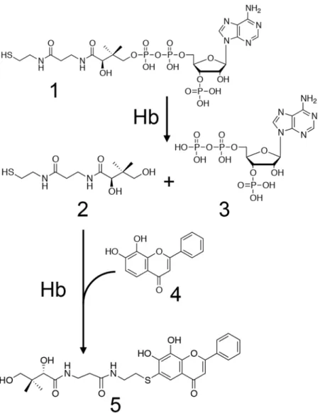
show that heme-containing catalysts including hemoglobin (Hb), peroxidase and catalase add each of thiols (e.g., pantetheine, glutathione and cysteine) to lavonoids. Moreover, we report that Hb has the ability to cleave CoA to yield pantetheine and 3′-phospho-ADP (Fig. 1). We propose a new function of Hb.
Results
Detection of a novel metabolite of 7,8-dihydroxylavone.
In order to detect and identify a novel metabolite of a lavonoid, we incubated cell-free extracts prepared from the liver of a pig with 7,8-dihydrox-ylavone (DHF) for 10 h at 37 °C. Ater incubation of the reaction mixture, however, no reaction product was observed on LC/MS analyses. Next, we added each of AMP, ADP, ATP, NADH, NADPH, NAD+, NADP+, FAD,FMN, inositol, thiamine·HCl, pantothenic acid, vitamin B6, and CoA (Fig. 2) to the above reaction mixture at a
inal concentration of 1 mM. Ater incubation for 7 h at 37 °C, the products that could be derived from 7,8-DHF were formed when CoA was added to the reaction mixture (Fig. S1). One of the reaction products gave a [M-H]−
ion at m/z 529.1631, which is in good agreement with the molecular formula [C26H29N2O8S]−. his molecular
formula corresponded with that of the pantetheine conjugate of 7,8-DHF. Unexpectedly, these results indicate that CoA was used as a substrate but not as a cofactor, being degraded into pantetheine and adducted to 7,8-DHF by a 7,8-DHF-converting enzyme.
Puriication and identiication of the 7,8-DHF-converting enzyme.
hrough the puriication steps described under “Methods”, the 7,8-DHF-converting enzyme was puriied over seven-fold, with a yield of 0.230%,www.nature.com/scientificreports/
from the liver of a pig (Table S1). he puriied enzyme gave two bands on SDS-PAGE, which corresponded to molecular masses of 15 and 16 kDa (Fig. S2).
In order to identify the 7,8-DHF-converting enzyme, partial N-terminal amino acid sequencing was carried out. he 20 amino acid sequences of the two subunits were as follows: VLSAADKANVKAAWGIVGGQ (15 kDa small subunit) and VHLSAEEKEAVLGLWGKVNV (16 kDa large subunit). A search with the BLAST program revealed that these N-terminal amino acid sequences exhibited high similarity (95% and 100%, respectively) with those of hemoglobin (Hb) subunit alpha and beta of Sus scrofa (pig), respectively.
We then investigated whether the puriied Hb is identical to blood Hb (from the same pig) or not. Blood Hb was puriied from red blood cells of the pig with a HiPrep DEAE FF 16/10 column ater hemolysis (Table S2). he puriied blood Hb gave two bands on SDS-PAGE, and the apparent molecular mass of the blood Hb was the same as that of that from liver (Fig. S3). he molecular masses of the subunits of both proteins were also determined by MALDI-TOF/MS to be as follows: alpha subunit, 15065.991 (from liver) and 15065.619 (from blood); and beta subunit, 16064.434 (from liver) and 16064.710 (from blood). hese molecular masses show that both subunits of Hb from liver are identical to the corresponding subunits of Hb from red blood cells. Furthermore, the molecular masses of the heme extracted from Hb of blood and liver were determined by LC-ESI-MS as described under “Methods”, both hemes exhibiting a m/z value of 616.2 in the positive ion mode. his m/z value corresponded with that of protoporphyrin IX and iron16. herefore, the puriied enzyme from porcine liver was identiied as Hb,
which is a tetrameric protein comprising two alpha subunits and two beta subunits (α2β2).
By using the puriied Hb from liver, the product that shows the m/z value of 529.1631 was clearly formed (Fig. S4). Using CoA as a substrate, we next compared the activities of Hb from porcine blood and human blood with that of Hb from porcine liver (Table S3). Both of them exerted the activity, but it was lower than that of Hb from porcine liver.
Pantetheine as a substrate for Hb.
he results of mass spectrometry analysis described under “Detection of a novel metabolite of 7,8-DHF” indicated the reaction product was the pantetheine conjugate of 7,8-DHF. We thus tested pantetheine as a substrate for the Hb reaction. Pantetheine was prepared from pantethine (dimeric form of pantetheine) with a 4-fold higher concentration of DTT, and then puriied by HPLC. he reaction was carried out in the standard assay mixture using puriied pantetheine as a thiol substrate. In this reaction, the product exhibited the same m/z value and retention time ([M-H]− ion at m/z 529.2 and 8.4 min) as the productobserved when CoA was used as a substrate. On the other hand, the speciic activity of Hb increased to 1.01 µmol/ min/mg from 0.403 × 10⁻³µmol/min/mg by using pantetheine as a substrate instead of CoA (Table 1).
Structure determination of the pantetheine conjugate.
he pantetheine conjugate of 7,8-DHF was prepared and puriied as described under “Methods”. We determined its structure by NMR analysis (Fig. 3, Table S4 and Figs S5–9). Heteronuclear multiple bond coherence (HMBC) analysis revealed the correlation between H-2” of the pantetheine moiety and C-6 of the lavonoid moiety. his observation demonstrates that the thiol group of pantetheine was adducted to C-6 of 7,8-DHF.Degradation of CoA by Hb and Co-bound Hb.
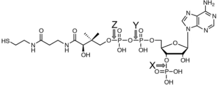
We then investigated the mechanism of degradation of CoA by Hb. When CoA was incubated with Hb, CoA was consumed and two products were observed (Fig. 4). he m/z values of the products were 506 [M-H]− and 277 [M-H]−, indicating that Hb cleaved the CoA at the Z Figure 2. he structure of CoA. Arrows X, Y, and Z indicate the cleavage sites in CoA for CoA phosphatase, dephospho-CoA pyrophosphatase and Hb, respectively.hiol donor Speciic activity (µmol/min/mg)
Glutathione 1.35 ± 0.12 Cysteine 1.29 ± 0.09 Pantetheine 1.01 ± 0.11 CoA 0.403 ± 0.033 (×10−3)
arrow in Fig. 2 to yield 3′-phospho-adenosine-5′-diphosphate (ADP) and pantetheine. In this reaction, the activ-ity of CoA degradation by Hb was measured to be 0.657 ± 0.105 × 10⁻³µmol/min/mg.
By LC/MS, we investigated this reaction by using CO treated Hb. he speciic activity of CO-bound Hb was 0.311 ± 0.016 × 10⁻³µmol/min/mg.
Efect of the redox state of heme on the activity of Hb.
We investigated the efects of the redox state and ligands on the activity of Hb. At irst, we examined the efects of ligands (i.e., KCN and CO) and redox rea-gents (i.e., dithionite (Na2S2O4) and potassium ferricyanide (K3[Fe(CN)6]), under aerobic and anaerobiccondi-tions. he spectra of the puriied Hb drastically changed on the addition of various compounds (Fig. S10). hese spectral shits coincided with those reported previously17. he addition of CO or dithionite decreased the activity
of Hb under anaerobic conditions. Also, the addition of potassium ferricyanide and KCN reduced the enzymatic activity by 60–80% under aerobic conditions (Table S5). hese results showed that dioxygenated (Fe2+-O
2) or
oxidized (Fe3+) heme was required to convert 7,8-DHF to the corresponding conjugate.
Efects of temperature and pH on the activity and stability of Hb.
he efects of pH and temper-ature on the activity of Hb were examined (Fig. S11). he optimum pH and tempertemper-ature were pH 8.5–9.0 and 45 °C, respectively. Hb was stable between pH 5.0 and pH 11.0, and under 50 °C.he stability of Hb was examined at various pH values. Ater Hb had been incubated at 25 °C for 30 min in 100 mM bufers, citrate/sodium citrate bufer (pH 3.0–6.5), Hepes/NaOH bufer (pH 6.5–8.0), Tris/HCl bufer (pH 8.0–9.0), NH4Cl/NH4OH bufer (pH 9.0–10.0), and NaHCO3/NaOH bufer (pH 10.0–11.0), an aliquot of
each enzyme solution was taken and assayed under the standard conditions. he activity of Hb was maintained even ater high pH (pH 11.0) treatment. his is in agreement with previously reported results showing that the extent of denaturation of a hemolysate was relatively low ater high pH (pH 12.5) treatment18.
he stability of Hb was examined at various temperatures. Ater Hb had been preincubated for 30 min in 100 mM Hepes-NaOH (pH 7.4), an aliquot of each solution was taken, and then the activity of Hb was assayed under the standard conditions. he activity of Hb was suddenly lost above 60 °C, which is in agreement with previously reported results showing that a rapid increase in the rate of denaturation of Hb was observed above 60 °C19.
Kinetic properties of Hb.
Using CoA and diferent concentrations of 7,8-DHF as substrates, a typical hyperbolic curve of pantetheine-conjugated product formation over substrate concentration was obtained, indi-cating that the reaction followed Michaelis-Menten-type kinetics (Fig. S12). he Km, Vmax, and kcat values were0.404 ± 0.081 mM, 0.466 × 10⁻³± 0.161 × 10⁻³µmol/min/mg, and 1.24 × 10⁻³ s−1, respectively.
Substrate speciicity.
he substrate speciicity of Hb toward various lavonoids was assayed by LC-ESI-MS in the standard assay mixture containing 2 mM CoA, 0.2–1 mM each lavonoid, and 1 mg/ml Hb. Among the tested lavonoids, quercetin, (+)-catechin, and naringenin were converted to the corresponding products, whichFigure 3. Determined structure of the pantetheine conjugate of 7,8-DHF.
www.nature.com/scientificreports/
exhibited the m/z values of pantetheine conjugates (Fig. 5). However, lavone, 7-hydroxylavone (8-dehydroxylated derivative of 7,8-DHF), apigenin (3-dehydroxylated derivative of quercetin), (−)-epicatechin (diastereomer of (+)-catechin), 4′,5-dihydroxylavone (3,7-dehydroxylated derivative of quercetin), and 4′-hydroxylavone were inert as substrates for Hb. his indicates that the number and position of the hydroxyl group of a lavonoid is unrelated to the activity of Hb. On the other hand, Hb did not convert isolavones (daidzein and genistein) into pantetheine conjugates. When pantetheine, glutathione, and cysteine were used as substrates instead of CoA, Hb was found to act on a much broader range of lavonoids including isolavones (Table S6).
Flavonoid-converting activity of heme-containing enzymes.
We investigated the lavonoid-converting activity of the heme-containing enzymes, peroxidase and catalase. We incubated each of these enzymes with a thiol donor and 7,8-DHF and then analyzed the resultant reaction mixture by LC/MS. Both heme-containing enzymes showed lavonoid-converting activity when glutathione, cysteine or pantetheine were used as a thiol donor. While the conjugation-activity of Hb was higher than those of peroxidase and catalase when glutathione and cysteine were used as a substrate, that of catalase was higher than those of Hb and peroxidase when panteth-eine was used as a substrate (Table 2).The efects of ROS scavengers on the activity of Hb.
We detected non-enzymatic and time-dependent production of H2O2 in the presence of 7,8-DHF (Fig. S13), and the addition of H2O2 to the reaction mixtureincreased the activity 2.25-fold (Table S7). In a previous study, H2O2 was proposed to degrade Hb and to release
the iron ion from Hb, which reacts with H2O2 to form reactive •OH20. We then investigated the involvement of
ROS in the 7,8-DHF-converting activity of Hb. We measured the 7,8-DHF-converting activity of Hb in the pres-ence of various scavengers for such reactive species (i.e., H2O2, •OH, O2− and 1O2). In this experiment, the tested
ROS scavengers increased the activity of Hb (Table S8).
Discussion
In humans, lavonoids have several beneicial efects due to such as their antioxidant, anti-inlammatory, antitu-mor, and estrogenic activities. Because of these attractive functions, many lavonoids have been searched for and characterized in plants.
Figure 5. Substrate speciicity of Hb. he reaction was carried out in the modiied standard assay mixture; various lavonoids were used as substrates in place of 7,8-DHF at a concentration of 0.2 mM. ND, no product could be detected. ***he pantetheine conjugate was detected, but the peak intensity was too weak to calculate the speciic activity. he experiments were carried out three times independently.
Heme-containing protein
Speciic activity (µmol/min/µmol heme)
Glutathione Cysteine Pantetheine CoA
Hb (Porcine blood) 101 ± 13 99.1 ± 8.3 81.1 ± 5.4 0.033 ± 0.002 Peroxidase (Horse radish) 76.4 ± 8.6 54.6 ± 4.9 76.8 ± 3.8 N.D. Catalase (Bovine liver) 9.77 ± 2.5 13.4 ± 1.8 154 ± 15 N.D.
In animals, dietary lavonoids are absorbed from the intestine and converted into their conjugated metabolites in the epithelial cells by enzymes such as uridine-5′-diphosphoglucuronosyltransferase, phenolsulfotransferase, catechol-O-methyltransferase, and glutathione S-transferase. he resultant conjugated metabolites and each of the corresponding aglycones are excreted into the digestive tract or low into the portal vein through speciic transporter systems. Flavonoids absorbed from the intestine are transferred to and also metabolized in the liver. hen, some resulting metabolized lavonoids are transferred from the liver to the bloodstream, the others being mixed with bile and returned to the intestinal tract via the enterohepatic circulation21. Studies on the metabolic
fate of lavonoids are important to understand the bioavailability and function of a lavonoid. However, there are lavonoids of which the metabolic pathways have not been clariied such as 7,8-dihydroxylavone (7,8-DHF).
7,8-DHF exerts a protecting efect on neurons22, improves memory23, antagonizes depression24, and functions
as a vasorelaxing and hypertensive agent25. Although these beneicial efects have attracted interest, the
metabo-lism of 7.8-DHF has never been investigated.
In this study, we explored novel metabolism of a lavonoid in mammals, using pig liver and 7,8-DHF as the enzyme source and substrate, respectively. At last, we successfully identiied CoA as another substrate for pantetheine-conjugating activity of cell-free extracts toward 7,8-DHF. Surprisingly, we found that Hb, which is usually considered as an oxygen transporter, catalyzed this conjugation reaction. We clariied that the novel Hb-catalyzed reaction consists of CoA degradation into pantetheine and 3′-phospho-ADP and synthesis of pan-tetheine conjugated lavonoids, which are novel compounds (Fig. 3).
As found in a previous study, the NH2 group of the Val-1(β), Lys-82(β) and Lys-144(β) of human Hb attacks
the carbon of the ester bond (-COO-) of the methyl acetyl phosphate to yield acetylated Hb26. his is a type of
chemical modiication of Hb and such covalent chemical modiication inhibits the sickling of red blood cells27.
In another study, enzyme-like activities of Hb were found. For example, hydroxylase-28, hydrolase-29, oxygenase-,
peroxidase- and catalase activities30,31 have been reported. Among these reactions, only the hydroxylation
reac-tion was analyzed kinetically and it was shown to be a Michaelis-Menten-type reacreac-tion. In our study, the hydro-lytic cleavage of CoA and conjugation of the resulting pantheteine to 7,8-DHF showed typical Michaelis-Menten kinetics, and Km for 7.8-DHF was calculated to be 0.4 mM. To the best of our knowledge, conjugating activity
of Hb toward lavonoids has never been reported. In Hb, the degradation of CoA into pantetheine would be a rate-limiting step, because the speciic activities of Hb for pantetheine conjugation and that for CoA hydroly-sis were 1.01 ± 0.11 µmol/min/mg and 0.657 ± 0.105 × 10⁻³µmol/min/mg, respectively. In the common
deg-radation pathway for CoA, CoA is irst dephosphorylated (Fig. 2, arrow X), and then cleaved (Fig. 2, arrow Y) into 4′-phosphopantetheine and the resulting dephosphorylated adenosine moiety by CoA phosphatase and dephospho-CoA pyrophosphatase, respectively32. On the contrary, we found that Hb catalyzed the degradation
of CoA at the cleavage site between the pantetheine moiety and phosphate (Fig. 2, arrow Z). his cleavage site is diferent from those for the CoA-degrading reactions catalyzed by the above enzymes. Moreover, carbon mon-oxide (CO) did not inhibit this reaction catalyzed by Hb, demonstrating that the hydrolysis of CoA was carried out independent of heme. his is the irst example that Hb showed the enzymatic activity regardless of the heme.
In general, heme functions as an active site in heme-containing enzymes. Because Hb has a heme in each subunit, we assumed that heme is involved in the catalysis of the 7,8-DHF-converting reaction. To examine this speculation, we investigated the efects of redox reagents and exogenous ligands on the 7,8-DHF-converting activity of Hb. CO is known to bind to the ferrous iron (Fe2+) of Hb with much higher ainity than O
2, i.e., about 210-fold higher33. On the other
hand, potassium cyanide (KCN) speciically binds to the ferric iron (Fe3+). Enzyme assays under these conditions have
revealed that reduced and dioxygenated (Fe2+-O
2) or oxidized (Fe3+) heme was required for the 7,8-DHF-converting
activity of Hb with CoA as a substrate. In the blood circulatory system, Hb with Fe2+-O
2 and Fe3+ accounts for a large
portion of total Hb. his suggests that Hb could exert lavonoid-converting activity under physiological conditions. In previous studies, Hb was found to catalyze hydroxylation and peroxidation of aniline29 and anthracene31, respectively.
Both reactions are suggested to be catalyzed by heme in Hb. his means the pocket around heme is enough wide for a lavonoid (whose size is similar to the above aromatic compounds) to bind. herefore, the reaction of pantetheine conjugation to 7,8-DHF possibly proceeds on the heme-binding site. However, it is not clear that the space around the heme of Hb is wide enough for binding of CoA, because CoA is bigger than 7,8-DHF. We assume that the hydrolysis of CoA would proceed on a site other than the heme-binding site of Hb. In heme-containing enzymes, reactions in which hydrogen peroxide acts as a mediator are also known34. In our study, however, reactive oxygen species (ROS) scavenger
experiments showed that ROS including hydrogen peroxide are not involved in this reaction.
We here propose a possible reaction mechanism of pantetheine conjugation ater CoA hydrolysis by anal-ogy with the hydroxylation catalyzed by cytochrome P450s. Cytochrome P450s catalyze the hydroxylation of substrates ater the formation of the key intermediate Compound I (Fe4+•=O). he heme iron of cytochrome
P450 binds to O2, and then forms Compound I via electron transfer from NAD(P)H to the oxygen atom in the
P450-catalyzed reaction. We speculate that Hb catalyzes the pantetheine conjugation of a lavonoid via the forma-tion of the same intermediate Compound I using pantetheine as an electron donor instead of NAD(P)H. A possi-ble reaction mechanism of the pantetheine conjugation catalyzed by Hb is as follows: (i) the oxygen of oxygenated ferrous heme (Fe2+-O
2) of Hb is reduced by pantetheine (in the case of ferric heme [Fe3+], it is reduced to ferrous
[Fe2+] by thiol, and then the resulting Fe2+ binds to O
2 before the reduction by pantetheine described above), (ii)
through protonation of the resulting reduced oxygen (Fe2+-O-O−), the intra-molecular O-O bond is cleaved to
yield Compound I (Fe4+•=O) with heme, (iii) Compound I abstracts the proton of C-6 of 7,8-DHF, and (iv) the
resulting electrophilic C-6 is attacked by thiol to form a thiol conjugate. Induction of the 7,8-DHF-converting activity (2.25-fold) of Hb by H2O2 (Table S7) would support this hypothesis, because it is proposed that H2O2
reacts with Hb and forms a compound I-like ferryl derivative (Fe4+=O) with a radical site on a porphyrin or
protein35. Furthermore, peroxidase and catalase may also catalyze pantetheine conjugation of 7,8-DHF in similar
www.nature.com/scientificreports/
Interestingly, Hb was also able to conjugate flavonoids with glutathione as well as pantetheine (Table 1, Table S6). Although glutathione conjugation is an important metabolic reaction, in general, glutathione
S-transferase is the only known enzyme that catalyzes this reaction37. Expression of glutathione S-transferase is
induced by various compounds, such as aromatic compounds or isothiocyanates, that are recognized as substrates by this enzyme. Flavonoids also induce the expression of glutathione S-transferase38, but lavonoids cannot act
as substrates for glutathione S-transferase. It is proposed that lavonoids are inhibitors rather than substrates for glutathione S-transferase, because lavonoids bind to the cysteine residue near the active site of this enzyme39.
Although a glutathione conjugate of quercetin has been detected and identiied on LC-MS/MS in humans40,
glutathione S-transferase has not been identiied as the responsible enzyme because of this inhibitory efect of lavonoids. In the present study, we found that Hb had glutathione-conjugating activity. Taking into account the abundance of Hb in the liver, Hb may act as an alternative metabolic enzyme for glutathione conjugation of lavonoids ater absorption in the small intestine. In other words, Hb could make up for the lack of the ability of glutathione S-transferase as to lavonoids.
We also found that Hb can act on other lavonoids as well as 7,8-DHF (Fig. 5, Table S6). he inding that Hb can act on these lavonoids (quercetin, catechin and naringenin) shows that a wide range of lavonoids in dietary plants are recognized as substrates by Hb. Furthermore, this is the irst report showing that isolavones, whose glutathione or cysteine conjugates have never previously been reported, undergo pantetheine conjugation cata-lyzed by Hb. In general, glutathione conjugation of a variety of polyphenols frequently enhances redox activity, and the conjugates exhibit a wide array of cellular activities41. As pantetheine conjugation of lavonoids has never
been reported before, elucidation of the bioactivity and metabolic fate of the conjugates is expected.
Hb is the most studied protein in human history, in terms of physiological function and molecular structure. Although the primary role of Hb in vertebrates is to transport molecular oxygen, our indings are the irst to show that Hb can act as an enzyme that converts lavonoids into the thiol conjugates using CoA, pantetheine, glutathione, or cysteine as a substrate. Human Hb was also found to have 7,8-DHF-converting activity (Table S3). We thus suggest that the novel activity is common to Hb of both pigs and humans.
A recent hypothesis42 proposes that an oxygen-transport protein, hemocyanin, is an origin of tyrosinase and
catechol oxidase which catalyze oxidative reactions and share the distinct metal-containing structure of the active site with hemocyanin. However, the fact that hemocyanin also catalyzes tyrosinase- and catechol oxidase-like phenol oxidation indicates that the oxygen-transport protein can catalyze the oxidative reaction according to the substrate-binding geometry43. herefore it is not unreasonable that Hb, which is also a metal-containing oxygen
transport protein, oxidatively added each of the thiol substrates to 7,8-DHF in our study. On the other hand, it is very surprising that Hb catalyzes the hydrolysis of CoA into pantetheine and 3′-phospho-ADP independently of heme of Hb. his hydrolytic activity is a new function of Hb.
Whether the substrates used in this study are the physiological substrates for Hb or not, our discovery of the novel ability of Hb is important. Although the speciic activity of the Hb-catalyzed reaction is low, this does not rule out the possibility that the enzymatic activity of Hb, which exists abundantly in liver and blood, is physiolog-ically signiicant. As physiological importance of thiol conjugates of lavonoids was not investigated, elucidation of their efects on human is the next challenge. Our indings pave the way to further studies on this new function, in which heme or polypeptide regions in Hb are involved, and new physiological roles are revealed for this ubiq-uitous protein.
Methods
Matrix-assisted laser desorption ionization time-of-flight mass spectrometry (MALDI-TOF/MS) analysis, Quantiication of the reaction product, Structural analysis of the reaction product, Quantiication of H2O2
pro-duction in the presence of 7,8-DHF, Efects of various ROS scavengers on the 7,8-DHF-converting activity of Hb. Please see Supplementary Methods.
Materials.
7,8-Dihdroxylavone, 7-hydroxylavone, 4′-hydroxylavone, lavone, quercetin, naringenin, and 4-aminoantipyrine were purchased from Tokyo Kasei Kogyo Co., Ltd. (Tokyo, Japan). (+)-Catechin, pantethine, superoxide dismutase (from bovine liver), hemoglobin (from humans), and carboxypeptidase B were purchased from Sigma (Missouri, USA). 4′,5-Dihydroxylavone was purchased from Alfa Aesar (Massachusetts, USA). CoA and peroxidase (from horseradish) were purchased from Oriental Yeast (Tokyo, Japan). Catalase (from bovine liver) was purchased from Wako Pure Chemical Industries, Ltd. (Osaka, Japan). Resource Q, HiPrep DEAE FF 16/10, and a low molecular weight standard kit were obtained from GE Healthcare (Buckinghamshire, UK). Bio-Scale mini cartridges CHT Type I 40 µm media (5 ml) were obtained from Bio-Rad (California, USA). he “Protease inhibitor cocktail for use with mammalian cell and tissue extracts” was purchased from Nacalai Tesque (Kyoto, Japan). N-Ethyl-N-(2-hydroxy-3-sulfopropyl)-3,5-dimethoxyaniline was purchased from Dojindo (Kumamoto, Japan). All other chemicals used were from commercial sources and of analytical grade. Porcine liver and blood were collected from the same individual.LC-ESI-MS analysis was performed with a Nexera X2 system (Shimadzu, Kyoto, Japan) equipped with a Cosmosil
πNAP column 4.6 × 150 mm (Nacalai Tesque). HRMS analysis was performed with a UPLC/Synapt G2 HDMS (Waters, Massachusetts, USA) equipped with an ACQUITY UPLC BEH C18 column (Waters).
Puriication of the 7,8-DHF-converting enzyme from porcine liver.
All puriication procedures were performed at 0–4 °C. Cell-free extracts were prepared as described above and fractioned with ammonium sulfate (40–60% saturation), followed by dialysis against 20 mM Tris-HCl bufer (pH 8.0). Each dialyzed solution was applied to a Resource Q column (6 ml) equilibrated with the same bufer. Protein was eluted from the column by increasing NaCl linearly from 0 to 1 M in the same bufer. he active fractions were collected and then dialyzed against 1 mM potassium phosphate bufer (pH 7.5). he protein solution was applied to a CHT Type I column equilibrated with the same bufer. he protein was eluted by increasing the concentration of the potassium phos-phate bufer linearly from 0 to 500 mM. he active fractions were collected and then dialyzed against 20 mM Tris-HCl bufer (pH 8.0). he protein solution was applied to a Resource Q column (6 ml) equilibrated with the same bufer. Protein was eluted from the column by increasing NaCl linearly from 0 to 1 M. he active fractions were collected and then dialyzed against 10 mM Hepes-NaOH bufer (pH 7.4). he homogeneity of the puriied protein was conirmed by SDS-PAGE. he relative molecular mass of the enzyme subunit was determined from the relative mobility of marker proteins on SDS-PAGE, phosphorylase b (97 kDa), BSA (66 kDa), ovalbumin (45 kDa), carbonic anhydrase (30 kDa), soybean trypsin inhibitor (20.1 kDa), and alpha-lactalbumin (14.4 kDa).Enzyme assays.
All of the reactions were performed under linear conditions with an appropriate amount of protein and a suitable reaction time, unless otherwise noted. he standard assay mixture comprised 100 mM Hepes-NaOH bufer (pH 7.4), 1 mM 7,8-DHF (in DMSO), 2 mM CoA, and an appropriate amount of enzyme in a total volume of 200 µl. his standard assay mixture was used for the assaying of 7,8-DHF-converting activity, with the exception of investigation of the substrate speciicity, for which the concentration of lavonoids was 0.2 mM. In these standard assays, the reaction was started by addition of the enzyme and carried out at 37 °C. he reaction was stopped by the addition of 200 µl acetonitrile to the reaction mixture, and a supernatant was obtained by centrifugation (20,630 ×g, 10 min).One unit of the 7,8-DHF-converting activity was deined as the amount of the enzyme that catalyzed the production of 1 µmol pantetheine conjugate per hour under the standard assay conditions. he concentration of the pantetheine conjugate was calculated from a standard curve established from the peak area of the puriied pantetheine conjugate of 7,8-DHF in a mass chromatogram (m/z 529.2 [M-H]−). Speciic activity is expressed
as units per milligram of protein. he protein concentrations were determined with a protein assay kit (Nacalai Tesque) using bovine serum albumin as the standard, as in the method of Bradford44.
Flavonoid-converting activity of heme-containing enzymes.
Heme contents of heme-containing enzymes were analyzed with a pyridine hemochrome method45. An extinction coeicient value for heme Bpyri-dine hemochrome of 34.4 mM−1 cm−1 at 557 nm was used46. Each heme-containing enzyme was added to the
standard assay mixture. he inal concentration of each heme-containing enzyme was 25 µM. Ater incubation at 37 °C, reaction mixtures were analyzed by LC/MS.
Substrate speciicity.
he following lavonoids were examined as to substrate speciicity at inal con-centrations of 0.2–1 mM: quercetin, genistein, daidzein, apigenin, naringenin, (+)-catechin, (−)-epicatechin, (+)-taxifolin, 7-hydroxylavone, 4′, 5-dihydroxylavone, 4′-hydroxylavone, and lavone. Each of these lavonoids was added to standard assay mixture instead of 7,8-DHF. he production of the pantetheine conjugates was detected by LC-ESI-MS.References
1. Vaishnav, P. & Demain, A. L. Unexpected applications of secondary metabolites. Biotechnol. Adv.29, 223–229 (2011).
2. Agati, G., Azzarello, E., Pollastri, M. & Tattini, M. Flavonoids as antioxidants in plants: Location and functional signiicance. Plant Science196, 67–76 (2012).
3. Del Rio, D. et al. Dietary (poly)phenolics in human health: structures, bioavailability, and evidence of protective efects against chronic diseases. Antioxid. Redox Signal18, 1818–1892 (2013).
4. Middleton, E. Jr., Kandaswami, C. & Theoharides, T. The effects of plant flavonoids on mammalian cells: implications for inlammation, heart disease, and cancer. Pharmacol. Rev.52, 673–751 (2000).
5. Hollman, P. C. H. Absorption, bioavailability, and metabolism of lavonoids. Pharm. Biol.42, 74–83 (2004).
6. Day, A. J. et al. Human metabolism of dietary lavonoids: identiication of plasma metabolites of quercetin. Free Radic. Res.35, 941–952 (2001).
7. Perez-Vizcaino, F., Duarte, J. & Santos-Buelga, C. he lavonoid paradox: conjugation and deconjugation as key steps for the biological activity of lavonoids. J. Sci. Food Agric.92, 1822–1825 (2012).
8. Terao, J., Murota, K. & Kawai, Y. Conjugated quercetin glucuronides as bioactive metabolites and precursors of aglycone in vivo.
Food Funct.2, 11–17 (2011).
9. Loke, W. M. et al. Quercetin and its in vivo metabolites inhibit neutrophil-mediated low-density lipoprotein oxidation. J. Agric. Food Chem.56, 3609–3615 (2008).
10. Stefen, Y., Gruber, C., Schewe, T. & Sies, H. Mono-O-methylated lavonols and other lavonoids as inhibitors of endothelial NADPH oxidase. Arch. Biochem. Biophys.469, 209–219 (2008).
11. Lodi, F. et al. Glucuronidated and sulfated metabolites of the lavonoid quercetin prevent endothelial dysfunction but lack direct vasorelaxant efects in rat arota. Atherosclerosis204, 34–39 (2009).
12. Hassaninasab, A., Hashimoto, Y., Tomita-Yokotani, K. & Kobayashi, M. Discovery of the curcumin metabolic pathway involving a unique enzyme in an intestinal microorganism. Proc. Natl. Acad. Sci. USA108, 6615–6620 (2011).
13. Kumano, T., Fujiki, E., Hashimoto, Y. & Kobayashi, M. Discovery of a sesamin-metabolizing microorganism and a new enzyme.
www.nature.com/scientificreports/
14. Doi, S. et al. Discovery of piperonal-converting oxidase involved in the metabolism of a botanical aromatic aldehyde. Sci. Rep.6, 38021 (2016).
15. Nishiyama, T., Hashimoto, Y., Kusakabe, H., Kumano, T. & Kobayashi, M. Natural low-molecular mass organic compounds with oxidase activity as organocatalysts. Proc. Natl. Acad. Sci. USA111, 17152–17157 (2014).
16. Huang, L. & Colas, C. & Ortiz de Montellano, P. R. Oxidation of carboxylic acids by horseradish peroxidase results in prosthetic heme modiication and inactivation. J. Am. Chem. Soc.126, 12865–12873 (2004).
17. Sowole, M. A., Vuong, S. & Konermann, L. Interactions of hemolgobin and myoglobin with their ligands CN−, CO, and O 2
monitored by electrospray ionization-mass spectrometry. Anal. Chem.87, 9538–9545 (2015).
18. Rieder, R. F. Hemoglobin stability: observations on the denaturation of normal and abnormal hemoglobins by oxidant dyes, heat, and alkali. J. Clin. Invest.12, 2369–2376 (1970).
19. Di Domenico, R. & Lavecchia, R. hermal stability of human haemoglobin in the presence of sarcosine and sorbitol. Biotechnol. Lett. 22, 335–339 (2000).
20. Puppo, A. & Halliwell, B. Formation of hydroxyl radicals from hydrogen peroxide in the presence of iron. Biochem, J.249, 185–190 (1988).
21. Santos-Buelga, C., Escribano-Bailon, M. T. & Lattanzio, V. Recent advances in polyphenol research, Vol. 2. 305–306 (Wiley-Blackwell, 2010).
22. Jang, S. et al. A selective TrkB agonist with potent neurotrophic activities by 7,8-dihydroxylavone. Proc. Natl. Acad. Sci. USA107, 2687–2692 (2010).
23. Spencer, J. P. E. he impact of fruit lavonoids on memory and cognition. Br. J. Nutr.104, S40–S47 (2010).
24. Cazorla, M. et al. Identiication of a low-molecular weight TrkB antagonist with anxiolytic and antidepressant activity in mice. J. Clin. Invest.121, 1846–1857 (2011).
25. Huai, R. et al. Vasorelaxing and antihypertensive efects of 7,8-dihydroxylavone. Am. J. Hypertens.27, 750–760 (2014).
26. Ueno, H., Pospischil, M. A. & Manning, J. M. Methyl acetyl phosphate as a covalent probe for anion-binding sites in human and bovine hemoglobins. J. Biol. Chem.264, 12344–12351 (1989).
27. Manning, J. M. Covalent inhibitors of the gelation of sickle cell hemoglobin and their efects on function. Adv. Enzymol. Relat. Areas Mol. Biol.64, 55–91 (1991).
28. Mieyal, J. J., Ackerman, R. S., Blumer, J. L. & Freeman, L. S. Characterization of enzyme-like activity of human hemoglobin. J. Biol.
Chem.251, 3436–3441 (1976).
29. Elbaum, D. & Nagel, R. L. Esterase activity of hemoglobin. J. Biol. Chem.256, 2280–2283 (1981).
30. Paco, L. et al. Catalase-like activity of bovine met-hemoglobin: interaction with the pseudo-catalytic peroxidation of anthracene trace in aqueous medium. Biotechnol. J.4, 1460–1470 (2009).
31. Smith, M. J. & Beck, W. S. Peroxidase activity of hemoglobin and its subunits: Efects thereupon of haptoglobin. Biochim. Biophys. Acta147, 324–333 (1967).
32. David, G. Metabolism of sulfur compounds. (ed. David, G.) 10–11 (Academic Press, 1975). 33. Blumenthal, I. Carbon monoxide poisoning. J. Roy. Soc. Med.94, 270–272 (2001).
34. McCue, J. M., Driscoll, W. J. & Mueller, G. P. Cytochrome c catalyzes the in vitro synthesis of arachidonoyl glycine. Biochem. Biophys.
Res. Commun.365, 322–327 (2008).
35. Bell, S. R. & Groves, J. T. A highly reactive P450 model compound I. J. Am. Chem. Soc.131, 9640–9641 (2009). 36. Gebicka, L. & Banasiak, E. Flavonoids as reductants of ferryl hemoglobin. Acta Biochim. Pol.56, 509–513 (2009).
37. Strange, R. C., Spiteri, M. A., Ramachandran, S. & Fryer, A. A. Glutathione-S-transferase family of enzymes. Mutat. Res.482, 21–26 (2001).
38. Boušová, I. & Skálová, L. Inhibition and induction of glutathione S-transferases by lavonoids: possible pharmacological and toxicological consequences. Drug Metab. Rev.44, 267–286 (2012).
39. van Zanden, J. J. et al. Inhibition of human glutathione S-transferase P1-1 by the lavonoid quercetin. Chem. Biol. Interact.145, 139–148 (2003).
40. Hong, Y. & Mitchell, A. E. Identiication of glutathione-related quercetin metabolites in humans. Chem. Res. Toxicol.19, 1525–1532 (2006).
41. Monks, T. J. & Lau, S. S. he pharmacology and toxicology of polyphenolic-glutathione conjugates. Annu. Rev. Pharmacol. Toxicol. 38, 229–255 (1998).
42. Ginsbach, J. W. et al. Structure/function corelations among coupled binuclear copper proteins through spectroscopic and reactivity studies of NspF. Proc. Natl. Acad. Sci. USA109, 10793–10797 (2012).
43. Decker, H. & Tuczek, F. Tyrosinase/catecholoxidase activity of hemocyanins: structural basis and molecular mechanism. Trends Biochem. Sci.25, 392–397 (2000).
44. Bradford, M. M. B. A rapid and sensitive method for the quantitation of microgram quantities of protein utilizing the principle of protein-dye binding. Anal. Biochem.72, 248–254 (1976).
45. Fuhrop, J. H. & Smith, K. M. Porphyrins and Metalloporphyrins. 755–869 (Elsevier, 1975).
46. Fruk, L., Kuhlmann, J. & Niemeyer, C. M. Analysis of heme-reconstitution of apoenzyme by means of surface plasmon resonance.
Chem. Commun.2, 230–232 (2009).
Acknowledgements
Porcine liver and blood were supplied by Dr. Masaya Katsumata (National Institute of Livestock and Grassland Science, Ikenodai, Tsukuba, Ibaraki, Japan; his present ailiation is Department of Veterinary Medicine, Azabu University, Kanagawa, Japan).
Author Contributions
T.N., T.K., and M.K. designed the research; T.N. and T.K. performed the research; T.N., T.K., Y.H., and M.K. analyzed the data; and T.N., T.K., and M.K. wrote the paper.
Additional Information
Supplementary information accompanies this paper at https://doi.org/10.1038/s41598-018-19585-7.
Competing Interests: he authors declare that they have no competing interests.
Open Access This article is licensed under a Creative Commons Attribution 4.0 International License, which permits use, sharing, adaptation, distribution and reproduction in any medium or format, as long as you give appropriate credit to the original author(s) and the source, provide a link to the Cre-ative Commons license, and indicate if changes were made. he images or other third party material in this article are included in the article’s Creative Commons license, unless indicated otherwise in a credit line to the material. If material is not included in the article’s Creative Commons license and your intended use is not per-mitted by statutory regulation or exceeds the perper-mitted use, you will need to obtain permission directly from the copyright holder. To view a copy of this license, visit http://creativecommons.org/licenses/by/4.0/.