Comparative transcriptome analysis for immune response against fungal infection in Drosophila virilis.
Yosuke Seto
Department of Biological Sciences Tokyo Metropolitan University
2013
CONTENTS
Page
General introduction 1
Literature cited 4
Abstract 7 Introduction 8
Materials and Methods 12
Results 23 Discussion 33 Conclusion 41
Literature cited 42
Tables 51 Figures 74 Appendix-I 96 Appendix-II 103 Acknowledgements 111
1
GENERAL INTRODUCTION
A major goal in evolutionary biology is to uncover the mechanisms of phenotypic evolution.
It is considered that many phenotypic characters have been evolved to adapt to various environmental conditions by natural selection (Darwin 1859). So far, molecular evolutionary biologists have tried to understand the adaptive evolution at molecular level using theoretical and experimental analyses (Hughes and Nei 1988, Nielsen and Yang 1998, Suzuki and Gojobori 1999, Smith and Eyre-Walker 2002, Yokoyama et al., 2008). In these studies, it was postulated that changes of nucleotide or protein sequence causing functionary alteration of the gene are the most reflect the adaptive evolution. A representative example of the theoretical studies is that focused on evolution of immune systems. Hughes and Nei (1988) revealed that the rate of nonsynonymous substitution (dN) was significantly higher than the rate of synonymous substitution (dS) in antigen recognition sites (ARS) of major histocompatibility complex (MHC) genes by comparing protein-coding region of MHC genes among mammals, whereas dN for non-ARS region was significantly lower than dS. From this results, they claimed that the ARS have been evolved under positive selection and the highly polymorphism of ARS have been maintained by overdominant selection, based on the prediction from the neutral theory of molecular evolution (Kimura 1983). Similarly, many immune-related genes involving in innate immune system are shown to be rapidly evolving compared to non-immune-related genes (Schlenke and Begun, 2003, Sackton et al., 2007, Obbard et al., 2009). These observations were explained by coevolutionary interactions between hosts and pathogens, so-called “arms races”
(Dawkins and Krebs 1979). Under this conception, compared to non-immune-related genes, immune-related genes are expected to be rapidly evolving or to have elevated polymorphism to maintain various alleles to cover ever-changing pathogens (Schlenke and Begun, 2003, Sackton
2
et al., 2007, Obbard et al., 2009). These studies were focused on to detect evolutionary traces from the primary structure of DNA or protein sequences.
However, Wilson, Maxson and Sarich (1974) proposed that phenotypic evolution have more arisen from changes of gene regulatory system than from changes of protein function.
Consistently, in recent years, it has been revealed that changes in gene expression pattern play an important role in phenotypic evolution, e.g., novelty of pigmentation pattern on Drosophila wings generated by changing spatial expression pattern of yellow (Gompel et al., 2005), changes in butterfly eyespots on the wings by changes in Distal-less expression pattern (Beldade, Brakefield and Long 2002) and changes in beak morphology in Darwin’s Finches generated by gene expression changes of BMP4 (Abzhanov et al., 2004). In Drosophila immune system, similar situation was also reported. Sackton and Clark (2009) found that the expression patterns of antimicrobial peptide (AMP) genes against bacterial infection by septic injury were different between two Drosophila species, Drosophila melanogaster and D. virilis. Although they suggested that this difference in the immune-response was due to different ecological traits of the two species, they did not clarify the relationship between the phenotype and the gene expression pattern.
D. melanogaster feeds on fermented or rotting fruits, which mainly harbor Baker’s yeast, Saccharomyces cerevisiae, whereas D. virilis feeds on slime flux and decaying bark of trees, on which a variety of yeasts and filamentous fungi thrive (Carson 1971, Throckmorton 1975, Weber, Davoli and Anke 2006, Weber 2006). From this difference in the natural habitat, D.
virilis is supposed to have a higher risk of the infection by a variety of fungi. However, according to 12 Drosophila species genomes analysis, D. virilis does not have the antifungal peptide, Drosomycin (Sackton et al., 2007), which is known to be an essential AMP in antifungal immune system of D. melanogaster (Lemaitre et al., 1996, Tzou, Reichhart and
3
Lemaitre 2002). This raises the question about the immune mechanism contributed to the antifungal resistance of D. virilis, which is thought to be an important factor for understanding the adaptive evolution of D. virilis to its habitat in moldy environment. To answer this question, I investigated the immune gene response to the fungal infection of D. virilis to clarify what immune system of D. virilis has evolved to defend against fungal infection.
My comparative transcriptome analysis revealed that many immune-related genes, such as AMP genes and immune-induced molecule (IM) genes, showed extensively different expression pattern between D. melanogaster and D. virilis in response to the infection of Penicillium fungus. Furthermore, I found a possibility that unknown immune-related genes have been recruited in antifungal immune system of D. virilis during its evolution. This D. virilis-specific immune gene response may contribute to the observed high resistance to the fungal infection.
My results provide an important example for understanding the mechanism of phenotypic evolution by gene expression changes proposed by Wilson, Maxson and Sarich (1974).
4
Literature cited
Abzhanov A., M. Protas, B. R. Grant, P. R. Grant and C. J. Tabin, “Bmp4 and Morphological Variation of Beaks in Darwin's Finches,” Science, vol. 305, no. 5689, pp. 1462-1465, 2004.
Beldade P, P. M. Brakefield and A. D. Long, “Contribution of Distal-less to quantitative variation in butterfly eyespots,” Nature, vol. 415, no. 6869, pp. 315-318, 2002.
Carson H. L., “The ecology of Drosophila breeding sites”, Harold L. Lyon Arboretum Lecture Number Two, University of Hawaii, pp. 1-28, 1971.
Darwin C., "On the origin of species by means of natural selection,"John Murray, London, 1859.
Dawkins R. and J. R. Krebs, “Arms races between and within species,” Proceedings of the Royal Society of London B Biological Sciences, vol. 205, no. 1161, pp. 489-511, 1979.
Gompel N., B. Prud’homme, P. J. Wittkopp, V. A. Kassner and S. B. Carroll, “Chance caught on the wing: cis-regulatory evolution and the origin of pigment patterns in Drosophila,”
Nature, vol. 433, no. 7025, pp. 481-487, 2005.
Hughes, A. L., and M. Nei, “Pattern of nucleotide substitution at major histocompatibility complex class I loci reveals overdominant selection,” Nature, vol. 335, no. 6186, pp.
167-170, 1988.
Janeway C. A. Jr, P. Travers, M. Walport, et al., “Immunobiology: The Immune System in Health and Disease,” 5th edition, New York, Garland Science, 2001.
Kimura M., “The neutral theory of molecular evolution,” Cambridge University Press, Cambridge, 1983
Lemaitre B., E. Nicolas, L. Michaut, J. M. Reichhart and J. A. Hoffmann, “The dorsoventral regulatory gene cassette Spatzle/Toll/cactus controls the potent antifungal response in
5
Drosophila adults,” Cell, vol. 86, no. 6, pp. 973-983, 1996.
Nielsen, R. and Z. Yang, “Likelihood models for detecting positive selected amino acid sites and applications to the HIV-1 envelope gene,” Genetics, vol. 148, no. 3, pp. 929-936, 1998.
Obbard, D. J., J. J. Welch, K. W. Kim and F. M. Jiggins, “Quantifying Adaptive Evolution in the Drosophila Immune System,” PLoS Genetics, vol. 5, no. 10, Article ID e1000698, 2009.
Sackton T. B., B. P. Lazzaro, T. A. Schlenke, J. D. Evans, D. Hultmark and A. G. Clark,
“Dynamic evolution of the innate immune system in Drosophila,” Nature Genetics, vol. 39, no. 12, pp 1461-1468, 2007.
Sackton T. B. and A. G. Clark, “Comparative profiling of the transcriptional response to infection in two species of Drosophila by short-read cDNA sequencing,” BMC Genomics, vol. 10, article 259, 2009.
Schlenke T. A. and D. J. Begun, “Natural Selection Drives Drosophila Immune System Evolution,” Genetics, vol. 164, no. 4, pp. 1471-1480, 2003.
Smith N. G. C and A. Eyre-Walker, “Adaptive protein evolution in Drosophila,” Nature, vol.
415, no. 6875, pp. 1022–24, 2002.
Suzuki, Y. and T. Gojobori, “A method for detecting positive selection at single amino acid sites,”
Molecular Biology and Evolution, vol. 16, no. 10, pp. 1315–1328, 1999.
Throckmorton L. H., “The phylogeny, ecology and geography of Drosophila,” in Invertebrates of Genetic Interest, R. C. King (ed), Plenum Press, New York, pp. 421-469, 1975.
Tzou P., J. M. Reichhart and B. Lemaitre, “Constitutive expression of a single antimicrobial peptide can restore wild-type resistance to infection in immuno-deficient Drosophila mutants,” Proceedings of the National Academy of Sciences of the United States of America, vol. 99, no. 4, pp. 2152-2157, 2002.
Weber R. W. S., P. Davoli and H. Anke, “A microbial consortium involving the astaxanthin
6
producer Xanthophyllomyces dendrorhous on freshly cut birch stumps in Germany”, Mycologist, Vol. 20, no. 2, pp. 57-61, 2006.
Weber R. W. S., “On the ecology of fungal consortia of spring sap-flows”, Mycologist,Vol. 20, no. 4, pp. 140-143, 2006.
Wilson A. C., L. R. Maxson and V. M. Sarich, “Two Types of Molecular Evolution. Evidence from Studies of Interspecific Hybridization,” Proceedings of the National Academy of Sciences of the United States of America, vol. 71, no. 7, pp. 2843-2847, 1974.
Yokoyama S., T. Tada, H. Zhang and L. Britt, “Elucidation of phenotypic adaptations:
Molecular analyses of dim-light vision proteins in vertebrates,” Proceedings of the National Academy of Sciences of the United States of America, vol. 105, no. 36, pp.
13480-13485. 2008
7
ABSTRACT
The innate immune system of Drosophila is activated by ingestion of microorganisms. D.
melanogaster breeds on fruits fermented by Saccharomyces cerevisiae, whereas D. virilis breeds on slime flux and decaying bark of tree housing a variety of bacteria, yeasts and molds. In this study, it is shown that D. virilis has a higher resistance to oral infection of a species of filamentous fungi belonging to the genus Penicillium compared to D. melanogaster. In response to the fungal infection, a transcriptome profile of immune-related genes was considerably different between D. melanogaster and D. virilis: the genes encoding antifungal peptides, Drosomycin and Metchnikowin, were highly expressed in D. melanogaster whereas the genes encoding Diptericin and Defensin were highly expressed in D. virilis. On the other hand, the immune-induced molecule (IM) genes showed contrary expression patterns between the two species: they were induced by the fungal infection in D. melanogaster but tended to be suppressed in D. virilis. Our transcriptome analysis also showed newly predicted immune-related genes in D. virilis. These results suggest that the innate immune system has been extensively differentiated during the evolution of these Drosophila species.
8
1. INTRODUCTION
In natural environments, Drosophila species feed and breed on fermenting fruits, slime fluxes on decaying parts of tree, etc., where biochemical processes of bacteria and fungi are extremely active (Carson 1971, Throckmorton 1975, Markow and O'Grady 2007). Therefore, Drosophila species are exposed to a huge number of microorganisms throughout their developmental stages. Feeding on decaying or fermented materials results in the ingestion of a wide variety of microorganisms in their digestive organs. Recent studies on larval immune response of D. melanogaster to oral infection of bacteria and fungi showed that the fat body mediated systemic immune response including antimicrobial peptide (AMP) production was triggered by infections of gram-negative bacterial species such as Pseudomonas entomophila and Erwinia carotovora carotovora 15 (Ecc15) and of a dimorphic fungal species, Candida albicans (Basset et al., 2000, Vodovaret al., 2005, Glittenberg et al., 2011).
In the expression of AMP genes, two major signaling pathways, Toll and Imd pathways, play a critical role. The Toll pathway is especially important in immune response to infection of fungi and gram-positive bacteria (Lemaitre et al., 1996, Rutschmann, Kilinc and Ferrandon 2002). After beta-(1.3)-glucans and Lys-type peptidoglycans, which are components of cell wall of fungi and gram-positive bacteria, are recognized by the gram-negative bacteria binding protein 3 (GNBP3) and peptidoglycan-recognition protein-SA (PGRP-SA), the Toll pathway is triggered by cleavage and binding of the ligand, Spatzle, to lead to degradation of Cactus, an inhibitor of NF-kappaB like transcription factor. The degradation allows NF-kappaB (NF-kB) like transcription factor, Dif and Dorsal, to translocate into nucleus and activate the transcription of a set of target genes. On the other hand, the Imd pathway has a key function in immune response to infection of gram-negative bacteria. After DAP-type peptide glycan, which is a component of cell wall of gram-negative bacteria, is recognized by peptidoglycan-recognition
9
protein-LC (PGRP-LC), a transcription factor, Relish, is phosphorylated and cleaved into the active form. As the result, expressions of a group of target genes are triggered (Ferrandon et al., 2007, Lemaitre and Hoffmann 2007) (Figure 1). In addition to these two pathways, JAK/STAT and JNK pathways are also important for immune response to infection of microorganisms in Drosophila (Boutros, Agaisse and Perrimon 2002, Agaisse and Perrimon 2004, Delaney et al., 2006, Lemaitre and Hoffmann 2007). The JAK/STAT signaling pathway mainly regulates phagocytosis, hemolymph coagulation and melanization (Agaisse and Perrimon 2004).
AMPs are cationic small secretory peptides that exhibit a wide range of activities against bacteria, fungi and/or viruses, playing an essential role in the innate immune system of Drosophila (Lemaitre and Hoffmann 2007). To date, seven AMP families, i.e., Attacin, Cecropin, Defensin, Diptericin, Drosocin, Drosomycin and Metchnikowin, have been identified in Drosophila melanogaster (Lemaitre and Hoffmann 2007). According to Sackton et al. (2007), it was indicated by their sequence analysis of the 12 Drosophila genomes that only the species belonging to the melanogaster species group of the subgenus Sophophora had Drosomycin genes. Drosomycin is known to be a major antifungal peptide (Fehlbaum et al., 1994, Lemaitre et al., 1996, Tzou, Reichhart and Lemaitre 2002). This suggests that antifungal immune response varies among different Drosophila species and attacks from different bacteria and/or fungi might have produced different immune responses in Drosophila. Therefore, it is hypothesized that the differences in the environmental factors caused the difference in the immune system.
For instance, D. virilis feeds and breeds on slime flux and decaying bark of trees, which are infected by various bacteria, yeasts and molds. Indeed, many yeasts other than Saccharomyces cerevisiae and filamentous fungi, such as Xanthophyllomyces dendrorhous, Cryptococcus spp., Fusarium spp., etc., have been isolated from slime flux and decaying wood (Weber, Davoli and
10
Anke 2006, Weber 2006), whereas S. cerevisiae solely ferments various fruits, which D.
melanogaster thrives on (Carson 1971, Throckmorton 1975, Markow and O'Grady 2007). From this difference in the microbial community in host materials of D. virilis and D. melanogaster, it is conceivable that D. virilis is exposed to a wider variety of fungi and therefore D. virilis has a higher resistance to fungi compared to D. melanogaster. To test this hypothesis, I examined the immune response of D. virilis and D. melanogaster to a fungus species belonging to the genus Penicillium. Since Penicillium species are commonly found in both slime flux and rotting fruits (Coates and Johnson 1997, Peterson, Bayer and Wicklow 2004), both D. virilis and D.
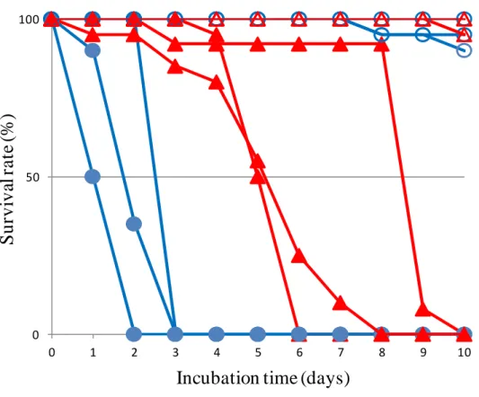
melanogaster likely have high risk of Penicillium infection throughout their developmental stages. To measure resistance of D. virilis and D. melanogaster to the fungal infection, adult flies of these species were reared on the culture medium that Penicillium fungi grew. The results showed that D. virilis adult flies survived more than two times longer than D. melanogaster flies (Figure 2), suggesting that D. virilis has a higher resistance to Penicillium infection. This higher antifungal activity without having Drosomycin motivated us to investigate the immune system of D. virilis.
In this study, to clarify the immune mechanism responsible for the higher antifungal resistance of D. virilis, larval immune response to the fungal infection between D. virilis and D.
melanogaster were compared by means of comparative transcriptome analyses. Using a Roche 454 GS Junior sequencer, I examined the transcriptome of fat body and salivary gland of 3rd-inster larvae with and without infection of a Penicillium species. Genes showing different expression pattern in response to the fungal infection between D. virilis and D. melanogaster were extracted and compared. These genes included those encoding AMPs and
‘immune-induced molecule (IM)’. Extensive differences were observed in the expression pattern of already known AMP and IM genes between D. virilis and D. melanogaster.
11
Additionally, two potential AMP genes were newly identified from function-unidentified genes.
Furthermore, three novel putative immune-related genes were identified: the products of them had a homology to an IM, Ras-like GTP binding protein Rho1 involved in many signaling pathways and Ficolin-2 binding to a cell wall component of bacteria and fungi, respectively.
12
2. MATERIALS and METHODS
2.1 Measurement of antifungal resistance
Twenty to twenty five adult flies 1-day after eclosion were reared at 25 ºC on a cornmeal-malt medium (50 g cornmeal, 50 g malt powder, 40 g dried brewer’s yeast, 50 g sucrose, 5 ml propionic acid and 5 g agar in 1 liter water) with and without Penicillium fungi. The medium containing Penicillium fungi was prepared by inoculating a small amount of spores of a Penicillium species (identified by its nucleotide sequence of 18S RNA gene) onto the cornmeal-malt medium and incubated at 20 ºC for a week or more until the surface was completely covered by the growing fungi. After the flies were transferred onto the medium with or without fungi, the number of flies alive was counted every day. To measure the resistance to the infection of the Penicillium species, the 50% lethal time (LT50) was estimated by the generalized linear method implemented in R version 2.15.2 software (R Development Core Team 2008). These processes were independently replicated three times.
2.2 Induction of gene expression by fungal-infection
A small amount of Penicillium’s spores were inoculated and cultured on a sabouraud dextrose agar (SDA) medium (10 g peptone, 40 g Dextrose and 15 g agar in 1 liter water) at 20 °C for several days until the fungi grew to cover the surface of medium. To prepare the fungus infected larvae, twenty 3rd-instar larvae of D. virilis or D. melanogaster were reared on the fungus-covered SDA medium for 12 hours at 20 °C. The induction of AMP genes is usually
13
detected in three hours after the infection and continued at least 24 hours at 25 °C (Vodovar et al., 2005, Glittenberg et al., 2011). However, I reared the larvae at 20 °C to postpone their pupation. The response to the fungal infection was confirmed by the raised expression level of the Metchnikowin gene (Mtk) (known antifungal AMP gene) measured by RT-PCR and only the induction confirmed samples were used for the transcriptome sequencing described in the next section. As the control, the naïve larvae were prepared by rearing with the same condition on fungus-free SDA medium.
2.3 Transcriptome sequencing
I analyzed transcriptome of larval fat body and salivary grand. This is because all AMPs were shown to be expressed in fat body and a major antifungal AMP, Drosomycin, was highly expressed in larval salivary gland in D. melanogaster (Tzou, De Gregorio and Lemaitre 2002).
Larval fat bodies and salivary glands dissected from twenty fungus infected or naïve 3rd-instar larvae were pooled and the total RNA was extracted from these fat bodies and salivary glands by acid-guanidium phenol-chloroform (AGPC) method (Chomczynski and Sacchi 1987). Then, mRNA was isolated by using Dynabeads mRNA purification kit (Invitrogen) according to the supplier’s instruction. The complementary DNA (cDNA) library was constructed according to the Roche GS Junior cDNA rapid library preparation protocol with a modification to keep short molecules expected for AMP genes. The double-stranded cDNA was synthesized by using cDNA synthesis system (Roche Diagnostics) with random hexamer primers. The resultant cDNA was purified by using AMPure XP kit (Agencourt) and the end-polished cDNA fragments were ligated with the FAM-labeled RL adaptor included in Lib-L GS FLX Titanium Rapid
14
Library Preparation kit (Roche Diagnostics). The adaptor-ligated cDNA was then purified by using Agencourt AMPure XP system and finally eluted in 50 μl TE buffer. The cDNA solution was then concentrated by extracting with the equal volume of 2-butanol twice and subsequently with diethyl ether to remove the residual 2-butanol. Instead of the sizing procedure described in the standard protocol, I conducted 2% agarose-gel electrophoresis, excised the gel section containing 200 bp to 1 kb DNA fragments and extracted the cDNA using High Pure PCR Clean-up kit (Roche diagnostics). The quality and quantity of the cDNA was evaluated by using QuantiFluor™-P Handheld Fluorometer (Promega) and Agilent 2100 Bioanalyzer High Sensitivity DNA kit (Agilent Technologies). The pyrosequencing was conducted by using a 454 GS junior sequencer after the emulsion PCR according to manufacturer’s instructions (Roche diagnostics).
2.4 Gene prediction for pyrosequencing reads
All the sequence reads obtained from a 454 GS Junior sequencer were filtered by the shotgun full processing of GS Run Processor application with the default setting. The filtered pyrosequencing reads of D. melanogaster and of D. virilis were queried to the complete mitochondrial genome sequence of D. melanogaster (Flybase genome database release 5.46, ftp://ftp.flybase.net/genomes/) and that of D. virilis (NCBI; gi 190710421), respectively, by using the stand-alone BLAST 2.2.25+ software (Altschul et al., 1990, Camacho et al., 2009) to remove the reads derived from mitochondrial genes. The reads that did not hit the mitochondrial genome sequence were then queried to D. melanogaster ribosomal RNA (rRNA) sequences (NCBI; gi 158246) to remove the reads from rRNA. To identify the gene, from which each read
15
derived, each read was queried against the Flybase D. virilis database release 1.2 or D.
melanogaster database release 5.46 downloaded from Flybase FTP site (ftp://ftp.flybase.net/genomes/), depending on which species it was derived from. Using the stand-alone BLAST 2.2.25+ software, I first queried against the CDS database and the reads that did not hit were subsequently queried against gene and transcript databases (Figure 3a).
Finally, the reads that did not hit any target were used for further analyses to search for novel immune -related genes as explained later in the section 2.6
For the genes identified in the D. virilis genome, most of them have different names from their orthologues in the D. melanogaster genome. In this study, however, I used the gene names of D. melanogaster for both species for the ease of comparison between species. The correspondence of gene ID between the two species was according to the 12 Drosophila genome analyses (ftp://ftp.flybase.net/genomes/12_species_analysis/clark_eisen/homology/) (Drosophila 12 Genomes Consortium 2007). For genes that have multiple IDs corresponding to multiple copies in either or both species, one-to-one correspondence of homologue between the two species was determined by TBLASTN search with the translated protein sequence of D.
virilis gene as the query against the D. melanogaster CDS database. Whether a gene is immune-related or not was determined by referring to the list of Drosophila immune-related genes (Sackton et al., 2007). (Figure 3b)
The D. virilis genes of unknown function, which did not have homologue in the D.
melanogaster genome, were further BLAST searched for their homologues in other organisms’
genomes (http://blast.ncbi.nlm.nih.gov/) (Altschul et al., 1990). In this homology search, only the genes, for which the number of reads was significantly different between fungus infected and naïve larvae, were used. For the genes that did not hit any homologue in any organism (D.
virilis-specific genes), their functions were predicted by using domain and motif search
16
programs available in NCBI Conserved Domain Database (http://www.ncbi.nlm.nih.gov/Structure/cdd/cdd.shtml) and Pfam (http://pfam.sanger.ac.uk/) (Figure 3b). When any conserved domain or motif was not predicted, the presence of signal peptide was predicted by using SignalP (v4.0) (Petersen et al., 2011) and ProP (v1.0) (Duckert, Brunak and Blom 2004) programs as a criterion to consider the possibility of antimicrobial peptide. For the candidates with putative signal peptide, the molecular weight, net charge and structural features were computed by using JEMBOSS (v1.5) program (Carver and Bleasby 2003). Finally, from the amino acid sequence of putative mature peptide after removal of the putative signal peptide, the possibility of antimicrobial peptide was examined by AMP prediction web programs, AntiBP2 (Lata, Mishra and Raghava 2009), CAMP (Thomas et al., 2010) and AMPA (Torrent, Nogués and Boix 2009).
2.5 Estimation of gene expression level
To estimate the expression level of each gene, the total number of reads to hit the gene in the BLAST search was counted (Figure 3b). To calibrate the difference in transcript length among different genes, the number of reads counted was then standardized to be the number of reads per site per million reads (RPSM) as follows.
RPSM = Number of reads / Total number of reads / Transcript length × 1,000,000
I further normalized RPSM to take the difference in total gene expression level between the samples into account and computed Trimmed Mean of M values (TMM) (Robinson and
17
Oshlack 2010), using TCC package implemented in R version 2.15.2 software (R Development Core Team 2008, Sun et al., 2013). For each gene, the TMM for the fungus infected larvae was compared to that for the control naïve larvae to quantify the extent of gene expression change in terms of the induction coefficient (IC) as follows.
IC = TMM of the infected larvae / TMM of the naïve larvae
To test a statistical significance of the induction, the difference in the number of actual reads was compared between the fungus infected and naïve larvae. In this test, ribosomal protein L32 (RpL32) and glyceraldehyde 3 phosphate dehydrogenase (GAPDH) genes were used as endogenous control genes. Although Actin was also a well-known endogenous control gene, Actin was reported to play an important role in phagocytosis against fungi in Drosophila S2 cell (Stroschein-Stevenson et al., 2006) and that the expression of an actin gene (Act42A) of D.
melanogaster 3rd-instar larvae was induced by Saccharomyces cerevisiae contained in the culture medium (Gershman et al., 2007). Indeed, the expression of D. melanogaster Act42A was not detected in the control naïve larvae but in the fungus infected larvae (the number of reads was 6 and TMM = 0.0619). Therefore, only RpL32 and GAPDH genes were used as the endogenous control genes in this study. Since the homogeneity of the numbers of reads for the two genes between the fungus infected and the naïve larvae was statistically supported (P = 0.14 in D. virilis and P = 0.51 in D. melanogaster by Fisher’s exact test, Supplementary Table 1), the total number of reads derived from the two genes was used as the number of reads for the endogenous control genes. Finally, the difference in the number of reads between the fungus infected larvae and the naïve larvae was tested on the 2x2 contingency table with the numbers for the endogenous control genes by Pearson's chi-square test or Fisher’s exact test dependent
18
on whether the minimum number of reads was five or more or not.
2.6 Prediction of new immune-related genes in D. virilis
The pyrosequencing reads which were derived from the fungus infected D. virilis but not matched any known gene were subjected to predict a new gene (Figure 3c). These pyrosequencing reads were mapped to the D. virilis genome sequence by Newbler GS reference mapper software (Roche Diagnostics) with the default parameter setting designated for CDS sequences to obtain continuous transcript sequences. Since the median length (192 bp) of the obtained contigs was similar to that (230 bp) of 3’-UTR of D. melanogaster (Sackton and Clark 2009), many contigs might not include protein coding region at all. Therefore, for each contig, the corresponding genome sequence plus 250 bp each of its upstream and downstream flanking regions were extracted to build a query sequence to search for new gene. All the query sequences obtained were subjected to BLASTX search against Swissprot protein database downloaded from the Uniprot web site (http://www.uniprot.org/downloads) with the condition of e-value <= 1E-05. For the identified putative genes, the difference in the number of reads was statistically tested between the fungus infected and the naïve larvae in the same way as that for the known genes described above and if the number of reads was significantly different, then the gene ontology was analyzed by STRAP software (v1.1.0.0) (Bhatia et al., 2009).
2.7 Pyrosequencing and data analyses of oligo-capped full length cDNA
19
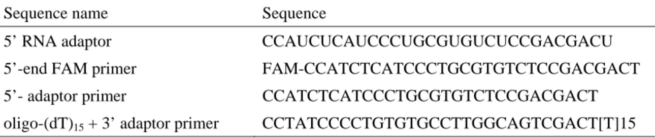
The 5’-end sequences of the new immune-related genes described in section 2.3 was determined by the BAP-TAP method (Maruyama and Sugano 1994, Suzuki et al., 1997) (Figure 4). Total RNA extraction and mRNA purification from twenty Penicillium-fungus infected 3rd-instar larvae of D. virilis were performed by the same way as described in section 2.3. The purified mRNA was treated by 2 U bacterial alkaline phosphatase (BAP) (Nippon Gene) in a 50 μl mixture containing 10 mM Tris-HCl and 0.1 mM MgSO4 at 37 °C for 1 hour. After the reaction, BAP was removed by Phenol-Chloroform purification. The BAP treated mRNA was then treated with 45 U tobacco acid pyrophosphatase (TAP) (Nippon Gene) in a 50 μl mixture containing 5 mM Sodium Acetate (pH 5.5), 0.5 mM EDTA (pH 8.0), 1 mM 2-Mercaptoethanol at 37 °C for 1 hour and then TAP was removed by Phenol-Chloroform purification The BAP-TAP treated mRNA was ligated with 400 ng 5’ RNA adaptor designed for 454 GS Junior sequencer (Table 1) by 40 U T4 RNA ligase in 50 μl mixture containing 50 mM Tris-HCl (pH 7.5), 10 mM MgCl2, 10 mM dithiothreitol (DTT), 1 mM ATP and 0.01 % bovine serum albumin (BSA) at 16 °C for 3 hours. The resultant oligo-capped mRNA was purified by Phenol-Chloroform extraction, and then treated with 2 U DNase I (Invitrogen) in 20 μl of 20 mM Tris-HCl (pH 8.4), 2 mM MgCl2, 50 mM KCl at room temperature for 15 minutes. The reaction was terminated by adding 2 μl 25 mM EDTA and incubated at 65 °C for 10 minutes.
The first-strand cDNA of the oligo-capped mRNA was synthesized in a 40 μl mixture containing the DNase I-treated oligo-capped mRNA, 50 mM Tris-HCl (pH 8.3 at room temperature), 1X First-Strand buffer (75 mM KCl, 3 mM MgCl2), 2.5 μM oligo-(dT)15 added 3’
adaptor primer (Table 1), 0.5 mM dNTP mix, 5 mM DTT and 400 U SuperScript III Reverse Transcriptase (Invitrogen). The oligo-capped mRNA, the oligo-(dT)15 + 3’ adaptor primer and the dNTP mix was firstly mixed and incubated at 65 °C for 5 minutes. Then, the primer mixed reaction was placed on ice more than 2 minutes. Finally, the first-strand buffer and the reverse
20
transcriptase were added into the primer mixed reaction and incubated at 50 °C for 50 minutes.
The reverse transcription reaction was terminated by placing at 70 °C for 15 minutes.
Double-stranded oligo-capped cDNA was synthesized by using 1.25 U Taq polymerase in a 50 μl mixture containing 1X PCR buffer (50 mM KCl, 10 mM Tris-HCl (pH 8.5), 2 mM MgCl2
and 0.001 % gelatin), 0.2 mM dNTP mix, 250 nM FAM-labeled 5’ adaptor primer and the first-strand cDNA as the template (Table 1). The reaction was conducted by incubation at 95 °C for 2 minutes followed by 20 cycles of 95 °C for 15 seconds and 68 °C for 2 minutes. Then, the 3’ adaptor primer was added into the reaction to amplify the synthesized double-stranded oligo-capped cDNA by PCR. The PCR amplification was performed with 25 cycles of 95 °C for 15 seconds, 68 °C for 2 minutes, and a final extension at 72 °C for 5 minutes. To purify the PCR products, the 3-fold volume of Binding Buffer (5 M Guanidine Thiocyanate; 100 mM Tris-HCl (pH 6.6) and 10 μl silica particles (5 μm diameter) suspended in 0.01 N HCl were added into the PCR products. The mixture was incubated at room temperature for 5 minutes and centrifuged at 12,000 rpm for 1 minute. After the supernatant was removed, the precipitated silica particles were washed twice with Wash Buffer (10 mM Tris-HCl (pH 7.5), 100 mM NaCl : Ethanol = 1 : 4). Finally, the purified PCR products (oligo-capped cDNA library) were eluted in 50 μl TE buffer. Concentrating, sizing and 454 GS Junior sequencing of the PCR products were conducted by the same way as described in section 2.3 with 5’ adaptor primer (Figure 4).
The obtained 5’-end-enrichd pyrosequencing reads were assembled by using Newbler GS De Novo Assembler software (Roche Diagnostics) with the default parameter setting designated for CDS sequences. The nucleotide sequence of each contig obtained in section 2.3 was queried against the assembled 5’-end-enriched sequences by using the stand-alone BLAST 2.2.25+
software (Camacho et al., 2009). In the obtained full length cDNA, the protein-coding region was predicted by using getorf program implemented in EMBOSS (Carver and Bleasby 2003).
21
Finally, the secondary structure of the obtained protein sequence was predicted by using Disulfind and Jpred programs (Ceroni et al., 2006, Cole, Barber and Barton 2008).
2.8 Real-time reverse transcriptase PCR (RT-PCR)
The total RNA was extracted by AGPC method (Chomczynski and Sacchi 1987) from pooled fat bodies, salivary glands and guts dissected from ten fungus infected or naïve 3rd-instar larvae.
The first-strand cDNA was synthesized from 1 μg of total RNA by the same way as described in section 2.7 except that the reaction was conducted in a half volume of mixture and 2.5 μM oligo-(dT)28 primer was used for the first-strand cDNA synthesis instead of the oligo-(dT)15 + 3’
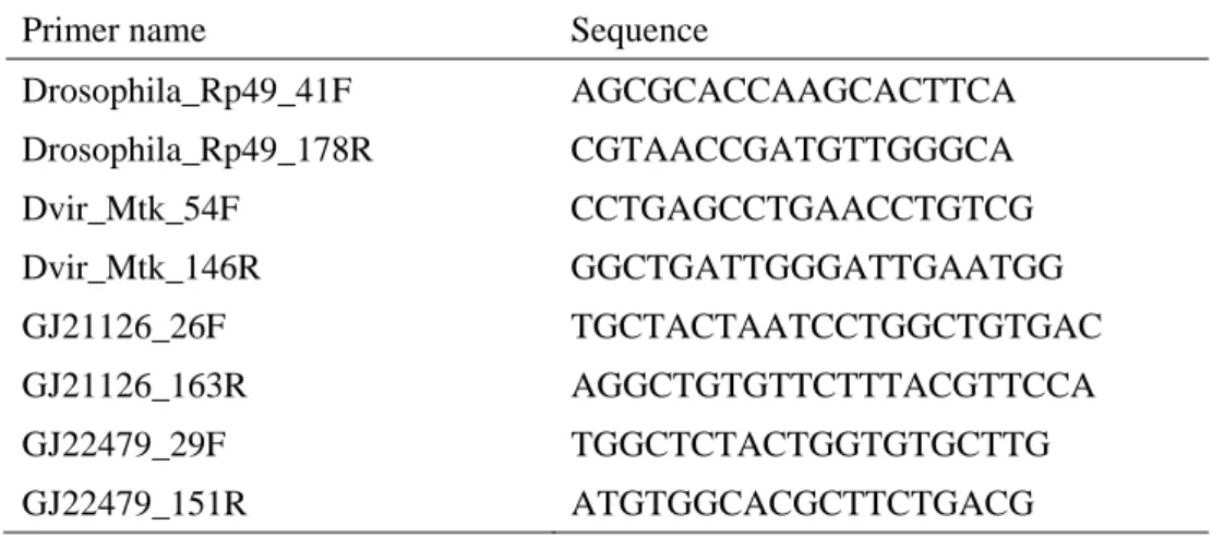
adaptor primer. The same reaction without the reverse transcriptase was conducted to verify the absence of genomic DNA. Real-time RT-PCR was conducted by using StepOne PLUS real-time PCR system (Applied Biosystems). The amplification of the PCR product was detected by SYBR Green I (Camblex Bio Science, Rockland). Primers were designed for the amplicon size to be less than 150 bp. The primers were listed in Table 2. In this analysis, RpL32 was used as the endogenous control to normalize gene expression level. An 20 μl PCR mixture was contained 1X PCR buffer (50 mM KCl, 10 mM Tris-HCl (pH 8.5), 2 mM MgCl2 and 0.001 % gelatin), 0.2 mM dNTP Mix, 250 nM gene-specific forward and reverse primers, one 20 thousandth diluted SYBR Green I, and 0.67 U Taq polymerase. The real-time RT-PCR amplification was conducted under the condition of 95 °C for 15 seconds followed by 40 cycles of 95 °C for 15 seconds, 62 °C for 20 seconds and 72 °C for 20seconds. The melting-curve analysis was then performed under the condition of 95 °C for 15 seconds, 60 °C for 1 minutes and then slow heating at 0.3 °C per second up to 95 °C. The obtained gene expression levels
22
were compared by the comparative Ct analysis method (Livak 1997) between the fungal infected and naïve samples. The gene expression level was measured in three biological repetitions with two technical replications.
23
3. RESULTS
3.1 Difference in antifungal resistance between D. virilis and D. melanogaster
To compare antifungal resistance between D. virilis and D. melanogaster, adult flies of these species were reared on a culture medium harboring Penicillium fungi and their survival time was measured. The results showed that the D. virilis flies survived more than two times longer than the D. melanogaster flies did (Figure 2); the average 50% lethal times (LT50) of D. virilis and D. melanogaster flies were 6.04 days and 1.75 days, respectively, whereas their survival time on the normal culture medium without fungi was much longer (LT50 >> 10 days). This suggests that D. virilis has a higher resistance to the infection of the Penicillium species than D.
melanogaster at the adult stage.
3.2 Summary of transcriptome analysis
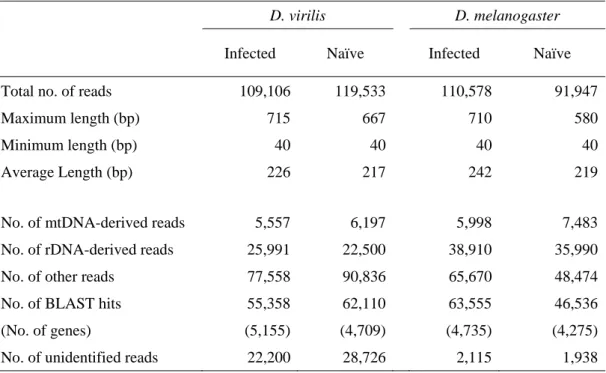
Many AMP genes encode relatively short peptides less than 100 amino acids long. Therefore, to avoid the loss of sequences derived from such short transcripts, the 454 GS junior sequencing was adjusted for cDNA library containing cDNA fragments longer than 200 bp long, whereas the standard sizing procedure selects DNA fragments of 600 - 900 bp long on average by removing those shorter than 350 bp long to be less than 10%. This resulted in 109,106 reads with the average length of 226 bp and 119,533 reads with the average length of 217 bp from the fungus infected and the naïve (uninfected) D. virilis larvae, respectively (Table 3). On the other hand, 110,578 reads with the average length of 242 bp and 91,947 reads with the average length
24
of 219 bp were obtained from the fungus infected and the naïve (uninfected) D. melanogaster larvae, respectively (Table 3).
After removing the reads derived from mitochondrial genes and rRNA genes, the total numbers of the remaining reads were 77,558 and 90,836 for the fungus infected and naïve D.
virilis larvae, respectively, and 65,670 and 48,474 for the fungus infected and naïve D.
melanogaster larvae, respectively. They were thought to be derived from mRNA transcribed from nuclear protein-coding genes. For 55,358 and 62,110 out of the 77,558 and 90,836 reads, respectively, I found BLAST hits for 5,155 and 4,709 genes, respectively, in D. virilis, whereas for 63,555 and 46,536 out of the 65,670 and 48,474 reads, respectively, I found BLAST hits for 4,735 and 4,275 genes, respectively, in D. melanogaster. It is noteworthy that the numbers of the remaining reads for D. virilis were 22,200 (fungus infected) and 28,726 (naïve), which were more than ten times as many as the corresponding 2,115 (fungus infected) and 1,938 (naive) for D. melanogaster (Table 3).
3.3 Expression pattern of immune-related genes
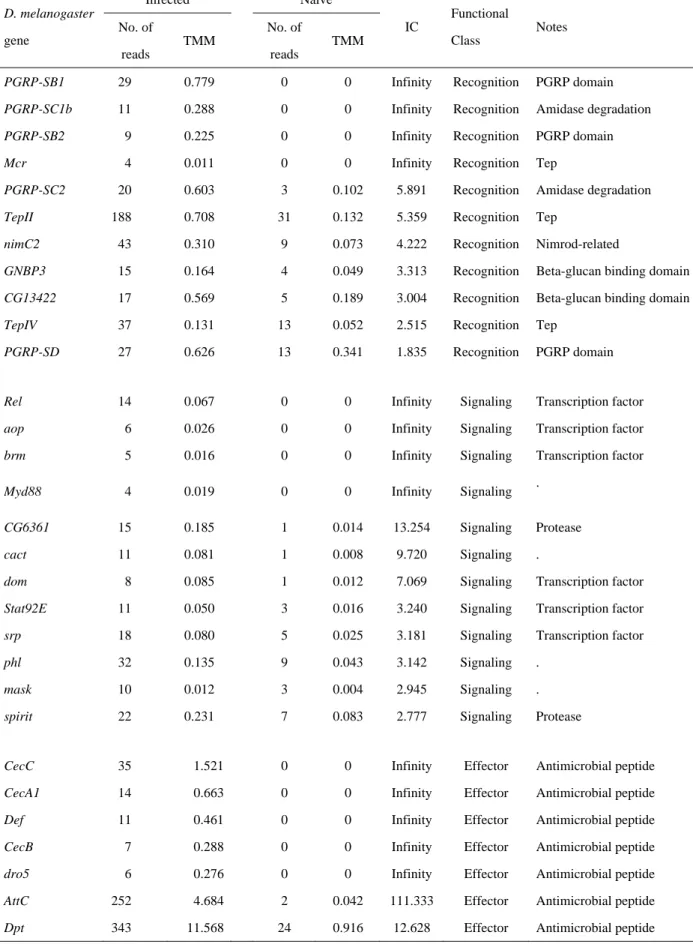
According to Sackton et al. (2007), innate immune system is categorized into three functional classes, ‘recognition’, ‘signaling’ and ‘effector.’ In the D. virilis transcriptome analysis, 128 immune-related genes were detected, in which 23, 68 and 37 were assigned to recognition, signaling and effector classes, respectively (Table 4, Supplementary Table 2 and Figure 5). In the case of the D. melanogaster transcriptome, 129 immune-related genes were detected, in which 28, 62 and 39 genes were assigned to recognition, signaling and effector classes, respectively (Table 5, Supplementary Table 3, Figure 5). Among the immune-related
25
genes, many of recognition and signaling class genes expressed in the fungus infected larvae were present in both D. virilis and D. melanogaster (Figure 5). In the recognition class genes, PGRP-SA, PGRP-LC, PGRP-LE and GNBP3 involved in Toll and Imd pathways were expressed in both species (Figure 1, Supplementary Tables 2 and 3). The expression of genes for nimrod and complement-like proteins called thioester-containing proteins (TEPs), which activate cellular immune response such as phagocytosis, were also detected in both species.
Among the TEP genes, TEPII (IC = 5.359, P = 4.68E-22) and TEPIV (IC = 2.515, P = 8.24E-05) were significantly up-regulated in D. melanogaster (Table 5, Supplementary Table 3), whereas the expressions of their homologs in D. virilis were not induced by the fungal infection (Table 4, Supplementary Table 2). I also detected the genes for negative regulators of systematic immune response, such as PGRP-SC1a, PGRP-SC2 and PGRP-LB (Mellroth, J. Karlsson and Steiner 2003, Bischoff et al., 2006, Zaidman-Remy et al., 2006, Paredes et al., 2011), as well as the genes for activators. Consistent with the expression of these recognition class genes, the expressions of signaling class genes, e.g., Myd88, Rel, STAT92E, hep, etc., involved in Toll, Imd, JNK and JAK/STAT pathways, were also detected in both species (see Tables 4 and 5, Supplementary Tables 2 and 3 for details).
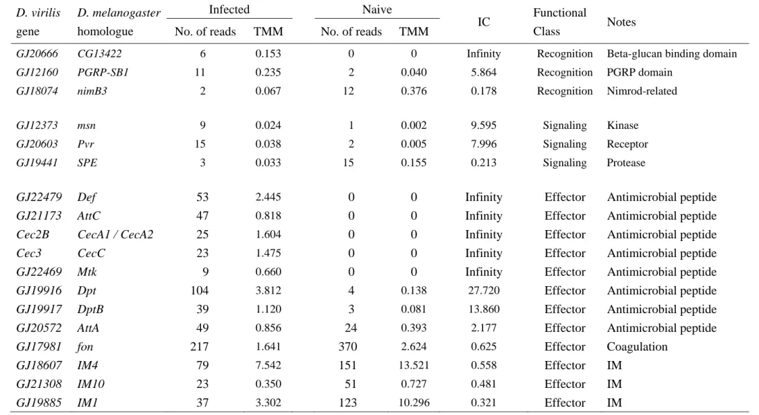
3.4 Between-species differences in the expression pattern of effecter class genes
Since the effectors directly function against infected microbes, in this study I focus on the response of the effector class genes to the Penicillium infection to elucidate the differences in the antifungal resistance between D. melanogaster and D. virilis. In contrast to the shared expression pattern between the species observed in the recognition and signaling class genes,
26
substantial differences in the expression pattern were observed in the effector class genes.
AMPs are known to be a major effector that has a critical role in the innate immune system of Drosophila (Tzou, Reichhart and Lemaitre 2002). In D. melanogaster, 20 AMP genes belonging to seven AMP gene families have been found, whereas 15 AMP genes belonging to five AMP gene families have been identified in D. virilis (Drosocin and Drosomycin in D.
melanogaster are missing in D. virilis) (Sackton et al., 2007). In both D. virilis and D.
melanogaster, many AMP genes (11 of 15 in D. virilis and 14 of 20 in D. melanogaster) were expressed in the fungus infected larvae (Tables 4 and 5, Supplementary Tables 2 and 3). In D.
virilis, genes encoding Diptericin (GJ19916, TMM = 3.812), Defensin (GJ22479, TMM = 2.445) and Cecropin (Cec2B, TMM = 1.604 and Cec3, TMM = 1.475) showed high TMM and Diptericin (GJ19916) was most highly expressed in the fungus infected larvae (Table 4). In contrast, the expression level of Metchnikowin (GJ22469), which was the only known antifungal peptide in D. virilis, was not so high (TMM = 0.660; Table 4). In contrast, Drosomycin (Drs) and Metchnikowin (Mtk), which were known as antifungal peptide genes, were most strongly expressed in the fungus infected D. melanogaster larvae (TMM = 23.817 and 23.719, respectively), followed by Diptericin (Dpt, TMM = 11.568), Attacin (AttC, TMM = 4.684) and Drosocin (Dro, TMM = 4.237) (Table 5). Among the Drosomycin gene family, only Dro5 responded to the fungal infection, suggesting that D. melanogaster uses the specific Drosomycin gene copy against the Penicillium species. However, the expression level of Dro5 was 100-fold lower than that of Drs (TMM = 0.276) (Table 5). These observations indicate substantial differences in the AMP usage between the species, i.e., against the fungal infection, Diptericin, Defensin and Cecropin were the three major AMPs in D. virilis, whereas Drosomycin and Metchnikowin were the two major AMPs in D. melanogaster (Figure 6).
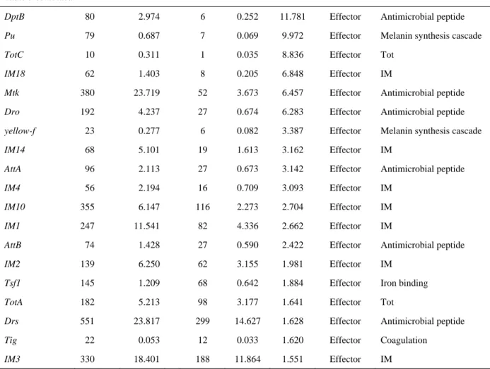
Among other effector class genes, the immune-induced molecule (IM) genes showed
27
distinct expression pattern between the species. The IM genes are known as the genes induced by bacterial or fungal infection in D. melanogaster. However, their functions mostly have not been characterized. In this study, 10 IM genes were identified to be expressed in the fungus infected D. melanogaster larvae and five of them, IM1, IM4, IM10, IM14 and IM18 were significantly up-regulated by 2-fold or more (Table 5 and Supplementary Table 3). For most of the D. melanogaster IMs, their expressions tended to be induced by the fungal infection. On the other hand, five IM genes, IM1 (GJ19885), IM4 (GJ18607), IM10 (GJ21308, GJ21309) and IM23 (GJ22454), were identified to be expressed in D. virilis, but their expression tended to be down-regulated by the fungal infection (Table 4, Supplementary Table 2). Especially, the expressions of IM1 (GJ19885), IM4 (GJ18607) and IM10 (GJ21308) were significantly reduced by the fungal infection by half or less (Table 4). These differences in the expression pattern may indicate that IMs play separate roles in the immune response to fungal infection in D.
melanogaster and D. virilis.
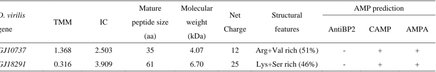
3.5 Novel AMP genes in the annotated D. virilis genes
Using the BLAST search against all the known D. melanogaster genes, I could not find the homologues for three D. virilis annotated genes significantly up-regulated by the fungal infection. They were GJ10737 (IC = 2.503, P = 0.0037), GJ11722 (IC = 3.198, P = 0.032) and GJ18291 (IC = 3.909, P = 0.047). Additional queries to orthologue database (orthoDB:
http://cegg.unige.ch/orthodb6) (Waterhouse et al., 2012) and the non-redundant gene database in the NCBI BLAST web server failed to find any known gene, suggesting that they were D.
virilis-specific genes. Although I further searched for annotated domains and motifs in the
28
expected products of these genes using the domain and motif search programs on NCBI Conserved Domain Database and Pfam, no conserved domain or motif was predicted. However, using SignalP (v4.0) (Petersen et al., 2011), ProP (v1.0) (Duckert, Brunak and Blom 2004) and JEMBOSS (v1.5) (Carver and Bleasby 2003) programs, the expected products of GJ10737 and GJ18291 were predicted to be secretory peptides having propeptide sequences and positively charged mature peptide (Table 6). These features are commonly found in AMPs. Indeed, AMP prediction web programs, CAMP (Thomas et al., 2010) and AMPA (Torrent, Nogués and Boix 2009), predicted them to be AMPs, although another program, AntiBP2 (Lata, Mishra and Raghava 2009), did not (Table 6). These results suggested the possibility that D. virilis possesses unknown AMP genes functioning in its innate immune system.
3.6 Novel immune related genes in D. virilis
In our BLAST analysis described above, 22,200 and 28,726 pyrosequencing reads respectively from the fungal infected and naïve D. virilis larvae did not hit any known gene, whereas such reads were only 2,115 (infected) and 1,938 (naive) in D. melanogaster (Table 3). I hypothesized that this is because there were many unidentified genes in D. virilis. To examine whether or not these reads were derived from unidentified immune related genes, I assembled these reads by mapping each read onto the D. virilis genome sequence to make contigs. Then, I performed a BLASTX search against Swissprot protein database using each of these contigs as the query.
Out of the 22,200 reads, 21,488 (about 97%) were mapped onto the D. virilis genome sequence to be assembled to 3,269 contigs of the average length 237 bp in total (Figure 7). This
29
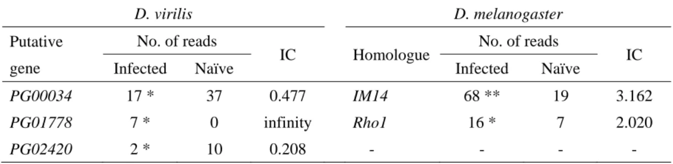
indicates that these reads were actually derived from transcripts of the D. virilis genome rather than possible contaminants and that there are unidentified transcription units potentially encoding polypeptide. Since most of the contigs were shorter than the median length of 3’-UTR of D. melanogaster genes, I extended each contig with 250 bp each of upstream and downstream genome sequences to make a query sequence subjected to the BLAST search against Swissprot protein database. As the result, I identified 620 putative genes in the 3,269 contigs. Among them, 27 putative genes showed a statistically significant difference in the number of reads between the fungus infected and naïve larvae. Three out of the 27 putative genes, PG00034, PG01778 and PG02420, were assigned to potential immune-related genes for subsequent GO analysis (Supplementary Table 4). PG00034 was homologous to IM14 of D.
melanogaster. Although the expression of IM14 was significantly up-regulated in D.
melanogaster (Tables 5 and 7), the expression of PG00034 was significantly down-regulated by the fungal infection in D. virilis. PG01778 was homologous to a Ras-like GTP-binding protein, Rho1, of D. melanogaster. This gene is known to play a role in regulating actin genes involved in phagocytosis (Hariharan et al., 1995, Magie et al., 1999, Greenberg and Grinstein 2002, Magie and Parkhurst 2005). The expression was observed only in the infected larvae in D.
virilis and induced by the fungal infection (IC = 2.020) in the D. melanogaster larvae, indicating that this gene was up-regulated by the fungal infection in both species. PG02420 was homologous to Ficolin-2 that binds to the cell wall component of bacteria and fungi (Ma et al., 2004, Endo, Matsushita and Fujita 2007), and the expression of PG02420 was significantly down-regulated in the infected D. virilis (IC = 0.208) (Table 7).
For the remaining 2,649 contigs, I did not find any homologue in Swissprot protein database. However, among the 2,649 contigs, the number of pyrosequencing reads was significantly different between the fungal infected and naïve larvae in 64 contigs and 26 of them
30
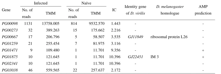
were considered to be up-regulated by the fungal infection. In order to predict the protein-coding region for these 26 contigs, I tried to determine the 5’-end sequence by the oligo-capping method adjusted for the use of 454 GS Junior sequencer. As the results, 50,573 reads with the average length of 190 bp were obtained from the infected D. virilis larvae, and these reads were assembled to construct continuous transcript sequences. Out of the 50,573 reads, 41,423 (about 82%) were assembled to 900 contigs (Table 8). Combining the 900 contigs with the assembled sequences in the section 2.3, I determined 5’-end sequence of 8 contigs. Two of them, PG00667 and PG01875, were identified to be GJ11849 and GJ22451 by NCBI BLAST analysis, respectively. GJ11849 was ribosomal protein L26 and GJ22451 was a homologue of IM3 of D. melanogaster. Interestingly, the expression of GJ22451 was up-regulated (IC = 10.396, Table 9), whereas the expressions of all other IM gene homologues were tend to be down-regulated in the fungal-infected D. virilis. In this study, it was predicted that GJ22451 encoded small secretary peptide having weak positive net charge (net charge = 0.5). In addition, GJ22451 was predicted to have two beta-sheets and to be stabilized by a disulfide-bridge between two cysteine residues (Figure 8). Since these features are found in some AMPs, whether GJ22451 could function as AMP was evaluated by using AMP prediction programs. As the result, AntiBP2 AMP prediction program predicted that GJ22451 was similar to beta-Defensin of mammals (Table 9).
Since the remaining 6 contigs did not show similarity to any known genes, I predicted the protein-coding region using getorf program implemented in EMBOSS (Carver and Bleasby 2003). As the result, it was predicted that PG01471 encoded a proline-rich and positively-charged secretory peptide, which is the features often observed in AMPs. Indeed, PG01471 was predicted to be AMP by AntiBP2 AMP prediction program (Table 9). Particularly, C-terminal region of the PG01471 was very similar to that of Metchnikowin (Figure 9). Since
31
any sequence showing homology to PG01471 was not found in other Drosophila genome sequences excepting D. mojavensis, PG01471 seemed be a lineage-specific AMP gene, which may contribute to different antifungal resistance between D. virilis and D. melanogaster (Figure 10).
3.7 Local expression of Defensin gene in response to the fungal infection
My transcriptome analysis indicated that D. virilis uses a specific Defensin in response to the fungal infection. However, while my transcriptome analysis particularly focused on systemic immune response to the fungal infection, local immune response is also important to defend from the infection (Tzou et al., 2000, Liehl et al., 2006). Therefore, I surveyed differences in expression responses of Metchnikowin and Defensin genes among fat body, salivary gland and gut of the fungal-infected D. virilis larvae by real-time RT-PCR. The analysis for fat body and gut was conducted three biological replications, whereas no replication was made for salivary gland, because the tissue is too small to extract enough amount of RNA. In the fat body, strong induction of Defensin gene (GJ22479) expression was observed in all replicates, consistent with the result of the transcriptome analysis. Contrary, the expression of Metchnikowin gene (GJ22469) was not always induced by the fungal infection (Figures 11). Additionally, expression level of GJ22479 was tended to be higher than that of Metchnikowin (GJ22469) (Figure 11). In the salivary gland, the expressions of both Metchnikowin (GJ22469) and Defensin (GJ22479) genes were induced by the fungal infection (Figure 12). The expression of another Defensin gene (GJ21126) was observed in neither the fat body nor salivary gland (Figures 11and 12). Interestingly, GJ21126 was locally expressed in the gut, and the expression
32
was tended to be up-regulated by the fungal infection, whereas the expression of GJ22479 gene was tended to be down-regulated by the fungal infection in the gut (Figure 13). Nevertheless, the expression level of GJ21126 was very low compared to that of GJ22479 even in the gut (Figure 13). These results support the hypothesis that D. virilis uses mainly one of the Defensin genes, GJ22479, against the fungal infection.
33
4. DISCUSSION
In this study, I first clarified that the antifungal resistance against Penicillium fungal infection is higher in D. virilis than in D. melanogaster. In general, adult flies of most Drosophila species are attracted to, feed and breed upon a variety of fermenting substances such as fallen fruit and flowers, slime fluxes of forest trees, decaying bark of trees, mushrooms, etc.
(Carson 1971). However, there are inter-species variations of the fermenting substances utilized by Drosophila species for feeding and breeding. For instance, D. virilis is known to feed on slime flux and decaying bark of tree harboring many yeasts and filamentous fungi, such as Xanthophyllomyces dendrorhous, Cryptococcus spp., Fusarium spp., etc. (Weber, Davoli and Anke 2006, Weber 2006), whereas D. melanogaster feeds on fermented fruits, which mainly harbor Baker’s yeast, Saccharomyces cerevisiae (Carson 1971, Throckmorton 1975, Markow and O'Grady 2007). The Penicillium species is ubiquitously and abundantly found in natural environment, where Drosophila species live, and grow on both decaying woods and fruits (Coates and Johnson 1997, Peterson, Bayer and Wicklow 2004). Therefore, both D. virilis and D. melanogaster are likely to be infected by them in nature during their life time. According to the theory of evolutionary adaptation, the higher antifungal resistance of D. virilis observed in this study (Figure 2) is expected to reflect the result of higher risk of the infection in their living environments over the evolutionary time compared to D. melanogaster. This raises the question of the immune mechanism attributed to the higher antifungal resistance of D. virilis, and it is thought to be a key factor for understanding the adaptive evolution of D. virilis to its habitat in moldy environment. To answer this question, I compared the immune response to the fungal infection between D. virilis and D. melanogaster by analyzing their transcriptome extracted from larval salivary gland and fat body. Although the antifungal resistance was compared at the
34
adult stage, I focused on the transcriptome at the larval stage. Since the larvae live and feed on fermented substances in their habitat environment and cannot escape from the surrounding microbes as the adults fly away, the larvae are consistently infected by microbes. Therefore, I assume that the resistance at the larval stage is more important for their adaptation to environment. Unfortunately, it was difficult to measure the antifungal resistance at the larval stage since the larvae became pupae within several days and some larvae avoided immediate infection of fungi by digging the medium deeply. Accordingly, my interpretation in the following is on the basis of the assumption that the resistance at the adult stage correlates with the resistance at the larval stage.
My comparative transcriptome analysis revealed that the genes involved in all major signaling pathways for immune response, i.e., Toll, Imd, JAK/STAT and JNK, were triggered by the infection of the Penicillium species in both D. virilis and D. melanogaster (Tables 4 and 5, Supplementary Tables 2 and 3). These pathways regulate humoral and cellular immune responses, such as AMP production, phagocytosis, etc. (Lemaitre and Hoffmann 2007, Agaisse and Perrimon 2004, Kallio et al., 2005). Among the signaling pathways, the Toll pathway plays an essential role against fungal infection in D. melanogaster (Lemaitre et al., 1996, Lemaitre, Reichhart and Hoffmann 1997). The Toll pathway regulates expressions of two antifungal peptides, Drosomycin and Metchnikowin (De Gregorio et al., 2002). Consistent with this fact, the expression levels of Drosomycin and Metchnikowin genes were highest in the fungus infected D. melanogaster larvae (Table 5). The response of these AMP genes to the infection of an entomopathogenic fungus, Beauvaria bassiana, was highest in adult D. melanogaster as well (De Gregorio 2001, Irving et al., 2001). Interestingly, seven genes encoding Drosomycin have been found in D. melanogaster genome (Drs, Drsl, Dro2, Dro3, Dro4, Dro5 and Dro6) (Sackton et al., 2007). Nevertheless, I found that only Drs and Dro5 were induced by the fungal infection
35
in the D. melanogaster larvae (Table 5). This specificity of the expression pattern was consistent with the result of the microarray analysis by De Gregorio et al. (2001), suggesting that the specific genes, Drs and Dro5, are used against the fungal infection at both larva and adult stages.
In contrast, any Drosomycin gene is absent in the D. virilis genome and the expression of the Metchnikowin gene (GJ22469) was not high (TMM = 0.660) compared to that of other AMP genes in the fungus infected D. virilis larvae (Table 4, Supplementary Table 2, Figure 6). This result was rather unexpected since Metchnikowin was the only known antifungal peptide in D.
virilis, suggesting that Metchnikowin of D. virilis does not compensate for the lack of Drosomycin. Since the comparison of D. melanogaster and D. virilis genomes revealed that Mtk is present as a single copy gene in both species (Sackton et al., 2007), it is implausible that D.
virilis has an additional copy of Mtk responsible for the observed higher antifungal resistance.
On the other hand, the genes encoding Diptericin (GJ19916), Defensin (GJ22479) and Cecropin (Cec2B and Cec3) were highly expressed (TMM = 3.812, TMM = 2.445, TMM = 1.604 and TMM = 1.475, respectively) in the fungus infected D. virilis larvae compared to other AMP genes (Table 4), suggesting a substantial difference in the AMP usage in response to the fungal infection between the two species and a possibility that Diptericin, Defensin and Cecropin have an antifungal function in D. virilis. The antifungal activity of Diptericin and Defensin against an ascomycete fungus, Fusarium oxysporum, has been reported, although they are not effective against other fungi (Neurospora crassa, Beauvaria bassiana and Aspergillus fumigatus) in D. melanogaster (Tzou, Reichhart and Lemaitre 2002). Comparing the Diptericin protein sequence of D. virilis to its orthologue in D. melanogaster, I found substantial amino acid differences (50-70%) (Figure 14). This may indicate the possibility that Diptericin of D.
virilis has a different activity spectrum against fungi from that of D. melanogaster, although the main activity of the latter is not antifungal but antibacterial (Wicker et al., 1990). In contrast,
36
amino acid sequences of mature peptide from Cec2B and Cec3 of D. virilis are almost identical (92.5-100%) to those of Cecropin of D. melanogaster, and the few amino acid substitutions observed are all conservative to maintain physicochemical properties of the peptide (Figure 15).
Therefore, it is likely that the functions of Cecropin are conserved in the two species. A notable difference was observed in the Defensin gene. Defensin is known to be an AMP of main specificity to gram-positive bacteria in D. melanogaster (Dimarcq et al., 1994). However, the Drosophila Defensin is classified into Defensin_2 superfamily (Pfam: PF01097), which has antifungal activity in mosquito (Anopheles gambiae) and sand fly (Phlebotomus duboscqi) (Vizioli et al., 2001, Boulanger et al., 2004). D. virilis has two Defensin genes (GJ21126 and GJ22479). The mature peptide sequence translated from GJ21126 is closely related to the D.
melanogaster Defensin gene as expected from their phylogenetic relationship of species, whereas the mature peptide sequence translated from GJ22479 is more similar to those of Anopheles gambiae (AgaDef) and Phlebotomus duboscqi (PduDef), which have antifungal activity (Figures 16 and 17). In my transcriptome analysis for fat body and salivary gland, I detected the expression of GJ22479 but not GJ21126 in response to the Penicillium infection.
This result was confirmed by real-time RT-PCR analysis. However, although the expression level was much lower than that of GJ22479, the expression of GJ21126 was detected in gut (Figure 13). This observation suggests the possibility that the functions of the two Defensin genes have been differentiated through D. virilis evolution. A possible speculation based on these observations is that Defensin functions differently as an antifungal peptide in D. virilis from that in D. melanogaster. Since the expression of these three AMPs are under the regulation of the Imd pathway rather than the Toll pathway (Imler and Hoffmann 2000, De Gregorio et al., 2002), this result suggests that the Imd pathway plays an important role in the response to the fungal infection in D. virilis, in contrast to the fact that the Toll pathway is more important to
37
regulate the Drosomycin genes as the antifungal response in D. melanogaster. Alternatively, the Diptericin, Defensin and Cecropin genes may be under the Toll pathway regulation in D. virilis.
To examine this possibility, I analyzed the upstream region of these genes to see differences in the binding sites of NF-kB-like transcription factors, DIF, Dorsal and Relish between D. virilis and D. melanogaster. In addition to these binding sites, I also compared the binding site of a GATA factor, Serpent, which regulates synergistically the expressions of AMP genes with the NF-kB-like transcription factors (Senger et al., 2004). Senger et al. (2004) discussed that the organizations of these transcription factor binding sites of AMP genes were related to whether the Toll or Imd pathway had main effect on their expression regulation. However, there was no clear difference in the number, position and direction of these binding sites, suggesting that the alternative possibility is not likely (Figures 18, 19 and 20, Appendices I and II).
A striking difference in the expression pattern was observed in the immune-induced molecule (IM) genes. The IM genes of D. melanogaster showed a similar expression pattern to that observed in the previous study conducted by De Gregorio et al. (2001). In this study, ten IM genes were expressed in the fungus infected D. melanogaster larvae and five of them, IM1, IM4, IM10, IM14 and IM18, were significantly up-regulated by 2-fold or more and down-regulated gene was not observed (Table 5, Supplementary Table 3). Similar inductions of IM genes were observed in adult flies by the infection of B. bassiana (De Gregorio 2001). This suggests that the IM genes play a similar role in antifungal immunity in larvae and adults of D. melanogaster and against Penicillium and Beauvaria fungi, although the function of the IM genes has not been characterized. However, the IM genes showed contrary expression pattern in D. virilis: the expressions of five IM genes, IM1 (GJ19885), IM4 (GJ18607), IM10 (GJ21308, GJ21309) and IM23 (GJ22454), detected in D. virilis were rather down-regulated by the fungal infection (Figure 6). Indeed, three of them, IM1 (GJ19885), IM4 (GJ18607) and IM10 (GJ21308),
38
showed statistically significant reductions (Table 4, Supplementary Table 2). This result suggests differences in the functions of IMs between D. virilis and D. melanogaster. In other words, the definition of immune-induced molecule (IM) holds true in D. melanogaster but not necessarily so in other Drosophila species. It can be speculated that D. virilis may have other immune-related genes that have the functions of IMs in D. melanogaster. Based on the comparative transcriptome analysis using bacterial-infected D. melanogaster and D. virilis flies, Sackton and Clark (2009) suggested that new components were recruited into the immune system of D. virilis. Therefore, my results as well as their observation motivated us to search for novel immune-related genes in D. virilis.
In our transcriptome analysis, I found that three D. virilis-specific genes were induced by the fungal infection and two of them, GJ10737 and GJ18291, were predicted to encode novel AMPs (Table 6). This suggests that D. virilis has acquired lineage-specific AMPs against fungal infection through its evolution. Since no orthologous sequences of these genes were found in other Drosophila genomes either, these genes seemed to be recruited to the D. virilis genome de novo. In addition to the fraction of these genes of unknown function, I also predicted new D.
virilis genes from the pyrosequencing reads that did not show any BLAST hit.
In our BLAST analyses of the pyrosequencing reads, approximately 30% of the reads from D. virilis did not hit any gene, whereas only 3-4% of the reads from D. melanogaster fell in the same situation (Table 3). This may suggest the possibility that many genes in the D. virilis genome have not been identified yet. Actually, I found 620 putative genes in 3,469 contigs and three of them, PG00034, PG01778 and PG02420, were predicted to be immune-related genes with expression level significantly changed by the fungal infection. PG00034 is homologous to IM14 and PG01778 is homologous to a Ras-like GTP-binding protein, Rho1, which regulates actin cytoskeletal organization (Hariharan et al., 1995, Magie et al., 1999) and is involved in
39
phagocytosis (Greenberg and Grinstein 2002, Magie and Parkhurst 2005) in D. melanogaster (Table 7). PG02420 is homologous to Ficolin-2 of Bos taurus. Ficolin binds to a cell wall component of bacteria and fungi and is involved in phagocytosis (Ma et al., 2004, Endo, Matsushita and Fujita 2007). Although the expression of the IM14 was significantly up-regulated by the fungal infection in the D. melanogaster larvae, the expression of PG00034 was significantly down-regulated as in the case of other homologues of IM genes in the D.
virilis larvae. Similarly, the expression of PG02420 was significantly down-regulated in the infected D. virilis larvae. On the other hand, the expression of PG01778 was significantly up-regulated by the fungal infection in D. virilis. For the remaining 2,649 contigs, I could not find any homologue in Swissprot protein database. This seems partly because many of them are too short to find a homology to a known gene, domain or motif in the homology search (Figure 7). Further experimental determination of their full length sequence is necessary for a better prediction of novel protein coding genes. From this perspective, I tried to determine the 5’-end sequence of these contigs using the oligo-capping method adjusted to 454 GS Junior sequencer.
As the result, I found two candidate genes, which encode potential AMP. One of them, GJ22451, is a homologue of IM3 of D. melanogaster. The expression of GJ22451 is exceptionally up-regulated in the fungal-infected D. virilis, whereas other IM genes tended to be down-regulated. GJ22451 is predicted to be similar to mammalian beta-Defensin by AntiBP2 prediction program (Table 9). The other candidate gene, PG01471, was predicted to encode a Metchnikowin-like proline-rich secretory peptide (Table 9, Figure 9). Although GJ22451 is present in the all 12 Drosophila species, homologue of PG01471 was not found in D.
melanogaster (Figure 10). This observation suggests that PG01471 may contribute to the higher antifungal resistance of D. virilis. Antifungal activity of PG01471 should be experimentally verified.
40
Our comparative transcriptome analysis revealed extensive differences in the immune response to the infection of Penicillium species between D. virilis and D. melanogaster at the transcriptome level. These results provide an important insight to the different role of immune system between ecologically diverged species. It is quite natural to consider that the observed differences resulted from evolutionary adaptation to their different habitat. This presumption should be further experimentally examined by the investigation of antimicrobial activities of AMPs, e.g., Diptericin and Defensin, to identify the component responsible for the higher antifungal resistance of D. virilis.