Acta med. Nagasaki. 140-155
Ultrastructural Study of Secretory Granules
in the Juxtaglomerular Cells
-Particularly on Formation and Extrusion-
Nobuo TSUDA*
Department of Pathology, Research Institute for Nuclear Medicine Nagasaki University School of Medicine,
Nagasaki, Japan
Received for publication, January 25, 1969
The secretory granules in the juxtaglomerular cells which are believed to be renin or renin-like substance are of experimental and clinical interest since they are related to hypertension and Na balance. Electron micro- scopic observations were made with rats on the mechanism of formation and extrusion of secretory granules upon giving stimulation to the juxta- glomerular cells by means of restricted salt diet and administration of diuretics. As to the formation of granules, rough-surfaced endoplasmic reticulum developed into Golgi apparatus, immature granules and finally mature granules. At least, one of the extrusion types was diacrine mecha- nism since there was observed "fading" of granule content while the limit membrane was maintained. In view of the fact that extrusion by diacrine mechanism is generally of the substance of low molecular weight, it is presumed that the content of JG cell might be prmitive renin of relatively low molecular weight or might possibly include some substance other than renin.
In 1939, GOORMAGHTIGH and GRIMSON suggested for the first time that renin is produced in the juxtaglomerular apparatus (JGA)191. Since then, many studies have been carried out concerning the relationship between the JGA and renin. One of the most demonstrative arguments in favor of GOORMAGHTIGH' S thesis was furnished by EDELMAN and HARTROFT who found that fluorescent anti-renin antibodies injected into the rabbit and dog were localized selectively in the juxtaglomerular cells (JG cells).
At present, the juxtaglomerular cell granule (JG cell granule) is generally believed to be renin-like substance"',2119281,43' Since the JG cell granules are formed and discharged in the arteriolar wall'", and can be identified by light microscopy by means of special staining," ,411 and electron microscopy",") their relation with hypertension 0,121,341 35,45' and Na balance 121 is of morphological interest.
*津 田 暢 夫
This paper deals with electron microscopic observations on the mechanism of formation and extrusion of JG cell granules in rats upon restriction of salt intake and administration of diuretics as the form- ation and extrusion of JG cell granules are slow in normal untreated animals.
MATERIALS AND METHODS
A) Animals
A total of nineteen female Wistar rats, each weighing 170 to 280 gm., were used for the present experiment.
The rats were classified into the following three groups:
I) Control group
Six rats were allowed free access to food and tap water till the time of sacrifice.
II) Group treated with acetazolamide (Diamox)
Five rats were given daily stomach-tubings of 60 mg. of acetazol- amide per 100 gm. of animal weight for seven days, and were main- tained on relatively low salt rat pellets (Orientaru K~bo Kc gyo K . K . )
and distilled water. They were sacrificed the day after the last tubing.
III) Group treated with furosemide (Lasix)
Eight rats were given daily intraperitoneal or intravenous injection
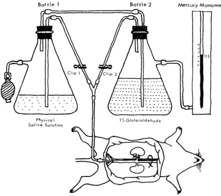
Fig. 1 Diagram of apparatus for pefusion fixation
of 0.1 mg. of furosemide per 100 gm. of rat weight for seven days, and were maintained on relatively low salt intake as group II. They
were sacrificed the day after the last injection.
B) Perfusion fixation"' (Fig. 1)
All animals were anesthetized with ether or nembutal. Blood sample for electrolyte evaluation was collected by a 5 ml cylinder from the
inferior abdominal aorta. The cylinder was exchanged for fixative tube,
and the main right renal vessels were clipped devoid of perfusion of
physical saline solution and glutaraldehyde. The left renal vein was
incised and the abdominal aorta was ligated at its superior portion
while physical saline solution was perfused for approximately 30 sec-
onds. Then, 1% buffered, isotonic glutaraldehyde fixative was perfused
for about 20 minutes. During this period, the clipped right kidney was removed. In the perfusion of both physical saline solution and glutar-
aldehyde, two bottles were maintained at the pressure of about 120 mm.
of mercury using a rubber bulb.
C) Preparation of sections for light microscopy and electron microscopy
For light microscopy, the tissue of the right kidney was fixed in Helly's fluid, embedded in paraffin, and sections were stained with
hematoxylin and eosin stain, and with Bowie's stain.
Material of the left kidney for electron microscopy was fixed in buffered 1.2% osmic acid, dehydrated in a graded series of acetones,
and embedded in Epon 812.
Blocks were cut on a Porter-Blum ultramicrotome MT-2. Sections
were stained on the grids with uranyl acetate and lead citrate. They were photographed in JEM 6C electron microscope.
D) Electrolyte evaluation
Serum sodium and potassium were evaluated with Hitachi's flame photometer FPF-2A.
RESULTS
The values of serum sodium, serum potassium and juxtaglomerular
index (JGI)24) by the group were as shown in Table I.
Table I Serum Electrolyte and JGla
No. of Rats I Serem Nad mEq/ l I Sarum Kd mEq/ 1 JGld
I Control 6 139.0_-2.02 4.35 r0.13 31.0±1.3
11 RLSDb+DWc +Acetazolamide 5 141.5+0.93 4.04--0.11 4'0.01--7.1
III RLSD+DW +Furosemide 8 138.61-0.76 3.29 32.71--1.7
a Juxtaglomerular Index
b Relatively low salt diet (Orientaru Kobo Kogyo K.K.) c Distilled water
d Mean ± standard error
Serum Na and JGI showed no statistical difference in value among the three groups (P>0.05) while the value of serum K was lower in Group III than in the other two (P<0.001, a = 0.05).
The vascular lumen and renal tubular lumen of non-perfusion fix- ation samples were collapsed but those of perfusion fixation samples remained open though the interstitium was narrowed (Fig. 2, 3).
Granular precipitates were observed in the area from the Bowman's capsule to the renal tubule in non-perfusion fixation samples of Group II but they were washed away and were not observed in perfusion fixation samples (Fig. 2, 3).
In Group II and III, particularly in the latter, cloudy granular swellwing was observed in the epithelial cells of the renal tubule (Fig. 4).
In electron microscopy, JG cell granules were located in the epi- thelioid cells of the renal arteriolar wall. The epithelioid cells showed the character of smooth muscle cells with the myofibril and dense attachment (Fig. 5, 6).
The JG cells were generally remarkable in the development of Golgi apparatus and demonstrated in the Golgi sac some materials of ap- proximately identical electrcn density to JG cell granule (Fig. 7).
Mature granules being round or oval in shape were surrounded by a layer of limit membrane, and the content of granule manifested crystalline structure of ""dot pattern" or "band pattern 113) (Fig. 5 to 12).
The crystalline structure showed periodicity in one direction (Fig. 8, 9) or in two directions (Fig. 10) according to the direction of cut surface of granule. Some mature granules 'lacked part of the limit membrane"' (Fig. 6) or had double membrane 331 (Fig. 10).
In Group II, small granules (daughter granules) were seen on the surface of mature granule. Electron density was low inside the daugh- ter granules but contrarily high at the periphery (Fig. 9, 11). It is unknown whether the daughter granules have any relation to the formation and extrusion of granules.
Some masses of the size of mature granule or smaller lacking the limit membrane and of the same electron density as the JG cell granule were observed in the vicinity of mature granules (Fig. 6, 8, 9, 11, 12 Ms). Some of those masses demonstrated cystalline structure (Fig. 9 MS I).
In Group II and III as well as in normal rats (Fig. 5), several mature granules were fused into a giant granule (Fig. 9, 11)12)34' The electron density of granule content decreased at the periphery of granule while the limit membrane was maintained (peripheral fading) (Fig. 10) and the decrease of electron density extended diffusely to the entier granule (diffuse fading) (Fig. 11, 12).
DISCUSSION
There are several methods for the grading of JGA granulation such as juxtaglomerular index (JGA) by HARTROFT24), index of hyperplasia and hypertrophy by RAPP39), juxtaglomerular cell count (JGCC) by TURGEON and SOMMERS3)45), a modification of JGCC by CELORIA and PUCHEII), and OKAMOTO's index38) . The JGI was adopted in this paper. Howeve, some investigators insist that JGI does not reflect increase in number of cells and the presence of those evidentally bright and hypertrophied cells without large granules which are frequently observed in abnormal kidneys45). In the JGA of sodium-depleted animals, JGI generally in- creasesll),12),24>,43) In this experiment, relatively low salt diet, distilled water and diuretics were used but sufficient hyponatremia could not be effected in rats. Accordingly, increase of JGI was not noted. How- ever, since the JGA was stimulated by low salt diet and diuretics, the stimulation to the JGA might have been indicated by other grading methods though it failed to appear as an increase of the JGI.
Recently, CELORIA and PUCHE stated that no correlation exists be- tween the JGI and plasma Na concentration but that a significant nega- tive correlation is found between the JGI and logarithm of the urinary Na excretion, and paid attention to the urinary Na excretion rather than plasma Nall)
Perfusion fixation by glutaraldehyde provides good specimens for electron microscopy37'. However, it is inappropriate for observation of precipitates since precipitates in the urinary tubules are washed out by perfusion. In electron microscopy, the JG cell shows myofibril and dense attachment and transition from the smooth muscle ce11331,42) JG cell granules are generally present in the afferent arteriole but oc- casionally in the efferent arterioles'), lacis cells33), or mesangial cells 15), all in the vicinity of the glomerular hilus. It is noteworthy that pre- sence of JG cells in the arteriole away from the glomerular hilus was reported by SHIMAMURA41).
BARAJAS considers that the crystalline pattern (or band pattern as named by BIAVA) exists as Golgi apparatus or protogranule which has immature character or is on the way of formation2' .
Immature granules are present near the Golgi apparatus developed in the JG cell. Like other protein secreta, the JG cell granules may be formed from rough-surfaced endoplasmic reticulum through the Golgi apparatus") 30),31) 34) Thus, it may well be said that granule formation is made in the space surrounded by the membrane.
CHANDRA et al stated that JG cell granules are formed directly from the reticulum according to their understanding that the endoplasmic reticulum and JG cell granules are fused"'. However, the granules which are said to be fused are mature and not in the course of maturation.
The electron dense mass indicated by Ms in the figure is not a
granule but a mass of secretory substance since the limit membrane is not present311. For the same reason, the mass is not being formed nor can be considered as mature granule but properly considered to be in the course of extrusion. This is equivalent to KUROSUMI's Excrusion type V (diacrine mechanism) subtype 131) . Discharge of such masses from the cytoplasm of JG cells into the intercellular space has been observed also by LEE et a134). Some of the electron dense masses pre- sumably in the course of extrusion indicate crystalline pattern. If the crystalline pattern is of immature nature as stated by BARAJAS, it would signify that immature masses at the stage of formation are also ex- truded.
Another possible type of extrusion is that the granule extrudes its content after having undergone peripheral fading and then diffuse fading while maintaining the limit membrane. This is equivalent to KuROZUMI's Extrusion type V subtype 2. A schemic diagram of for- mation and extrusion of JG cell granules in consideration of the theories of the above-mentioned investigators is shown in Fig. 13.
In view of the fact that the mechanism of extrusion type V is of the materials of relatively low molecular weight32 and that the molecu-
lar weight of renin is currently said to be 43, 000, the content of JG cell granule might be primitive renin of relatively low molecular weight or might possibly include some substance other than renin.
SUMMARY
1) The rats treated with relatively low salt diet and diuretics showed no difference from the control group in the value of serum Na and JGI but a decrease in the value of serum K.
2) The 1% glutaraldehyde perfusion fixation employed in this study proved to be outstanding for electron microscopy but unsuitable for observation of materials in the renal tubule as it washes out precipi-
tates therein.
3) The JG cells showed the character of smooth muscle cells with the myofibril and dense attachment, and were remarkable in the development of Golgi apparatus.
4) Mature granules were surrounded by the limit membrane and the content showed dot pattern or band pattern. Some granules lacked part of the limit membrane or had double membrane.
5) In the group treated with acetazolamide, daughter granules were observed.
6) Secretory masses were observed in the cytoplasm, which are con- sidered to be in the course of extrusion. Some of the masses showed crystalline structure.
7) One of the types of extrusion of granule content in the cytoplasm was diacrine mechanism in which the electron density decreased at
the periphery of granule while the limit membrane was maintained (peripheral fading) and further decreased diffusely in the entire granule (diffuse fading).
ACKNOWLEDGEMENT
The author wishes to express his cordial gratitude to his director, Professor Issei NISHIMORI, who gave kind guidance and review during this study. Gratitude is also due to Assistant Professor Shigeo TAKEBAYASHI of the Second Department of Pathology for the technical instruction and suggestions in electron microscopy.
REFERENCES
1) BARAJAS, L. and LATTA, H.: Lab invest 12: 257, 1963.
2) BARAJAS, L.: J Ultrastruct Res 15: 400, 1966.
3) BIAVA, C. and WEST, M.: Amer J Path 47: 287, 1965.
4) BIAVA, C. and WEST, M.: Amer J Path 49: 679, 1966.
5) BIAVA, C. and WEST, M.: Amer J Path 49: 955, 8966.
6) BIAVA, C. and WEST, M.: Lab Invest 15: 1902, 1966.
7) BOHLE, A.: Verk Deutsch Ges Path 43: 219, 1959.
8) BOUGHTON, R.M. and SOMMERS, S.C.: 1 Urol 89: 133, 1963.
9) BowiE, D. J.: Quart J Exp Physiol 25: 247, 1935.
10) CAJAL, S.R.: Trabajos Lab Invest Viol (Madrid) 12: 17, 1914.
11) CELORIA, G.C. and PUCHE, R.: Acta Physiol Lat Amer 17: 6, 1967.
12) CHANDRA, S., HUBBARD, J.C., SKELTON, F.R., BERNADIS, L.L., and KAMURA,
S.: Lab Invest 14: 1834, 1965.
13) DESPREZ, J.: Amer J Clin Path 18: 953, 1948.
14) DUNIHUE, F.W.: Amer J Path 23: 906, 1947.
15) DUNIHUE, F. W . and BOLDOSSER, W . G.: Lab Invest 12: 1228, 1963.
16) EDELMAN, R. and HARTROFT, P.M.: Circ Res 9: 1609, 1961.
17) FISHER, E.R. and KLEIN, H.Z.: Proc Soc Exp Viol Med 113: 37, 1963.
18) FISHER, E . R . , PEREZ-STABL, E. and PARDO, V.: Lab Invest 15: 1409, 1966.
19) GOORMAGHTIGH, N. and GRIMSON, K.S.: Proc Soc Exp Viol Med 42: 227, 1936.
20) GOORMAGHTIGH, N.: Proc Soc Exp Viol Med 42: 688, 1939.
21) GOORMAGHTIGH, N.: Rev Belge Sci Med 16: 65, 1945.
22) GRIFFITH, L.D., BULGER, R.E. and TRUMP, B.F.: Lab. Invest 16: 220, 1967.
23) GuILLAN, R.A., ZELMAN, S. and MURPHY, F.J.: Arch Path 84: 598, 1967.
24) HARTROFT, P.M., HARTROFT, W.S.: J Exp Med 97: 415. 1953.
25) HARTROFT, P.M.: J Exp Med 105 (Suppl. 6) : 501, 1957.
26) HARTROFT, W . S . and HARTROFT, P.M.: Fed Proc 20: 845, 1961.
27) HARTROFT, P.M.: Circ Res 12: 525, 1963.
28) H.ARTROFT, P.M., SUTHERLAND, L . E . and HA RTROFT , W . S .: Canad Med A: s j 90: 163, 1964.
29) KAUFMAN, W.: Amer J Path 17: 620, 1941.
30) KIRKMAN, H. and SEVERINGHAUS, A.E.: Anat Rec 70: 413, 1938.
31) KUROSUMI, K., YAMAGISHI, M. and SEKINE, M.: Z Zelljorsch 55: 297, 1961 32) KUROSUMI, K.: J Electron Micr 14: 12, 1965 (Japanese).
33) LATTA, H. and MAUNSBACH, A.B.: J Ultrastruct Res 6: 547, 1962.
34) LEE, J.C. , HURLEY, S. and HOPPER, J.: Lab Invest 15: 1459, 1966.
35) NISHIMORI, I.: Jap J Nephrol 7: 2, 1965.
36) NISHIMORI, I., TSUDA, N. and TAKESHITA, K.: Saisin Igaku 23: 1812, 1968 (Japanese).
37) MAUNSBACH, A.B.: J Ultrastruct Res 15: 242, 1966.
38) OKAMOTO, K.: Folia Endocr Jap 38: 782, 1962 (Japanese).
39) RAPP, J.P.: Amer J Physiol 206: 93, 1964.
40) SHIMAMURA, T. and SHAW, E . H .: S Dakota J Med 21: 33, 1968.
41) SHIMAMURA, T.: S Dakota J Med 21: 49, 1968.
42) TAKESHITA, K.: Arch pistol Jap 29: 237, 1968.
43) TOBIAN, L., THOMPSON, J., TWEDT, R. and JANECEK, J.: J Clin Invest 37: 660, 1958.
44) TOBIAN, L., JANECEK, J. and TOMBOULIAN, A.: Proc Soc Exp Riol Med 100:
94, 1959.
45) TURGEON, C. and SOMMERS, S.C.: Amer J Path 38: 227, 1961.
46) WILSON, W.: Anal Rec 112: 497, 1952.
EXPLANATION OF PHOTOGRAPHS Fig. 2 Rat treated with acetazolamide
A granule is noted on the afferent arteriolar wall (AA) at the glomerular hilus.
Distal convoluted tubules approach the afferent arteriole and become macula densa (MD). The renal tuft contains erythrocytes, and the Bowman's capsule and renal tubule contain precipitates.
Helly fixation, Paraffin embedding, Bowie stain Fig. 3 Rat treated with acetazolamide
In specimen processed by glutaraldehyde perfusion fixation, the renal tuft, Bow- man/s capsule and renal tubule are open, and the interstitium is narrowed. No ery- throcyte is present in the renal tuft and no precipitate in the renal tubule.
Glutaraldehyde perfusion fixation, Paraffin embedding, H & E stain Fig. 4 Rat treated with furosemide
The epithelial cells of collecting tube show cloudy swelling.
Helly fixation, H & E stain
Fig. 5 Normal rat
JG cells of the afferent arteriole. Developed Golgi apparatus, and myofibril and dense attachment are seen. Secretory granules are round or oval in shape. Fading granules are seen in normal rat as well (JGC1).
AL: arteriolar lumen MD: macula densa x6,000 Fig. 6 Rat treated with acetazolamide
Golgi apparatus (Ga) is noted in the JG cell. In the dilated Golgi sac, some substance with the same electron density as the content of JG cell granule is present.
E: endothelial cell x5,200
Fig. 7 Rat treated with acetazolamide
JG cell has myofibril (Fs) and dense attachment (Da). The content of mature granules shows dot pattern. Some granules (Gp) lack part of the limit membrane.
Secretory masses (Ms) totally lacking the limit membrane are also observed. x29,000 Fig. 8 Rat treated with acetazolamide
JG cell granule shows crystalline pattern (arrow). A secretory mass (Ms) with lower electron density than JG cell granule without the limit membrane is seen.
x 50, 0'00
Fig. 9 Rat treated with acetazolamide
Two granules are fused into one large granule (GI). Indicated by large arrow at the bottom is a small granule or daughter granule protruding on the surface of a granule. Several secretory masses (Ms) are present among mature granules and some of them have crystalline pattern (Ms small arrow). Dilatation of the endoplasmic reticulum is remarkable. x32,000
Fig. 10 Rat treated with acetazolamide
G1 shows crystalline pattern in two directions. G2 is low in electron density at the periphery showing peripheral fading. The limit membrane of G2 is partially double layered. x58,000
Fig. 11 Rat treated with acetazolamide
G1 is a large fused granule. In G2, the electron density of the content is decreased while the limit membrane is maintained (diffuse fading). The arrow indicates a daugh- ter granule.
Ms: secretory mass E : endothelial cell x32,000 Fig. 12 Rat trated with furosemide
Present at the central part of the photograph are granules at the stage of diffuse fading showing only the limit membrne. Secretory masses (Ms) are also noted. x
27,'000
ULTRASTRUCTURAL STUDY OF SECRETORY GRANULES
N. TSUDA
ULTRASTRUCTURAL STUDY OF SECRETORY GRANULES
N. TSUDA
ULTRASTRUCTURAL STUDY OF SECRETORY GRANULES
N. TSUDA
Fig. 13 Schematic Representation of Fomation and Extrusion of JG Cell Granules