Acta Med. Nagasaki 48: 107-116
Morphological Changes in Transplanted Solid Ehrlich Tumor of Mice after Transfer of Stimulated Spleen Cells
Ivan RAICHEV, Raina BORISSOVA
Department of Pathology, Pleven University School of Medicine, Pleven 5800, Bulgaria
Spleen cells from mice bearing solid Ehrlich tumor stimu- lated in vitro with Ehrlich-tumor extract or with PHA, IL-2 and tumor extract were transferred i.v. in mice-bearers of small transplanted Ehrlich tumor (Group I) and in tumor- free mice, followed by tumor transplantation (Group II).
Morphological findings for antitumor effect were found in tumors of 42.5% of experimental mice. Four criteria were established in this respect, including evidence for lymphocytic cytotoxity. Production of a factor inhibiting Ehrlich ascites-cell migration was found in a migration- inhibition assay from the stimulated spleen cells and spleen cells obtained from mice of Group I and II. The indirect data suggested that the transferred stimulated spleen cells (lymphocytes) propagated in lymphoid organs, particularly in the spleen of mice. Then T-cells, especially tumor-specific cytotoxic lymphocytes, migrated by circulation into the pe- riphery of the tumor nodule and influenced on tumor cells.
ACTA MEDICA NAGASAKIENSIA 48: 107-116, 2003
Key Words: solid Ehrlich tumor, mitotic index, apoptotic bod- ies, lymphocytic cytotoxicity, inhibitory factor
proved to be an appropriate model for investigation of the effect of adoptively transferred sensitized spleen cells. It is important for immunotherapy against the neoplasm to detect morphological findings connected with antitumor effect of transferred spleen cells stimu- lated with immunogenic Ehrlich-tumor extract.
The purpose of this study was to determine the morphological evidence suggesting for antitumor ef- fect against transplanted solid Ehrlich tumors in mice
in which spleen cells, obtained from Ehrlich-tumor bearing mice and stimulated with Ehrlich extract alone, or in combination with PHA and IL-2 were transferred.
Material and Methods Animals
inbred male %'_157B1_/6 mice, 10-12 weeks old, were obtained from the production colonies of the Bulgarian Academy of Science and were used in ex- periments according to the Institutional Guidelines of Pleven University School of Medicine.
Introduction
It has been shown that Ehrlich tumor can grow in almost any mouse strain in an ascitic or solid tumor, across histocompatibility barriers"", as it lacks H-2 antigens". On the other hand, Ehrlich cells possess an- tigens recognized as foreign in mice, which provoke an immune response after s.c. but not after i.p. inocu- lation of the living tumor cells". Subiza et al.2 ' re- ported that a nonspecific suppressor cell population (splenic natural suppressor cells) had been developed in the spleen of mice bearing solid Ehrlich tumor.
The transplanted solid Ehrlich tumor in mice
Address Correspondence: Ivan Todorov Raichev, M.D.
D.Sc. Liulin, 605 Street, bl.603, apt. 35, 1336 Sofia, Bulgaria.
TEL: 359 2 27 47 98 E-mail: [email protected]
Tumors and tumor-bearing mice
Solid Ehrlich tumor was established by s.c. inocula- tion of Ehrlich ascites tumor cells (2 x 10⁷) in the back of mice, and was maintained by serial s.c. transplantation of small pieces of tumor in mice. Animals carrying solid transplanted Ehrlich tumor of the 3rd to 8th pas- sage generations were used. Ehrlich ascites tumor was maintained by serial i.p. transplantation (1 x 10' cells) every 10 days in intact tumor-free mice.
Spleen cell suspensions
The spleen cell suspensions were prepared in RPMI 1640 (GIBCO) supplemented with 5% FCS, 2 mM L- glutamine, penicillin (100 U/ml) and streptomycin (100 ug/ml). The aseptically removed spleens from mice-bearers of growing tumors were pressed between
Ivan Raichev et al : Morphological Changes in Transplanted Solid Ehrlich Tumor of Mice after Transfer of Stimulated Spleen Cells
two sterile slides, and, after filtration, the spleen cells were freed of erythrocytes by treatment with Tris- buffered 0.83% ammonium chloride (pH 7.2). The sus- pensions were incubated in plastic flasks at 37°C for 35 min., and then were adjusted to a final concentra- tion of 4 x 10 6 cells/ml. A T-lymphocytes- rich fraction was obtained by passing spleen cell suspensions through a nylon wool column.
Preparation of an Ehrlich-tumor extract and stimulation of the spleen cells with it
Tumor extract was obtained by using an extraction procedure as it was described previously','). Briefly, ten milliliters of cold 3 M KCL in PBS (pH 7.2) was added to every 1 g of tissue of solid Ehrlich tumor for 16 hr. The mixture was centrifuged at 40,000 x g for 1 hr at 4°C. Further centrifugation of the mixture at 18,000 x g was also done. The supernatants were dia- lyzed against PBS, and the protein content was ad- justed to 0.30-0.40 mg protein/ml. Samples of 0.4 ml of spleen cell suspension, each containing 8 x 10 5 cells (2 x 10'/ml) were mixed with 80p 1 of Ehrlich extract and incubated for 24 hr at 37V in a 5% C02 atmos- phere. The mixture in approximately 1/10 of the total number of samples were centrifuged at 18,000 x g for 20 min at 4°C. The supernatants were decanted and then prepared for examination of migration inhibition activity as was described previously". The remaining samples were centrifuged at 400 x g for 1 0 min to preserve viability of the cells. The supernatants were decanted, passed through 0.45 fn m filters and investi- gated for migration inhibition, too. The spleen cells were harvested, washed and suspended in HBSS.
Complex stimulation of spleen cells was carried out in 24 flat-bottom well plates (Linbro, Flow Laboratories).
Spleen cell suspensions containing 8 x 10 5 cells in 0.4 ml of each well were supplemented with PHA (1%
v/v); 0.5 ml of the medium was added and incubated for 48 hr at 37 °C in a humid atmosphere of 5% CO2.
After the incubation with PHA a supernatant contain- ing T-cell growth factor (IL-2), 50% v/v, was added, approximately 10 U IL-2 in each well with 8 x 10' cells. This supernatant was obtained from spleen cells of exbreeder mice (1 x 10 6 cells/ml) stimulated with PHA (2,ug/ml) for 48 hr, and then depleted of PHA using chicken erythrocytes". After 4 days of culture the procedure was continued by adding the extract of solid Ehrlich tumor of 80,u1 to wells for 24 hr. The su- pernatants were decanted and after filtration were as- sayed for migration inhibition. The cells were har- vested, washed in HBSS and prepared for inoculation in mice.
Assessment of migration inhibition
Samples of 0.2 ml of Ehrlich ascites tumor cells (2 x 10' /ml), suspended in RPMI 1640 supplemented with 5% fetal calf serum and antibiotics were mixed with 0.1 ml of the supernatants of spleen cells stimulated as was indicated above, or not stimulated. The mix- tures were incubated for 2 hr at 37V in an atmos- phere of 5% CO2. Capillary tubes sealed at one end were filled with Ehrlich cells, and centrifuged at 250 x g. The portions with sedimented cells were cut off, placed in chambers filled with medium and incubated at 37°C for 24 hr. The areas of Ehrlich-cell migration were projected and measured by planimetry. The mi- gration index (MgI) was calculated according -to the following formula:
Average area of migration of Ehrlich cells incubated --with--the--supernatant
Average area of migration of Ehrlich cells incubated without the supernatant
Fraction of responding (positive) cultures (FrRCs) was calculated as number of responding cultures per total number of cultures. Cultures were designated as responding (positive) when the supernatants obtained from them show an inhibition effect (IE) against Ehrlich-cell migration (MgI<0.80). For example, FrRCs is 0.8 (8/10) in the presence of 8 responding (positive) cultures).
Inoculation of spleen cells in mice and formation of experimental groups
The stimulated spleen cells from tumor-bearing mice were concentrated in HBSS at 20-25 x 10' cells/ml.
Seventy animals were divided in three groups. In Group I the stimulated spleen cells were transferred by one or two i.v. injections through the tail vein in mice, bearing a small Ehrlich tumor, and in Group II in tumor-free mice in which the transplantation of tumor was accomplished 7-8 days after the inocula- tion. Twenty mice were in each group. According the preliminary experiments, the mice of Group I and II were treated predominantly with 35-40 x 10' stimu- lated spleen cells; ten mice-with 50-55 x 10' cells.
Previously, doses of 20-25 x 10' cells were used, too.
The mice of Group I were sacrificed by cervical dislo- cation after a mean term of 22.40±3.09 days of stimu- lated spleen cell transfer (34.86±4.13 days of tumor transplantation), and these of Group II- 34.45 ± 4.62 days after spleen cell transfer (27.85 ± 4.15 days of transplantation). Additionally, ten tumor-bearing mice treated with 35-40 x 106 stimulated spleen cells were killed within three to six days after the cell transfer.
Ivan Raichev et al : Morphological Changes in Transplanted Solid Ehrlich Tumor of Mice after Transfer of Stimulated Spleen Cells
As controls (Group III) were used 20 tumor-bearing mice untreated by transfer of stimulated spleen cells.
Histological examination
Small tissue slices were fixed in 10% neutral formalin (pH 7.0) for 24 hr and embedded in paraffin.
Deparaffinized sections 4 urn thick were stained by using the following procedures: hematoxylin and eosin (H&E), v. Gieson, Feulgen for DNA, methylgreen pyronin for RNA, PAS (Hotchkiss). The mitotic count was performed on H&E stained sections in the most mitotically active areas". High-power fields, limited by restrictive screen at 200 um x 200 um, were investi- gated by a Carl Zeiss microscope (Germany). Mitotic index (MI) was calculated as the number of mitoses per number of counted tumor cells x 100. The apoptosis, which is a mode of cell death, was estab- lished by the apoptotic cells and bodies. They were counted, in tumor slices stained with H&E and Feulgen. High-power fields, limited as is shown above, were randomly selected. The apoptotic index (AI) was assessed as a number of apoptotic cells and bodies per number of counted tumor cells x 100. Two thousand tumor cells were counted for determination the mi- totic and the apoptotic index.
The reactive changes in the T- and B-cell regions of spleens and lymph nodes of the mice were graded in three degrees (I-III) according to their size, cell activity and cell transformation.
Staining of cells in the smears and electron microscopy Smears from sediments were stained by the Giemsa method and by methylgreen pyronin for RNA. For electron microscopic examination pieces of tumor were fixed with 1.6% glutaraldehyde in 0.1 M phos- phate buffered saline (pH 7.4) at 4°C for 2 hr and with 2.5% for 1 hr, postfixed in 1 % osmium tetroxide, dehy- drated with ethanol and embedded in Durcupan (Fluka AG). Ultrathin sections were stained with uranyl acetate and lead citrate, and were investigated with an electron microscope Tesla B-500.
Statistics
Statistical analysis was performed by using the Student's t test, chi-square test and by determing of the correlation coefficient. The significance of differ- ence between the compared groups was defined with the Mann-Whitney U test. P<0.05 was considered to be significant for the two-tailed test.
Results
Morphological changes in the transplanted solid Ehrlich tumors suggestive for antitumor effect
In mice bearers of solid Ehrlich tumor treated with stimulated spleen cells we described morphological changes related to the suppression of growth and ex- tension of tumor, death of tumor cells (apoptosis and necrosis) and tumor regression. On the basis of these morphological changes we described four criteria for antitumor effect (a.e.) against the transplanted tumor of mice treated with stimulated spleen cells as follows:
1) Evidence of a small tumor nodule without exten- sion in surrounding tissues usually with central zone of necrosis, and preserved tumor cells peripherally showing decrease of their proliferation and an activity of apoptosis. 2) Infiltration of the periphery of tumors with lymphocytes and a few plasmocytes which was complete, continuous, engaging entire circumference of tumor nodule, or incomplete. 3) Invasion of immune lymphocytes among tumor cells with histological and ultrastructural data for lymphocytic cytotoxity. 4) Formation of a fibrous capsule in the periphery of tumor between the cells of infiltrate.
Morphological findings in the transplanted solid Ehrlich tumor, suggesting for a.e., were found in 42.5%
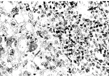
of mice of Group I and II treated by transfer of stimu- lated spleen cells (7 from 20 in Group I and 10 from 20 in Group II). In mice with such an effect small tumor nodule was found to be as big as grain lentil or pea, one was larger (5 x 6 mm). Histologically, tumor nodules were limited. Usually, around the area of ne- crosis tumor tissue was detected, sized between 250 p m and 600,u m wide, and rarely - 800,u m. It was composed mainly of mononuclear cells with pale nu- clei, but in some tumors numerous cells with hyperchromatic nuclei were found, as well as giant mono- and multinuclear cells in the proximity of ne- crosis. Infiltrate rich in lymphocytes and mixed with a few plasmocytes and macrophages was established in the periphery of the tumor (Fig. 1), and partially in- vaded tumor tissue. This infiltrate was mainly con- tinuous (complete) or, more rarely, incomplete engag- ing the half or two thirds of the circumference of the tumor nodule. Small blood vessels, often augmented in number, were found in this area (Fig. 2). Tumor cells in apoptosis and apoptotic bodies were observed among the invading lymphocytes and between pre- served tumor cells (Fig. 3). Focuses of necrosis were detected in proximity to the lymphocytic infiltrate.
The fibrous capsule formed in the periphery of tumor was mainly complete i.e. in the entire circumference of tumor, or incomplete. Evidence of proliferation of
Ivan Raichev et al : Morphological Changes in Transplanted Solid Ehrlich Tumor of Mice after Transfer of Stimulated Spleen Cells
Figure 1. Small limited tumor nodule with continuous lymphocytic infiltrate in its periphery. (H&E, x 50). Ehrlich tumor in a mouse of Group I with findings for a.e.
fibroblasts and formation of collagen fibers and their condensation among the infiltrate of lymphocytes were found, including by the ultrastructural examina- tion.
Four mice of Group II did not show a development of a tumor after the transplantation. In one mouse of Group I, bearing a small tumor and also treated with stimulated spleen cells, thirty days after transplanta- tion the tumor had completely regressed.
The data for a.e. in the transplanted tumor showed almost identical rate of cases among the mice treated with doses of 35 x 10' to 55 x 10' stimulated spleen cells. The previously examined doses of 20-25 x 10' proved to be with a low effectivity.
Morphological changes in the transplanted solid Ehrlich tumors without findings for antitumor effect
In these mice (13 in Group I and 10 in Group II), also treated with stimulated spleen cells like the mice with effect in the transplanted tumor and survived for the same periods, tumors were detected with the fol- lowing sizes: 0.5 x 1-1.5 cm, 1.5 x 2.5 cm. Histologically, the tumors were found to infiltrate the adjacent tis- sues. Peripheral cell infiltrate rich in lymphocytes oc- cupying as a tight strip from one third to half of the circumference of the tumor nodule was found in less than the half of the cases, but without evidence for lymphocytic cytotoxicity. An incomplete capsule was detected in a few tumors.
Figure 2. Cell infiltrate rich in lymphocytes in the periph- ery of tumor nodule. Small blood vessels (arrows) appeared among the infiltrate. (H&E, x 100). Ehrlich tumor in a mouse of Group II with findings for a.e.
Morphological changes in the transplanted solid Ehrlich tumors in untreated mice
The tumor-bearing mice untreated by transfer of stimulated spleen cells (Group III, controls) killed in the identical term of survival after transplantation as in mice of Group I and II showed tumors sized 1 x 1.5 cm, 1.5 x 2.5 cm and rarely 3 x 4 cm. Histologically, the tumor displayed a solid growth pattern and infil- trated fibrotic, adipose and muscle tissue. Fields of ne- crosis were found among the growing tumor tissue and in central region of tumor. Scanty infiltrate of lymphocytes together with formation of a partial cap- sule was detected in the periphery of the tumor only in two cases, but no morphological evidence for lymphocytic cytotoxicity was found.
Figure 3. Lymphocytes from peripheral infiltrate partially invading tumor tissue. Apoptotic cells with compaction of nuclear chromatin and apoptotic bodies are observed (ar- rows). (H&E, x 100). Ehrlich tumor in a mouse of Group I with findings for a.e.
Mitosis and apoptosis
Mitotic rate and apoptosis of tumor cells at the time of killing of the mice, expressed by mean value of MI and AI, are shown in Table 1. The mean MI in solid
Table 1. Mitotic rate and apoptosis in transplanted solid Ehrlich tumors of mice
Groups Mitotic Index (MI) Apoptotic Index (AI)
---
Tumors with Tumors without Tumors with Tumors without findings for findings for findings for findings for antitumor antitumor antitumor antitumor
effect effect effect effect
(1) (2) (3) (4)
---
I* 0.99±0.25 2.39±0.41 5.70±0.32 4.39±0.43
II*a` 1.22±0.29 2.45±0.27 4.06±0.72 4.18±0.43
III*** 2.48±0.44 4.47±0.38
*Mice bearing a small Ehrlich tumor in which stimulated spleen cells were transferred.
"Mice previously treated by transfer of stimulated spleen cells and then transplanted with tumor.
***Tumor-bearing mice untreated by transfer of stimulated spleen cells (controls). Values are mean ± SE.
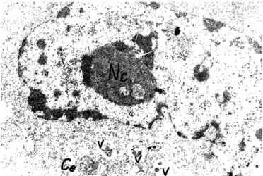
secondary lysosomes were detected (Fig. 4). Other cells showed enlarged nuclei with prevailing euchromatin and nucleoli with fibrilar centers. Multiple free ribosomes were found in the cytoplasm. These cells were characterized as actively synthesizing cells, par- ticularly in protein synthesis (Fig. 5). Cells with ex- posed ultrastructural features were found in the pre- served tumor tissue in small tumors of Group I and II with findings for a.e., and in tumor without such find- ings of the same two groups and of Group III.
Severe degenerative and necrotic changes in tumor cells akin to the irreversible hypoxic injuries were de- tected in all examined groups. Tumor cells in apoptosis in all groups were characterized by conden- sation and margination of nuclear chromatin, often
Ehrlich tumor of Group I and II with findings for a.e., column 1 in Table 1, was lower than the mean MI in tumors without finding for effect of the same groups, column 2 in Table 1, and in controls (Group III);
P<0.001. Concerning apoptosis, the mean AI in tumors of all groups (I, II and III) exceeded the mean MI;
P<0.001. The significance of the differences between the compared groups was determined by the Mann- Whitney U test, too (P<0.05). In tumors of Group I and II with findings for a.e. a few mitoses in one field (16 x 40) were counted, however there were fields without any mitoses. In tumors of Group I, II without findings for a.e. and Group III centers of proliferation with 8-14 mitoses in the high power field were found, mainly in the periphery of the tumor. The number of pathologic mitoses was high, at rate of 80.65% of all mitoses.
Apoptotic cells and numerous apoptotic bodies were detected everywhere in tumor tissue. Apoptotic cells were found to decrease in size with condensation of the eosinophilic cytoplasm, compaction and margination of nuclear chromatin. Small apoptotic bodies were in- gested by tumor cells. Between the cells, spherical globules (4-5 ,u m), basophilic, or acidophilic containing basophilic nuclear fragments, as well as large globules at the size 7-10 um, were observed. Around apoptotic bodies, often a clear space (halo) was seen.
Figure 4. Electron micrograph of two tumor cells, one is binuclear. Heterochromatin in the nuclei is shown as irregu- lar blocks. Mitochondria (arrows) are increased in number.
Original magnification x 3, 500. Ehrlich tumor in a mouse of Group II without findings for a.e.
Electron microscopic changes
In tumor cells, oval, elongated or irregular in shape nuclei was observed, with blocks of condensed chro- matin. In the cytoplasm, mitochondria, increased in
number, rough endoplasmic reticulum appeared as
small and tubule-like cisternae, ribosomes and
Figure 5. Electron micrograph of tumor cell with prevail- ing euchromatin in the large nucleus and big nucleolus (Nc).
Free ribosomes, vacuoles (V) containing residual bodies and two centrioles (Ce) are seen in the cytoplasm. Original magni- fication x 8,000. Ehrlich tumor in a mouse of Group II with findings for a.e.
Ivan Raichev et al : Morphological Changes in Transplanted Solid. Ehrlich Tumor of Mice after Transfer of Stimulated Spleen Cells
Figure 6. Ultrastructural findings for lymphocytic cytotoxicity: A. Two small lymphocytes close to a tumor cell in early apoptosis showing margination and condensation of nuclear chromatin (arrows). Vacuoles (V) in the cytoplasm containing resid- ual material. B. Small lymphocytes with primary lysosomes (Ly), one displaying membrane cell-to-cell contact (arrow) with tumor cell in apoptosis, revealing condensation of nuclear chromatin (arrows) and cytoplasm. C. Small lymphocyte contacts with cell in advanced apoptosis (A). Original magnifications: in A x 6,000, in B and C x 4,000. Tumor nodule in a mouse of Group I with findings for a.e.
forming crescent profile or its compaction as a dense masses with irregular outline, and condensation of or- ganelles of the cytoplasm. Apoptotic bodies containing masses of condensed chromatin and sometimes cytoplasmic organelles were seen.
Small lymphocytes were revealed in the peripheral cell infiltrate in tumor nodules of Group I and II with findings for a.e., as well as lymphocytes in activation.
Immunoblasts (stimulated lymphocytes) were also found. Evidence for lymphocytic cytotoxicity was de- tected by electron microscopy (Group I and II with findings for a.e.). Small lymphocytes were disposed in immediate proximity to apoptotic cells and bodies (Fig. 6 A). Cell-to cell contact of lymphocytes with tumor cells in apoptosis was found, too (Fig. 6 B, C).
The contacting lymphocytes contained in the cyto- plasm primary lysosomes increased in number and
grouped. Infiltration by lymphocytes in tumor nodule with the evidence for lymphocytic cytotoxicity was observed in some mice 6 days after transfer of stimu- lated spleen cells but not in mice three to five days after this transfer. In tumors of Group I and II with findings for a.e., small lymphocytes were detected in close proximity to small areas of necrosis.
Changes in the reactive structures of the spleen and lymph nodes
In the spleen of the tumor-bearing mice untreated by transfer of stimulated spleen cells (controls), par- ticularly in the periarterial (T) zone and the B zone of the Malpighian bodies lack of reaction, or rarely weakly expressed hyperplasia was observed. In some lymph nodes in the vicinity of tumors, the
paracortical areas were reacted - 450,u m in transver- sal size (grade I). Plasmocytes in the lower paracortex and in the medullary cords were 25-30% of the total number of cells (grade I, PL 1). In more of the mice of Group I and II with findings for a.e., reactive changes of mixed type, engaging T and B zones, were found in the spleens; T,B, and in one - T2B,. Isolated reactive changes in T zones (T,) and in B zones (B,) or lack of such changes were observed in some cases, too. In T1B,, periarterial (T) zones were 250-400,u m of transversal dimension, in T2B,- 450-550,u m, and B, zones (germi- nal centers) - 250-300 am. Small lymphocytes and scanty immunoblasts were detected in T zones. Centroblasts, centrocytes and macrophages ingested apoptotic bod- ies were found in the germinal centers. The mantle zone was well expressed. In the cords of the red pulp lymphocytes, macrophages, plasmocytes and megakaryocytes were revealed. The presence of the plasmocytes in the pulp was graded as 0 and grade I (Plo,1) - 10-20% of plasmocytes.
The examined tumor-draining lymph nodes in Group I and II with finding for a.e., situated nearby, close to the tumor, or in contact with it, displayed reactive changes mainly of the type T,B, with paracortex 400-4 50 um and germinal centers 250-300 gym. Plasmatization in the medullary cords was of grade 0 and I (Plo,,), and of grade II (P12) - 51-55% plasmocytes. Isolated T, hyperplasia was also established. Reactive changes of type T1B1, P11 in lymph nodes occurred in areas more distant from the tumor i.e. In the axiliary regions were detected, too. The paracortex was populated with lymphocytes, a few immunoblasts and interdigitating cells. Post-capillary venules previously with flattened endothelial cells, grade 1 in accordance with Syrj a nen 9' or with cuboidal endothelium (grade 2) were found.
Medullary sinuses contained lymphocytes and macro- phages. In mice without development of a tumor after transplantation, including the case with complete tumor regression, reactive changes of type TB, were established in the spleens. Lymph nodes found in the areas of transplantation showed reactive changes of type T1B1, Plt and of type T2 with paracortex 750,u m in one case. In the active paracortex, post-capillary venules of grade 2 were revealed.
Mice of Group I and II without findings for a.e.
demonstrated changes in the spleen mainly of type T1B1, Plo,1, but in others a reaction was absent. Lymph nodes situated near the tumor displayed changes of T1B1, T1 and of T2 (paracortex 720,u m) in one mouse;
Plo,1, rarely P12. In the paracortex small lymphocytes with various density, a few immunoblasts, interdigitating and histiocytic cells, and post-capillary venules of grade 1 and 2 were discovered.
Inhibition effect of the culture supernatants obtained from spleen cells of mice bearing solid Ehrlich tumor
The supernatants of spleen cells from mice bearing solid Ehrlich tumor non-stimulated with cancer ex- tract did not show an IE against migration of Ehrlich ascites tumor cells; mean MgI>0.80. On the contrary, the supernatants decanted from the samples and the wells inhibited Ehrlich-cell migration significantly, when the spleen cells were stimulated with Ehrlich extract, or with PHA, IL-2 and Ehrlich extract at al- most equal results received for the two ways of stimu- lation (Table 2); P<0.001. IE was also appeared when spleen cells were enriched with T lymphocytes and then stimulated with Ehrlich extract; MgI was 0.64±
0.03. According to the data of FrRCs, a correlation be- tween the IE showing production of a factor inhibit- ing Ehrlich-cell migration from the spleen cells of Ehrlich tumor-bearing mice, and the stimulation of these cells were found; correlation coefficient (r) =0.61, 0.60, 0.57.
Our preliminary experiments showed that the tumor extract, which could be found in a little amount in the supernatant containing inhibitory factor (IF), did not inhibit itself the migration of Ehrlich ascites cells in vitro. Identical results were received for the extract of the transplanted sarcoma induced in mice by 20- methylcholanthrene 5'.
Spleen cells obtained from mice treated by transfer of stimulated cells showed evidence of production of IF without to be stimulated with Ehrlich-tumor ex- tract in vitro. P<0.05 for Group I, and P<0.001 for Group II by comparison with the IE of the super- natants from unstimulated spleen cells (Table 2).
Production of this factor was not found in all treated
Table 2. Inhibition of Ehrlich-cell migration by culture super- natants of spleen cells from mice
MgI of culture supernatants of spleen cells from
Ehrlich-tumor Spleen cells were not Spleen cells were stimulated with bearing mice stimulated Ehrlich extract PHA, IL-2 and
Ehrlich extract
0.84±0.05 0.61±0.02 0.62±0.04
Mice treated by transfer of stimulated spleen cells
Group I* 0.75±0.08
Group II** 0.68±0.07
*Mice bearing a small Ehrlich tumor in which stimulated spleen cells were transferred.
**Mice previously treated by transfer of stimulated spleen cells and then transplanted with tumor. Values are mean ± SE.
mice (MgI- 0.63-0.83) and was not connected with the presence of findings for a.e. in the transplanted tu- mors.
In the smears obtained from suspension of spleen cells, stimulated with PHA, IL-2 and Ehrlich-tumor ex- tract, the following proportion of lymphoid cells were determined: small lymphocytes (6-7,u m) - 68.1 ± 1.51 %, middle-sized lymphocytes (8-10p m) - 20.52±0.45% and large lymphoid cells (blasts, immunoblasts) with pyroninophilic cytoplasm (18-25," m) - 10.88 ± 0.24%.
The rate of the lymphoid cells in the smears of spleen cells from intact mice was as followed: small lympho- cytes - 89.20 ± 1.06%, middle-sized lymphocytes - 8.65 ± 1.12% and large pyroninophilic cells 1.99 ± 0.32%.
Besides the lymphoid cells, a few plasmocytes and macrophages were found in the smears, because the suspensions were preliminarily incubated in plastic flasks.
Discussion
Previous investigations have shown that the regres- sion of localized and disseminated experimental tu- mors can be effectuated by adoptive transfer of spe- cifically sensitized lymphoid cells", 11,12.13' In the present study an adoptive transfer of spleen cells (lymphocytes) stimulated with Ehrlich-tumor extract or with PHA, IL-2 and tumor extract in mice-bearers of small trans- planted Ehrlich tumor (Group I) or in tumor-free mice with following transplantation (Group II) was accom- plished. The stimulation of spleen cells by Ehrlich ex- tract was in agreement with the report that Ehrlich murine tumor may be considered clearly immunogenic 2'.
Accentuating on the morphological changes in the transplanted solid Ehrlich tumors determined by mechanisms leading to blockage of growth and exten- sion of tumor nodule, we set four criteria for a.e.
These changes were interpreted together with the data for production of IF from the stimulated spleen cells. It was found the decrease of the proliferative ac- tivity in small tumor nodules of Group I and II with findings for a.e. in which mean MI was lower than this in tumors of the same groups (I and II) but with- out findings for a.e. and of Group III (Table 1). In this respect, it could be speculated on the effect of some cytokines: IFN y , TNF a / '9 , TGF p produced by cells of the infiltrate in the periphery of tumor, and proba- bly by tumor cells (TGF i) as antiproliferative agents for tumor cells"'. TGF beta precursors were detected in Ehrlich ascites cells and in ascitic fluid").
Our results showed an activity of apoptosis, which in Groups I, II, and III at the time of killing of the
mice, exceeded the mitotic activity of tumor cells.
According to data from literature, an influence of regulatory genes (oncogenes and oncosuppressor genes) as c-myc, bcl-2, p53 and ras could be suggested on this issue"'. The connection of the local hypoxia with apoptosis was considered, too. Graeber et all" has reported that in transplanted tumor highly apoptotic regions strongly correlate with hypoxic regions ex- pressing wild type p53.
The cell infiltrate rich in lymphocytes in the periph- ery of tumor nodules showed an evidence for lymphocytic cytotoxicity. Based on the ultrastructural findings, it was assumed that T-lymphocytes with cytotoxic activity, expressing as was shown elsewhere Lyt-2 phenotype"), killed tumor cells by inducing apoptosis, particularly through cell-to-cell contact.
Apoptosis induced by attack of lymphotoxin (TNF 9 ), secreted from stimulated T-lymphocytes 18), may be suggested in case of presence of the apoptotic bodies in the proximity of lymphocytes but without cell-to- cell contact with tumor cells. Focuses and small areas of tumor-cell necrosis nearby the lymphocytic infil- trate in tumors in Group I and II with findings for a.e.
were likely connected with the effect of TNF a / n 18,19'.
The proliferation of fibroblasts among the lympho-
cytes pho-cytes of the infiltrate with formation of collagen fi- bers, and in this way - a fibrotic capsule, was proba-
bly determined by stimulation of production of cytokines- FGFs, TGF 13 20' 2J, released from the cells of the infiltrate. Morphological changes for a.e. in trans- planted solid Ehrlich tumors were also obtained in exbreeder ICR mice treated with stimulated spleen cells (our unpublished data).
The stimulated spleen cells (lymphocytes) from Ehrlich-tumor bearing mice were immunologically ac- tive as they produced IF in the supernatants, sup- pressing the migration of Ehrlich ascites cells. The IF was produced by T-lymphocytes, as the spleen cells enriched with T-cells accomplished it. Previously, a production of IF was found from spleen cells of mice immunized with killed sarcoma cells and from eryth- rocyte rosette-forming cells, separated from peripheral blood mononuclear cells, in patients with gastric and breast cancer, when the cells were stimulated with tumor extract. Active fractions were isolated from crude supernatants in the range of molecular weight between 14 000 and 5 800 daltons and lower than 5 800 5, 6'. In the present study, there were data that the stimulation of spleen cells with PHA, IL-2 and Ehrlich extract caused an activation, partial blast- transformation and proliferation of T-lymphocytes.
The low concentration of IL-2 didn't favor generation of LAK cells22'.