Development of a Cre/lox-based multiple markerless gene disruption method for the extreme thermophile
Thermus thermophilus
防衛大学校理工学研究科後期課程
物質・基礎科学系専攻 高エネルギー・物質工学教育研究分野
外 川 陽 一 郎
平成30年3月
Table of Contents
Page
Introduction
··· 1Materials and methods
··· 5Bacterial strains and growth conditions ··· 5
Manipulation of DNA ··· 6
Preparation of T. thermophilus genomic DNA ··· 6
Plasmid construction ··· 7
Construction of a loxP–htk–loxP cassette ··· 11
Construction of a lox66–htk–lox71 cassette ··· 11
Construction of plasmids for gene disruption ··· 15
Construction of a cre-expressing plasmid··· 18
Transformation of T. thermophilus ···21
Gene disruption in T. thermophilus using the loxP–htk–loxP cassette ··· 21
Removal of the htk-selectable marker by Cre recombination ··· 21
Curing pSH-Cre from T. thermophilus ···22
Results
··· 23Gene disruption using the loxP–htk–loxP cassette ··· 23
Removal of the htk gene from the gene disruptant by Cre-mediated recombination ··· 26
pSH-Cre curing from the markerless gene disruptant ··· 28
Construction of a double markerless gene disruptant ··· 28
Construction of a triple markerless gene disruptant ··· 34
Markerless gene disruption by irreversible Cre/lox recombination using a lox66–htk–lox71 cassette ··· 37
Double gene disruption using a lox66–htk–lox71 cassette ··· 41
Discussion
··· 45Acknowledgements
··· 50References
··· 51Academic achievements
··· 561
Introduction
Cre/loxP site-specific recombination, which was first reported in 1981 (Sternberg and Hamilton 1981), has become a significant technology to manipulate DNA in vivo (Nagy 2000). This recombination is performed between two loxP sites using a 38-kDa Cre recombinase. loxP is a 34-bp consensus DNA sequence that has an 8-bp core spacer region, defining its orientation, flanked by two 13-bp palindromic sequences, which are the binding sites of Cre (Hoess and Abremski 1985). Cre-mediated recombination results in the excision of the DNA sequence between two loxP sites in the same direction, whereas it catalyzes inversion when the two loxP sites are in the opposite direction. The efficacy of the Cre/lox system in a broad spectrum of biological species and wide variety of applications has made this technology indispensable for in vivo genetic manipulation. This system enables various types of recombination, such as conditional recombination, intermolecular recombination, and time-and-space-specific recombination (Nagy 2000). An important application of this system is the removal of selectable markers after gene disruption, which enables the marker to be reused. Marker recycling is a crucial issue for researchers studying organisms that have a limited number of selectable markers, of which extreme thermophiles are a typical example. A shortage of selectable markers has been reported, even for the most studied extreme thermophile, Thermus thermophilus (T.
thermophilus).
T. thermophilus HB27 is a Gram-negative bacterium isolated from a Japanese hot spring, which grows optimally at high temperatures ranging between 65 and 72 °C (Oshima and Imahori 1974). Its natural competence (Koyama et al. 1986), relatively small genome size, consisting of a 1.89-Mb chromosome and 0.23-Mb plasmid pTT27, and available genome sequence (Henne et al. 2004) have made it suitable as a model organism to study thermophilic prokaryotes. The production of a yellow pigment (Oshima and Imahori 1974) and polyploidy,
2
harboring four to five copies of chromosomes in a cell (Ohtani et al. 2010), are also unique characteristics of T. thermophilus HB27. Due to the stability of its proteins and ease of purification, this extreme thermophile was also the focus of a structural genomics project (Yokoyama et al. 2000). Despite its ideal properties as a model organism, a genetic analysis similar to that for mesophiles has been difficult to perform due to the lack of selectable markers for T. thermophilus.
Since most of the mesophilic proteins used as selectable markers are sensitive to thermal denaturation at the optimal growth temperature for T. thermophilus, their thermostabilities have been improved by random mutagenesis. To date, three examples of antibiotic-resistant genes for kanamycin (Km) (Hoseki et al. 1999), hygromycin (Hm) (Nakamura et al. 2005), and bleomycin (Brouns et al. 2005) are available for T. thermophilus;
however, it is still a potential bottleneck in the genetic analysis of genes involved in functionally redundant pathways. Therefore, markerless gene disruption is an attractive strategy for research on T. thermophilus, and several methods that rely on counter-selection have been reported.
Among them, three methods require specific mutant backgrounds responsible for each counter-selectable marker as the parental strains, the ∆pyrE (Tamakoshi et al. 1999), ∆bgl (Angelov et al. 2013), or ∆crtB (Fujita et al. 2015) strain; however, recent studies have demonstrated that counter-selection strategies, in which the rpsL1 allele (Blas-Galindo et al.
2007), pheS allele (Carr et al. 2015), or codA gene of Thermaerobacter marianensis DSM 12885 (Wang et al. 2016) were used as counter-selectable markers, were applicable to the wild-type strain. When markerless gene disruptants were isolated as colonies resistant to each counter-selection agent, they contained spontaneous selection-resistant mutants depending on the respective background mutant frequencies, for example, 2.5 × 10⁻⁷ (Carr et al. 2015) and 10-6 to 10-7 (Blas-Galindo et al. 2007), and thus additional gene analyses are needed for these
3 methods, except for ∆crtB-based color-selection. These disadvantages are also a potential obstacle to the construction of multiple markerless gene disruptants because they may accumulate spontaneous drug-resistant mutations during repetitive counter-selection. Another strategy that does not require any selection process has recently been reported. In this method, markerless gene disruptants were identified by colony PCR on an enormous scale using the manual screening of colonies grown without selection after a conventional process of transformation, and, hence, it requires a large amount of work (Leis et al. 2014).
The objective of this study was to develop a novel markerless gene disruption method for T. thermophilus to overcome the limitations in the counter-selection strategies, namely, the requirement of specific mutant and/or the production of spontaneous selection-resistant mutants.
In this respect, I applied a Cre/lox system for removal a selectable marker after gene disruption.
A Cre/lox system has been used in a broad spectrum of biological species for genetic manipulations (Nagy 2000), however, it has not yet been examined in T. thermophilus. The activity of Cre recombinase has been experimentally confirmed up to 46 °C (Buchholz et al.
1996), which is slightly less than 50 °C, the minimum growth temperature of T. thermophilus HB27 (Ohtani et al. 2010). However, a CD spectral analysis showed that protein denaturation starts at approximately 54 °C (Buchholz et al. 1998). Assuming that Cre is still active at 50 °C, I examined the application of the Cre/lox system to markerless gene disruption in T. thermophilus.
In order to achieve this, two genetic tools, a loxP–htk–loxP cassette and the cre-expressing plasmid, pSH-Cre, were created. I found that the Cre/lox system was compatible with the proliferation of the T. thermophilus HB27 strain at the lowest growth temperature (50 °C), and confirmed that the Cre-mediated removal of the selectable marker, htk (Hoseki et al. 1999), from the polyploid genome was achieved by the introduction of pSH-Cre into a gene disruptant strain that was constructed by the insertion of the loxP–htk–loxP cassette into the TTC1535
4
gene. Moreover, I succeeded in establishing a triple gene disruptant strain without leaving behind a selectable marker. This is the first example of the disruption of three genes distantly located on the chromosome of T. thermophilus in the wild-type background. On the other hand, the Cre-mediated deletion and inversion of the chromosomal region between multiple loxP sites occurred in the process of the sequential disruption of multiple genes. In order to avoid these undesired chromosomal rearrangements, I created a lox66–htk–lox71 cassette that contained mutant lox sites (Albert et al. 1995), which allowed for the construction of a double gene disruptant without inducing the undesired deletion. My results showing chromosomal deletions (range, 0.7–122.9 kbp) and inversions (range, 34.5–88.4 kbp) also suggested that this system is applicable to the deletion or inversion of the targeted chromosomal region, which may enable the bioengineering of T. thermophilus on an unprecedented scale.
5
Materials and methods
Basic experiments were performed according to standard protocols described in Molecular cloning: a laboratory manual (Sambrook and Russell 2001).
Bacterial strains and growth conditions
All the T. thermophilus strains used in the present study are listed in Table 1, and were grown in PY medium (Ohta et al. 2006) at 70 °C unless otherwise stated. A total of 50 μg/ml of Km and/or 40 μg/ml of Hm was added to the medium when needed. Escherichia coli (E. coli) DH5α strain, which was used for genetic constructions, was grown at 37 °C in LB medium (Sambrook and Russell 2001). A total of 100 μg/ml ampicillin (Ap), 50 μg/ml Km, or 200 μg/ml Hm was added to the medium when required. In order to make agar plates, 1.5% (w/v) agar (Difco) was added to PY and LB media.
Table 1 T. thermophilus strains used in this study
Strain Genotype Reference
HB27 Wild type Oshima and
Imahori (1974)
ST1 ΔTTC1535KpnI::loxP–htk–loxP This study
ST1Δhtk ΔTTC1535KpnI::loxP This study
ST2 ΔTTC1535KpnI::loxP, ΔTTC1576::loxP–htk–loxP This study
ST2Δhtk ΔTTC1535KpnI::loxP, ΔTTC1576::loxP This study
ST2ΔhtkIN ΔTTC1535KpnI::loxP, ΔTTC1576::loxP , IN(TTC1535–1576) This study ST3 ΔTTC1454::loxP–htk–loxP, ΔTTC1535KpnI::loxP, ΔTTC1576::loxP This study ST3Δhtk ΔTTC1454::loxP, ΔTTC1535KpnI::loxP, ΔTTC1576::loxP This study
ST4 ΔTTC1535::lox66–htk–lox71 This study
ST4Δhtk ΔTTC1535::lox72 This study
ST5 ΔTTC1535::lox72, ΔTTC1537::loxP–htk–loxP This study
ST5Δhtk ΔTTC1535::lox72, ΔTTC1537::loxP This study
ST6 ΔTTC1535::lox72, ΔTTC1537::lox66–htk–lox71 This study
ST6Δhtk ΔTTC1535::lox72, ΔTTC1537::lox72 This study
ST7 ΔTTC1535::loxP–htk–loxP This study
ST7Δhtk ΔTTC1535::loxP This study
ST8 ΔTTC1535::loxP, ΔTTC1537::loxP–htk–loxP This study
6
Manipulation of DNA
The plasmids used in the present study were prepared from E. coli DH5α using the Wizard Plus SV Minipreps DNA purification system (Promega). DNA fragments were purified from agarose gels or the PCR reaction mixture using the Wizard SV Gel and PCR Clean-Up System (Promega). Three different DNA polymerases were used for PCR depending on the purpose of the experiment. GoTaq Green Master Mix (Promega) was used for the amplification of DNA fragments for TA cloning, a PCR analysis of T. thermophilus genomic DNA, and the amplification of DNA fragments for gene disruption. iProof High-Fidelity Master Mix (Bio Rad) was used to clone the genomic DNA fragments of T. thermophilus. Pyrobest DNA Polymerase (Takara) was used for inverse PCR during the construction of gene disruption plasmids. PCR was performed according to the manufacturer’s instructions. When the purified genomic DNA of T. thermophilus was used as a PCR template, the annealing temperature was set to either 60 °C or 65 °C depending on the Tm of the primers. In addition, Ribonuclease (DNase free) (Wako) and Hi-Di Formamide (Thermo Fisher Scientific) were added to the PCR mixture (final concentrations of 0.1 μg/μl and 2.0% (v/v), respectively) in order to improve the amplification of GC-rich DNA. PCR products were separated by 0.8% (w/v) agarose gel electrophoresis and stained with ethidium bromide.
Preparation of T. thermophilus genomic DNA
Genomic DNA was prepared from 2 ml of the overnight culture. The culture was centrifuged to pellet the cells. The pellet was resuspended in 1 ml of Pi buffer (33 mM Na2HPO4 and 33 mM KH2PO4) for washing and cells were re-pelleted. The pellet was resuspended in 300 µl of lysozyme solution (5 mg/ml lysozyme, 0.9% (w/v) glucose, 10 mM EDTA, and 25 mM Tris–
HCl pH8.0) and kept on ice for 10 min. Fifteen microliters of 10% (w/v) SDS was added, mixed
7 gently, and kept on ice for an additional 30 min. The chilled solution was then mixed thoroughly with an equal volume of phenol–chloroform. The mixture was centrifuged (5 min, 16,100 rcf, room temperature) and the supernatant was subjected to ethanol precipitation. Pelleted DNA was rinsed with 70% (v/v) ethanol, dried, and dissolved in 100 μl of TE buffer.
Plasmid construction
The plasmids and synthetic oligonucleotides used in this study are listed in Tables 2 and 3, respectively. Details on plasmid construction are described below.
8
Table 2 Plasmids used in this study
Plasmid Relevant characteristics Reference(s)
pUC18 Cloning vector Yanisch-Perron et al. (1985)
pBluescript SK(+) Cloning vector Short et al. (1988)
pCR2.1-TOPO Cloning vector Thermo Fisher Scientific
pGEM-T Easy Cloning vector Promega
pTAP60 htk, bla, repA of pTT8, replication origin of pUC18 Ohta et al. (2006)
pBSSK–loxP–RfA–loxP loxP–RfA–loxP Unpublished data, this study
pTN30 bla, supF, replication origin of pBR322 Hiratsu et al. (2013)
pT8H5-Pslp hph5, PslpA, replication origin of pUC19 Takayama et al. (2004); Nakamura et al. (2005) pSH-Cre hph5, PslpA-cre, repA of pTT8, replication origin of pBR322 This study
pUC18–loxP–htk–loxP BamHI-flanked loxP–htk–loxP This study
pUC18-lox66–htk–lox71 BamHI-flanked lox66–htk–lox71 This study
pTTC1454 pGEM-T Easy derivative, TTC1454 cloned by TA cloning This study pDELloxTTC1454 pTTC1454 derivative, ΔTTC1454::loxP–htk–loxP This study pTTC1535 pUC18 derivative, TTC1535 cloned at the EcoRI/HindIII site This study pDELloxTTC1535K pTTC1535 derivative, ΔTTC1535KpnI::loxP–htk–loxP This study pDELloxTTC1535 pTTC1535 derivative, ΔTTC1535::loxP–htk–loxP This study pDELmloxTTC1535 pTTC1535 derivative, ΔTTC1535::lox66–htk–lox71 This study pTTC1537 pUC18 derivative, TTC1537 cloned at the EcoRI/XbaI site This study pDELloxTTC1537 pTTC1537 derivative, ΔTTC1537::loxP–htk–loxP This study pDELmloxTTC1537 pTTC1537 derivative, ΔTTC1537::lox66–htk–lox71 This study pTTC1576 pUC18 derivative, TTC1576 cloned at the SmaI site This study pDELloxTTC1576 pTTC1576 derivative, ΔTTC1576::loxP–htk–loxP This study
8
9 Table 3 Synthetic oligonucleotides used in this study
Oligonucleotides Sequence (5' to 3')a Restriction site
M13-M4 GTTTTCCCAGTCACGAC -
M13-RV CAGGAAACAGCTATGAC -
104 AAAGGTACCTCTTGAGATCCTTTTTTTC KpnI
140 CTTCTATTCCTTTGCCCTCGGACGAGTG -
141 CAGATTCGGCCCAAGGTTTACAAAATCC -
146 ATGCGCCGGGAGATCCTGGTGGCGGCG -
149 GAAGATCTTGCGGCGGTCCTCCACCTG BglII
150 GAAGATCTTGAACCCCAGGGACCCCG BglII
161 TGGAGTTCAAGGTGCCCATCCGCAC -
162 GGCCTCCTGCCCCTCATGGTGAGCC -
165 CTCCAAGGTGGTCTTTGAGGTGCGG -
166 GAAGAGGTCCTCCAGGATTAGCTGC -
180 GGAATTCGGGAGCTTTTAGGGGTAGAGGTG EcoRI
181 CCAAGTACCTCCTGGAAGGGCTTAG -
182 GAAGATCTGCTGCAGGAGGACCTCGGAGAC BglII 183 GAAGATCTTCGCCCGGGAAAGCCCCAAGGAG BglII
193 GGGGTACCTCCCACCTCCCCCCGGG KpnI
194 TGCAGCTCGAGATGTCCAATTTACTGACCGTACACC XhoI 195 GCTCGCTCGAGCTAATCGCCATCTTCCAGCAGGCGC XhoI
196 GGGGTACCGCGGTGGCGGCCGCTCTAG KpnI
201 CATGGTGGAGCACAAGCGCCTCTTC -
206 TCGACTAGAATTCATGAAAGGACC EcoRI
207 CGGAATTCAAAATGGTATGCGTTTTGACAC EcoRI 211b CGGGATCCtaccgTTCGTATAGCATACATTATAC BamHI 212b CGGGATCCtaccgTTCGTATAATGTATGCTATAC BamHI 213 TAGAAGCTTTGTGAGCAAAAGGCCAGC HindIII
215 CGGGATCCTCGAGGTCGACGGTATCG BamHI
229 CGGAATTCTGCCAGGCCTCCACGGCTAG EcoRI
230 TGCTCTAGAAGCGCCCAAAGCCCAGGAG XbaI
231 GAAGATCTTGAAGCAGGCGCATCACGGCCAAG BglII 232 GAAGATCTTCGGGAGCTTTTAGGGGTAGAGGTG BglII
233 CCTTTACCCCCTTCTCCTCCTCCTC -
247 ACCGGGAGATGGCGAGGCTCAC -
lox1c AGCTTATAACTTCGTATAGCATACATTATACGAAGTTAT HindIII compatible
10
lox2c AGCTATAACTTCGTATAATGTATGCTATACGAAGTTATA HindIII compatible lox3c AATTATAACTTCGTATAGCATACATTATACGAAGTTATG EcoRI compatible lox4c AATTCATAACTTCGTATAATGTATGCTATACGAAGTTAT EcoRI compatible
n1 AGATCTTCTATTCCTTTGCCCTCGG BglII
n2 AGATCTCGGCCCAAGGTTTACAAAA BglII
n3 GTTAATCATGTTGGTTACGCTG -
n4 AAGCTTTTCCCCGAAAAGTGCCA HindIII
n5 TGGAGTTCAAGGTGCCCATCCG -
n6 AGCTTTGGTCCGAGAGGGTG -
n7 GAGCTCGGATCCCGTTGACG BamHI
n8 TGCAGCGGATCCAACATGATTAAC BamHI
n9 GACTTCGCCCTCAACATGGA -
n10 TTCTCCGCCTTGGTCTTGAG -
aRestriction sites used for cloning are underlined.
bRegarding the primers 211 and 212, the bases shown in bold lower-case letters indicate the regions specific to the mutant lox sites, lox71 and lox66, respectively.
cRegarding the primers lox1-4, the regions compatible with the restriction sites are underlined.
11
Construction of a loxP–htk–loxP cassette
pUC18–loxP–htk–loxP harboring a loxP–htk–loxP cassette (Fig.1a) was constructed as summarized in Fig. 2. The loxP sequences were obtained from pBSSK–loxP–RfA–loxP (unpublished work) consisting of a Reading Frame Cassette A (RfA) fragment (Invitrogen) sandwiched by two loxP sites oriented directly. In brief, the synthetic DNAs, lox1/lox2 for LoxPH and lox3/lox4 for LoxPRI, listed in Table 3 were annealed and ligated, respectively, into the HindIII site and EcoRI site of a pBluescript II SK (+) (pBSSK) (Short et al. 1988) derivative containing RfA at the EcoRV site of the vector to construct pBSSK–loxP–RfA–loxP. In the present study, RfA in the plasmid was replaced with the htk fragment. A 1.1-kbp DNA region, including htk, of pTAP60 (Ohta et al. 2006) was amplified by PCR using the primers n7/n8 and subcloned into pCR2.1-TOPO by TA cloning to generate pCR2.1-htk. The RfA fragment of pBSSK–loxP–RfA–loxP was removed by EcoRV digestion and the 1.1-kbp htk fragment treated by T4 DNA polymerase (Takara) after the BamHI digestion of pCR2.1-htk was inserted therein to generate pBSSK–loxP–htk–loxP. The loxP–htk–loxP cassette of pBSSK–loxP–htk–loxP was amplified by PCR using the primers M13-RV/215 to generate BamHI sites at both ends. The resultant 1.3-kbp PCR product was digested by BamHI and ligated to the corresponding site in pUC18 (Yanisch-Perron et al. 1985) to generate pUC18–loxP–htk–loxP. The BamHI-flanked loxP–htk–loxP cassette of the plasmid was used to construct the series of plasmids for gene disruption.
Construction of a lox66–htk–lox71 cassette
pUC18–lox66–htk–lox71 harboring a lox66–htk–lox71 cassette (Fig. 1a) was constructed as summarized in Fig. 2. pBSSK–loxP–htk–loxP was used as the template for PCR by the primers 206/211 and 207/212 in order to replace its loxP sites with lox71 and lox66, respectively. The
12
resultant PCR products, lox71–’htk and htk–lox66, were digested by EcoRI and BamHI and ligated to the corresponding sites of pUC18 and pBSSK, respectively. Both of the resultant plasmids were digested by HindIII and EcoRI, and a 0.4-kbp fragment from pUC18-(207/212) was ligated to the corresponding site of pBSSK-(206/211) in order to generate pBSSK–lox66–
htk–lox71 containing the lox66–htk–lox71 cassette. Since the transformation of E. coli DH5α with the ligation product was unsuccessful, it was used as a template for PCR by the primers M13-M4/M13-RV in order to amplify the lox66–htk–lox71 cassette. The 1.4-kbp PCR product was digested by BamHI and the resultant 1.2-kbp fragment was ligated to the corresponding site of pUC18 in order to construct pUC18–lox66–htk–lox71. A lox66–htk–lox71 cassette was prepared from this plasmid, and was also used to construct gene disruption plasmids.
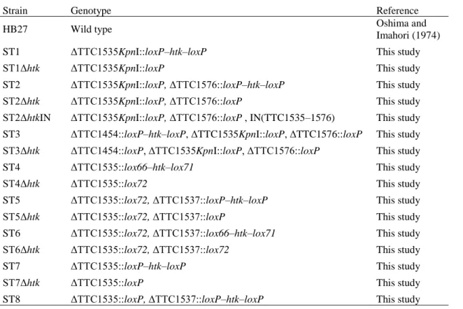
13 Fig. 1 Genetic tools for the application of the Cre/lox system to the markerless gene disruption in T. thermophilus HB27. a Structural features of the loxP–htk–loxP and lox66–htk–lox71 cassettes.
Shaded triangles depict the orientation of the lox sites, as described by Albert et al. (1995). The htk gene is a thermostable Km-resistant gene. b Structural features of the cre-expressing vector pSH-Cre. The cre gene is under the control of the strong PslpA promoter. The hph5 gene is a thermostable Hm-resistant gene. The repA region is the replication origin in T. thermophilus. The positions of the primers used for PCR analyses are indicated by filled triangles.
14
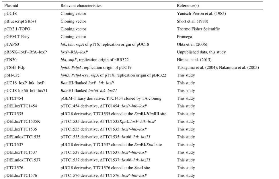
Fig. 2 Construction of plasmids containing a loxP–htk–loxP cassette or lox66–htk–lox71 cassette.
pUC18–loxP–htk–loxP contained a loxP–htk–loxP cassette, in which a Km-selectable marker, htk, is flanked by two directly oriented loxP sites. pUC18–lox66–htk–lox71 contained a lox66–
htk–lox71 cassette. lox66 and lox71 are mutant lox sites. The positions of the primers used for PCR are indicated by filled triangles.
15
Construction of plasmids for gene disruption
The plasmids for gene disruption were constructed as indicated in Fig. 3. In brief, genomic DNA fragments, which contain disruption target genes along with their up- and downstream flanking regions, were subcloned into the cloning vectors listed in Table 2. The disruption cassette, either loxP–htk–loxP or lox66–htk–lox71, was then inserted into the coding region of the target gene in order to construct gene disruption plasmids. The gene disruption constructs in the resultant plasmids were amplified by PCR using the primers M13-M4/M13-RV, which were purified from agarose gels and used to produce the gene disruptants of T. thermophilus.
16
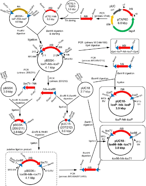
17 Fig. 3 Construction of gene disruption plasmids. Primers are shown in filled triangles. a pDELloxTTC1535K. A 1995-bp region of T. thermophilus HB27 genomic DNA containing the 978-bp TTC1535 gene was amplified by PCR using the primers 180/181. The EcoRI/HindIII digest of the PCR product (1849 bp) was subcloned into the corresponding site of pUC18. The resultant plasmid, pTTC1535, was cleaved at the unique KpnI site on the coding region of TTC1535, at which a KpnI-flanked loxP–htk–loxP cassette was inserted to generate pDELloxTTC1535K. The KpnI-flanked loxP–htk–loxP cassette was constructed by amplifying the loxP–htk–loxP fragment from pBSSK–loxP–htk–loxP (Fig. 2) using the primers M13-M4/196. The PCR product was digested by KpnI and the resultant 1254-bp fragment was used for the construction of the plasmid. b pDELloxTTC1576. A 1448-bp region of T.
thermophilus HB27 genomic DNA containing the 468-bp TTC1576 gene was amplified by PCR using the primers n9/n10. The PCR product was phosphorylated by T4 polynucleotide kinase (Toyobo) and ligated with SmaI-digested pUC18. The resultant plasmid, pTTC1576, was used as the template for inverse PCR with the primers 149/150, which have a BglII restriction site at their overhang. The resultant PCR product that lacks nucleotides (nt) 242 to 286 of the TTC1576 gene was digested by BglII (pTTC1576Δ242–286) and ligated with the loxP–htk–loxP cassette from BamHI-digested pUC18–loxP–htk–loxP to generate pDELloxTTC1576. c pDELloxTTC1454. A 1151-bp region of T. thermophilus HB27 genomic DNA containing the first 643 bp of the TTC1454 gene was amplified by PCR using the primers n5/n6. The PCR product was cloned into the pGEM-T Easy vector. The resultant plasmid, pTTC1454, was cleaved at the unique BamHI site on the coding region of TTC1454 and ligated with the loxP–htk–loxP cassette to generate pDELlox1454. d pDELloxTTC1535 and pDELmloxTTC1535. pTTC1535 was used as the template for inverse PCR using the primers 182/183, which have a BglII restriction site at their overhang. The resultant PCR product that lacks nt 119 to 418 of the TTC1535 gene was digested by BglII (pTTC1535Δ119–418) and ligated with the loxP–htk–loxP cassette and lox66–
htk–lox71 cassette to generate pDELloxTTC1535 and pDELmloxTTC1535, respectively. e pDELloxTTC1537 and pDELmloxTTC1537. A 1947-bp region of T. thermophilus HB27 genomic DNA containing the 651-bp TTC1537 gene was amplified by PCR using the primers 229/230. The XbaI/EcoRI digest of the PCR product was subcloned into the corresponding site of pUC18. The resultant plasmid, pTTC1537 was used as a template for inverse PCR using the primers 231/232, which have a BglII restriction site at their overhang. The resultant PCR product that lacks nt 177 to 456 of the TTC1537 gene was digested by BglII (pTTC1537Δ177–456) and ligated with the loxP–htk–loxP cassette and lox66–htk–lox71 cassette to generate pDELloxTTC1537 and pDELmloxTTC1537, respectively. Shaded triangles show the orientation of the lox sites. Restriction sites with underlines are introduced by PCR using primers with the restriction site at the overhang.
18
Construction of a cre-expressing plasmid
The cre-expressing vector pSH-Cre (Fig. 1b) was constructed as shown in Fig. 4. A 1.2-kbp DNA region, including the thermostable Hm-resistant gene, hph5 (Nakamura et al. 2005), of pT8H5-Pslp (Takayama et al. 2004; Nakamura et al. 2005) was amplified by PCR using the primers n1/n2 and subcloned into pCR2.1-TOPO by TA cloning to generate pCR2.1-hph5. The resultant plasmid was then digested by BglII and the hph5 fragment was ligated with BamHI-digested pTN30 (Hiratsu et al. 2013) to create pTN30-hph5. The repA region for the replication origin in T. thermophilus (Aoki and Itoh 2007) was amplified from pTAP60 by PCR using the primers n3/n4, and the resultant 2.8-kbp DNA fragment was subcloned into pCR2.1-TOPO by TA cloning. The resultant pCR2.1-repA was digested with HindIII and PstI, and the repA-containing 2.8-kbp fragment was ligated to the corresponding sites of pTN30-hph5.
The resultant plasmid, pKAT925 is an E. coli-T. thermophilus shuttle vector. Since hph5 is useful for plasmid maintenance in E. coli and T. thermophilus, the bla gene was removed from pKAT925. A 1.0-kbp region of pTN30 including pBR322 ori was initially amplified by PCR using the primers M13-M4/104. The PCR product was digested with KpnI and HindIII, which was then ligated with a 4.1-kbp fragment of KpnI/HindIII-digested pKAT925 to construct pKAT925s.
Regarding the expression of cre, the PslpA-cre region, in which cre is under the control of the strong T. thermophilus promoter PslpA (Faraldo et al. 1992), was cloned to pKAT925s. The 1.0-kbp ORF of the cre gene in the 705-Cre plasmid (GeneBridges, Germany) was amplified by PCR using the primers 194/195. The PCR product was digested by XhoI and cloned into the corresponding site of pBSSK to generate pBSSK-Cre. pT8H5-Pslp was digested with XbaI and SalI, and the 0.3-kbp fragment containing the promoter was ligated to the corresponding site of pBSSK-Cre to generate pBSSK-PslpA-Cre. PslpA-cre was amplified from
19 pBSSK-PslpA-Cre using the primers M13-M4/196. The resultant 1.4-kbp PCR product was digested by KpnI and ligated to the corresponding site of pKAT925s in order to construct pKAT925s-Cre. Since the supF gene was irrelevant to the purpose of this study, it was removed from pKAT925s-Cre. A 2.0-kbp region including cre and pBR322 ori in pKAT925s-Cre was PCR-amplified using the primers 195/213. The PCR product was digested with EcoRV and HindIII, and the resultant 1.5-kbp fragment was ligated with EcoRV/HindIII-digested pKAT925s-Cre to construct pSH-Cre.
20
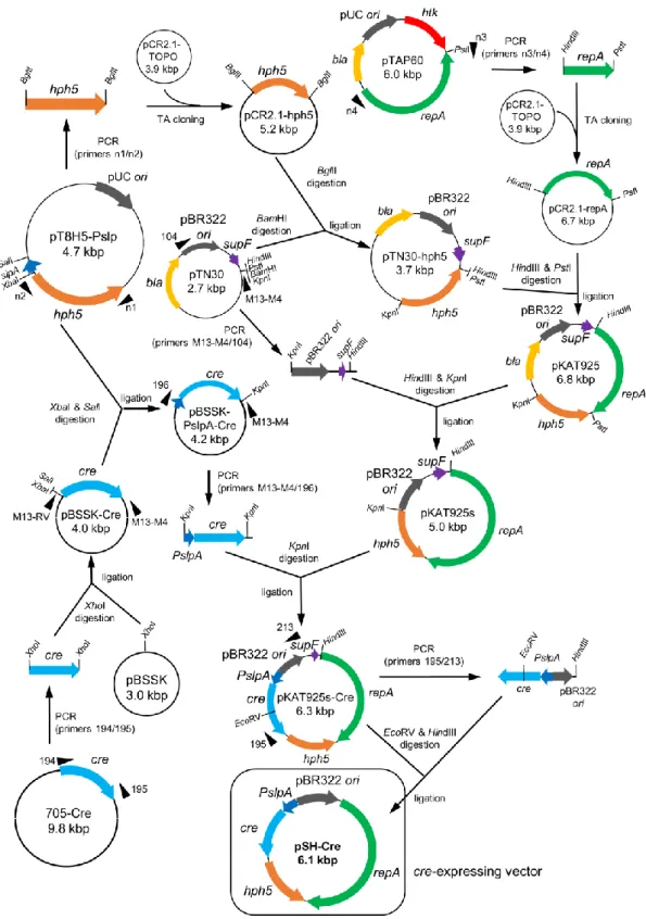
Fig. 4 Construction of the cre-expressing vector pSH-Cre. pSH-Cre is a E. coli-T. thermophilus shuttle vector containing the Hm-selectable marker, hph5. The cre gene is under the control of the strong T. thermophilus promoter PslpA. The positions of the primers used for PCR are indicated by filled triangles.
21
Transformation of T. thermophilus
The transformation of T. thermophilus was performed based on the protocol described by Hoseki et al. (1999). Transformants were selected on PY agar plates with appropriate antibiotics for an overnight incubation at 70 °C. In order to confirm transformation, selected colonies were streaked on the same plates and incubated overnight. Single-colony isolates were subjected to a genomic analysis to confirm transformation.
Gene disruption in T. thermophilus using the loxP–htk–loxP cassette
In the first step of markerless gene disruption, target genes were disrupted using the PCR product amplified from the gene disruption plasmids constructed for each disruption target gene (Fig. 3). Transformation was performed as described above. Transformants were selected by Km resistance, which was conferred by the htk gene in the loxP–htk–loxP cassette. In order to confirm gene disruption, the complete replacement of the target gene on multiple chromosomes with the disruption construct was identified by a PCR analysis using the primers specific to each disruption target gene.
Removal of the htk-selectable marker by Cre-mediated recombination
In order to remove htk from the chromosome, 50–150 ng of the Hm-selectable cre-expressing plasmid pSH-Cre was transformed into loxP–htk–loxP or lox66–htk–lox71 gene disruptant mutants. Transformation was performed as described above and the transformants were selected on PY/Km+Hm plates. In order to induce Cre/lox site-specific recombination for the removal of htk between two lox sites, single-colony isolates were re-streaked on PY/Hm plates and incubated at 50 °C for 5–7 days. The Hm-resistant colonies that grew were replica plated on PY and PY/Km plates and incubated at 70 °C overnight in order to select Km-sensitive candidates.
22
Km-sensitive clones were selected as candidates for markerless gene disruptants, and the corresponding colonies grown on PY plates were cultured in 3 ml of PY medium without antibiotics. Genomic DNA was prepared from the culture and the removal of htk was confirmed by a series of PCR analyses.
Curing pSH-Cre from T. thermophilus
pSH-Cre was removed from gene disruptants after the removal of htk had been confirmed by PCR. Gene disruptants harboring pSH-Cre were cultured at 70 °C overnight in 3 ml of PY medium without Hm. The culture was then diluted to 1:100 in 3 ml of fresh PY medium and grown overnight. The medium was preheated to 70 °C before the dilution to keep Cre inactive and prevent undesired recombination throughout the process. Subculturing was repeated twice, giving three overnight cultures, and 100 µl of the 10-6 dilution culture was spread on PY and PY/Hm agar plates and then incubated at 70 °C overnight. Four to 12 of the colonies grown on the PY plate were cultured at 70 °C overnight in 3 ml of PY medium. In order to confirm the absence of pSH-Cre, genomic DNA prepared from the culture was subjected to a PCR analysis using the primers 140/141 for the amplification of a 1.2-kbp DNA fragment for hph5 on the plasmid (Fig. 1b).
23
Results
Gene disruption using the loxP–htk–loxP cassette
In many biological species, a Cre/lox system has been applied for the removal of selectable markers after gene disruption; however, it has not yet been examined in extreme thermophiles.
In order to clarify whether this system is applicable to markerless gene disruption in T.
thermophilus, the most studied extreme thermophile, I constructed two genetic tools, a loxP–
htk–loxP cassette and pSH-Cre, respectively (Fig. 1a, b). The loxP–htk–loxP cassette was used to disrupt a target gene, which consisted of the thermostable Km-resistant gene, htk, flanked by two loxP sites in the same direction. Therefore, a gene disruptant was selected by Km resistance and subsequently transformed with pSH-Cre to remove htk via Cre-mediated recombination.
The cre gene in pSH-Cre is under the control of the strong PslpA promoter in T. thermophilus.
To examine the efficacy of the two genetic tools, a markerless gene disruption experiment targeting TTC1535, a chromosomal gene of T. thermophilus HB27, was performed.
In order to disrupt TTC1535 using the loxP–htk–loxP cassette, a gene disruption plasmid was constructed as summarized in Fig. 3a. The gene disruption plasmid, pDELloxTTC1535K, was used as a template for PCR to amplify a gene disruption construct, the 3143-bp DNA fragment containing ∆TTC1535KpnI::loxP–htk–loxP (Fig. 5a). The gene disruption of TTC1535 was performed by transforming the wild-type T. thermophilus HB27 strain with the resultant PCR product, as described in the Materials and methods. To confirm the disruption of TTC1535, genomic DNA was prepared from four of the Km-resistant clones, and subjected to a genomic PCR analysis using the primers 180 and 181. As shown in Fig. 6a, the TTC1535 region in multiple copies of chromosomal DNA was completely replaced by ∆TTC1535KpnI::loxP–htk–
loxP; a 2.0-kbp fragment (Fig. 5a) was amplified from wild-type genomic DNA (lane 1), whereas a 3.2-kbp fragment corresponding to the size of ∆TTC1535KpnI::loxP–htk–loxP (Fig.
24
5b) was amplified from the genomic DNA of all four transformants (lanes 2–5). The resultant TTC1535 disruptant, ∆TTC1535KpnI::loxP–htk–loxP, was designated as the ST1 strain.
Fig. 5 Description of the markerless gene disruption of TTC1535. a Disruption of TTC1535 by homologous recombination. The lower and upper parts represent the genomic TTC1535 region of the wild-type strain and the gene disruption construct for TTC1535, respectively. b and c represent the genomic TTC1535 region in ST1 and ST1∆htk, respectively. Primer 201 (thin arrow) was used for DNA sequencing. The positions of the primers used for PCR analyses are indicated by filled triangles.
25 Fig. 6 PCR analyses of the TTC1535 disruptants. The genomic regions analyzed by PCR are indicated on the left. Each primer set and the expected lengths (bp) of the objective PCR products are also indicated in parentheses. The sizes (kbp) of the PCR products are indicated on the right.
M indicates the lambda DNA/EcoRI+HindIII size marker. a Analysis of the gene disruption of TTC1535 using a loxP–htk–loxP cassette. The expected length of the PCR product for HB27 is 1995 bp. Lane 1 HB27, lanes 2–5 Km-resistant clones. b Analyses of the removal of htk from ST1. (Top) PCR analysis using the TTC1535-specific primers. The expected lengths of the PCR products for HB27 and ST1 are 1995 bp and 3243 bp, respectively. (Bottom) PCR analysis using the htk-specific primers. Lane 1 HB27; lane 2 ST1; lanes 3–8 Km-sensitive ST1 clones. c Analysis for the absence of pSH-Cre in ST1∆htk/pSH-Cre clones after three passages. The absence of pSH-Cre was examined by the amplification of hph5 on the plasmid. Lane 1 HB27;
lane 2 ST1∆htk/pSH-Cre; lanes 3–8 ST1∆htk candidate clones.
26
Removal of the htk gene by Cre-mediated recombination
In order to remove htk from chromosomal ∆TTC1535KpnI::loxP–htk–loxP, pSH-Cre was introduced into ST1. A CD spectral analysis previously indicated that the denaturation of Cre starts at approximately 54 °C (Buchholz et al. 1998). Therefore, I assumed that Cre-mediated recombination was accomplished at 50 °C, which is the minimum growth temperature of T.
thermophilus HB27 (Ohtani et al. 2010). Six colonies of ST1/pSH-Cre transformants were streaked on PY/Hm plates and incubated at 50 °C for five days to form large colonies for selection from each of the six sections. When they were restreaked on PY and PY/Km plates for an overnight incubation at 70 °C, all six clones grew normally on PY plates, but showed no or extremely poor growth on PY/Km plates. The removal of htk by Cre-mediated recombination was confirmed by a genomic PCR analysis for the six clones grown on PY plates (Fig. 6b, top);
a 2.1-kbp DNA fragment corresponding to the size of ∆TTC1535KpnI::loxP was amplified for all clones (lanes 3–8). Furthermore, a sequence analysis of the PCR product using primer 201 (Fig. 5c) identified the junction that was completely identical to the loxP sequence produced by Cre-mediated recombination of the loxP–htk–loxP cassette (Fig. 7a). These results clearly indicate that the removal of htk was successfully performed by Cre/lox site-specific recombination in vivo. Due to the polyploidy of T. thermophilus (Ohtani et al. 2010), the complete removal of htk from all copies of the chromosomal DNA of ST1/pSH-Cre was also confirmed by PCR with the htk-specific primers, 206/207 (Fig. 1a); an htk-derived DNA fragment was not detected for the six clones (Fig. 6b, bottom, lanes 3–8). These results indicate that the Cre/lox system worked efficiently in T. thermophilus cells at 50 °C, and I successfully performed the markerless gene disruption of TTC1535.
27 Fig. 7 Nucleotide sequences of loxP–htk–loxP and lox66–htk–lox71 cassettes after Cre-mediated recombination. a The loxP site produced by the Cre-mediated recombination of the loxP–htk–
loxP cassette. b The lox72 site produced by the Cre-mediated recombination of the lox66–htk–
lox71 cassette. Recombination between lox66 and lox71 generates lox72. The 34-bp lox sites are underlined, and each palindromic sequence for Cre binding is indicated by bold letters. The core spacer sequence is shown in lower case letters. Restriction sites are indicated on sequences.
28
pSH-Cre curing from the markerless gene disruptant
pSH-Cre curing from one of the six clones was performed in order to establish the
∆TTC1535KpnI::loxP strain, designated as ST1∆htk (Fig. 5c). One of the primary cultures of ST1∆htk/pSH-Cre was initiated in PY liquid medium and incubated at 70 °C without Hm. After three passages, it was plated on PY plates and incubated at 70 °C overnight. Six colonies were selected and their plasmid contents were analyzed by PCR with the hph5-specific primers, 140/141 (Fig. 1b); while an hph5-derived 1.2-kbp DNA fragment was amplified for ST1/pSH-Cre (Fig. 6c, lane 2), the fragment was not detected for the six clones examined (lanes 3–8). In addition, these clones were unable to grow on PY/Hm plates; therefore, I concluded that pSH-Cre was completely abolished and the ST1∆htk strain was successfully established.
These results demonstrate the efficacy of the two genetic tools, the loxP–htk–loxP cassette and pSH-Cre, in the disruption of the chromosomal genes of T. thermophilus without leaving the selectable marker, htk, in the polyploid genome.
Construction of a double markerless gene disruptant
Second gene disruption was performed for ST1∆htk in order to investigate the capability of the Cre/lox system in multiple markerless gene disruption. The next target, TTC1576, was located approximately 34.3 kbp from TTC1535 (Fig. 8a). The gene disruption plasmid for TTC1576 was constructed as described in Fig. 3b. Due to the lack of an appropriate restriction site in TTC1576 ORF, 45 bp of the coding region (between base positions 242 and 287) was replaced by the BglII site using inverse PCR to insert the loxP–htk–loxP cassette. The resultant plasmid, pDELloxTTC1576, was used as a template for PCR in order to amplify the gene disruption construct, ∆TTC1576::loxP– htk–loxP. The transformation of ST1∆htk was performed using the purified PCR product, and I confirmed the complete replacement of the multiple copies of genomic TTC1576 by the gene
29 disruption construct using a PCR analysis for all three of the resultant Km-resistant transformants examined (Fig. 9a, lanes 2–4). The resultant double disruptant (∆TTC1535KpnI::loxP, ∆TTC1576::loxP–htk–loxP), designated as ST2, was transformed with pSH-Cre to remove htk from the loxP–htk–loxP cassette. Five independent ST2/pSH-Cre transformants were incubated at 50 °C to remove htk, as described in the Materials and methods.
I found that all transformants were converted to Km-sensitive clones and their genomic DNAs were prepared for a PCR analysis. As shown in Fig. 9b (∆TTC1576::loxP), a 1.9-kb DNA fragment corresponding to the size of ∆TTC1576::loxP was specifically amplified from all Km-sensitive clones (lanes 2–6). The loss of htk in all clones was also confirmed by PCR with htk-specific primers (Fig. 9b, htk, lanes 2–6). Therefore, I concluded that the removal of htk from the polyploid genome of the five ST2/pSH-Cre clones was successfully accomplished, and the resultant markerless double disruptant was designated as ST2∆htk/pSH-Cre. The genotype of the ST2∆htk strain was (∆TTC1535KpnI::loxP, ∆TTC1576::loxP).
While Cre-mediated recombination between two loxP sites in the same direction resulted in the removal of the sequence in the middle, it led to the inversion of the sequence between the two oppositely oriented loxP sites (Nagy 2000) such as those inserted in TTC1576 and TTC1535 in the chromosome of ST2∆htk (see Fig. 8b); therefore, the genomic DNAs of the five ST2∆htk/pSH-Cre clones were subjected to an additional PCR analysis for the amplification of DNA fragments specific to the two junctions of the inversion between the two loxP sites in ∆TTC1535KpnI::loxP and ∆TTC1576::loxP, which was hereafter referred to as IN(TTC1535–1576) (Fig. 8c). As shown in Fig. 9b (IN(TTC1535–1576) left junction and right junction), 2.4-kb and 1.6-kb DNA fragments corresponding to the left and right junctions, respectively, were amplified for all five clones (lanes 2–6), indicating the occurrence of IN(TTC1535–1576).
30
Since the amplification of 1.9-kbp fragment corresponding to ∆TTC1576::loxP (Fig.
9b, ∆TTC1576::loxP, lanes 2–6) indicated the presence of the chromosome without IN(TTC1535–1576) in the genomic DNAs of the five ST2∆htk/pSH-Cre clones, two possibilities were proposed: the chromosome with and without IN(TTC1535–1576) co-existed in a single cell of ST2∆htk/pSH-Cre or the culture of ST2∆htk/pSH-Cre clones consisted of a mixture of cells with the two individual genotypes. Even in the former case, cultivation without selection was previously proven to be effective for chromosome segregation in T. thermophilus (Ohtani et al. 2010). Thus, one of the ST2∆htk/pSH-Cre clones was subjected to the plasmid curing process, as described in the Materials and methods, for the isolation of ST2∆htk without IN(TTC1535–1576) and pSH-Cre. After three passages, a culture was plated on PY plates for an overnight incubation at 70 °C. Genomic DNA was prepared from six independent colonies, and a series of PCR analyses was performed. As a result (Fig. 9c), the DNA fragments derived from the left and right junctions of IN(TTC1535–1576) were specifically amplified for three clones (lanes 2, 5, and 7), whereas the 1.9-kb DNA fragment derived from genomic DNA without IN(TTC1535–1576) was specifically amplified for the remaining clones (lanes 3, 4, and 6). In addition, the 1.6-kb DNA fragment amplified by PCR using the primers 165/180 was confirmed to contain the right junction of IN(TTC1535–1576) by sequencing the PCR product using the primer 146 (Fig. 8c). These results indicate that the segregation/isolation of the two individual genotypes was achieved by the plasmid curing process. The absence of pSH-Cre among them was also confirmed by a PCR analysis (Fig. 9c, hph5, lanes 2–7), and thus the double markerless gene disruptant, (∆TTC1535KpnI::loxP, ∆TTC1576::loxP) without IN(TTC1535–
1576) was established. The clones were conclusively designated as ST2∆htk, whereas the clones possessing IN(TTC1535–1576) were designated as ST2∆htkIN. In this experiment, I demonstrated that two distantly located genes were disrupted without leaving a selection marker
31 gene using the Cre/lox-based system, and also found the inversion of a 34.5-kbp chromosomal region between them.
32
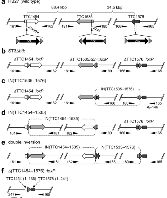
Fig. 8 Schematic representations of the chromosomal location of the three disruption target genes and possible Cre-mediated chromosomal rearrangements. a HB27 (wild-type). The physical distances between the genes are shown above the map. The disruption cassette was inserted at the KpnI and BamHI sites of TTC1535 and TTC1454, respectively. In order to insert the cassette in the TTC1576 ORF, inverse PCR was performed to replace 45 bp of the coding region by a BgIII site (see Fig. 4). b ST3∆htk. Triple markerless gene disruptant without any chromosomal rearrangements. c IN(TTC1535–1576). Inversion between the two loxP sites in TTC1535 and TTC1576. d IN(TTC1454–1535). Inversion between the two loxP sites in TTC1454 and TTC1535. e Double inversion. Co-occurrence of IN(TTC1535–1576) and IN(TTC1454–1535). f ∆(TTC1454–1576)::loxP.
Deletion of the chromosomal region between the two loxP sites in TTC1454 and TTC1576. Numbers in parentheses are the base positions of each coding region. The positions of the primers used for PCR analyses are indicated by filled triangles. The underlined primers in a are located outside of each cloned region in the gene disruption construct. Primers 146 and 233 (thin arrows) in c and f, respectively, were used for DNA sequencing. Shaded triangles show the orientation of the loxP sites.
33 Fig. 9 PCR analyses of the double gene disruptants. The genomic regions analyzed by PCR are indicated on the left. Each primer set and the expected lengths (bp) of the objective PCR products are also indicated in parentheses. The sizes (kbp) of the PCR products are indicated on the right. M indicates the lambda DNA/EcoRI+HindIII size marker. a Analysis of the gene disruption of TTC1576 using a loxP–htk–loxP cassette. The expected length of the PCR product for HB27 is 1846 bp. Lane 1 HB27, lanes 2–4 Km-resistant clones. b Analyses of the removal of htk and predicted junctions of inversion in Km-sensitive ST2 clones. Lane 1 HB27; lanes 2–6 Km-sensitive ST2 clones; lane 7 ST2. c Analyses for the absence of pSH-Cre and segregation/isolation of the ST2∆htk strain. Lane 1 HB27; lanes 2–7 ST2∆htk candidate clones isolated from a third-passage culture of ST2∆htk/pSH-Cre; lane 8 ST2/pSH-Cre.
34
Construction of a triple markerless gene disruptant
To further elucidate the efficacy of the Cre/lox-based system, triple gene disruption was performed for ST2∆htk in the same manner. The next target gene TTC1454 was located approximately 86.8 kb from TTC1535 (Fig. 8a). The ST2∆htk strain was transformed with the gene disruption construct, ∆TTC1454::loxP–htk–loxP, which was amplified from pDELloxTTC1454 (Fig. 3c). As shown in Fig 10a, all of the four Km-resistant transformants examined were identified as TTC1454 disruptants by a PCR analysis using primers 161/162 (lanes 2–5). The triple gene disruptants were designated as the ST3 strain and their genotype was (∆TTC1454::loxP–htk–loxP, ∆TTC1535KpnI::loxP, ∆TTC1576::loxP). ST3 was transformed with pSH-Cre to remove htk by Cre-mediated recombination. Five independent ST3/pSH-Cre transformants were incubated at 50 °C, as described in the Materials and methods.
I found that all transformants were converted to Km-sensitive clones (#1–5) and their genomic DNAs were prepared for a PCR analysis. As shown in Fig. 10b (∆TTC1454::loxP), a 1.9-kb DNA fragment corresponding to the size of ∆TTC1454::loxP was specifically amplified from them, except for clone 4 (lane 5). Since the PCR analysis with htk-specific primers resulted in no detectable DNA band for any clone (Fig. 10b, htk, lanes 2–6), the Km-sensitive clones were tentatively designated as ST3∆htk/pSH-Cre. The genotype of the ST3∆htk strain was (∆TTC1454::loxP, ∆TTC1535KpnI::loxP, ∆TTC1576::loxP).
Similar to the double disruptant, I investigated Cre-mediated chromosomal rearrangements by a series of PCR analyses using sets of primers to amplify each junction of possible inversions and a deletion (Fig. 8c–f). The results shown in Fig. 10b indicated that each multicopy chromosome of clones 3 (lane 4) and 4 (lane 5) was composed of the respective single genotype. Clone 3 was identified as my objective genotype (∆TTC1454::loxP,
∆TTC1535KpnI::loxP, ∆TTC1576::loxP) because a 2.1-kbp DNA fragment derived from
35 genomic DNA without any chromosomal rearrangements was specifically amplified by PCR only when using the primers 180/181 (lane 4). On the other hand, a 1.9-kbp DNA fragment derived from the right junction of the inversion, IN(TTC1454–1535), (Fig. 8d), was specifically amplified for clone 4 (lane 5). The genomic DNA of the other clones was a mixture of all possible patterns of chromosomal rearrangements because, in addition to the 2.1-kbp DNA fragment (no inversion), the junctions of each inversion and large chromosomal deletion described in Fig. 8 c-f were amplified for all of these clones (Fig. 10b, lanes 2, 3, and 6). These results indicate that the Cre/lox system has the potential to induce the inversion (88.4 kbp) and deletion (122.9 kbp) of chromosomal regions in T. thermophilus HB27. In fact, the 1.8-kb DNA fragment amplified by PCR using the primers 165/247 (Fig. 10b, ∆(TTC1454–1576)::loxP, lanes 2, 3, and 6) was confirmed to contain the junction of the 122.9-kbp deletion, ∆(TTC1454–
1576)::loxP, by sequencing the PCR product using the primer 233 (Fig. 8f). The pSH-Cre curing of clone 3 was performed in the same manner, and the absence of pSH-Cre was confirmed for 6 independent clones by a PCR analysis (Fig. 10c, lanes 3–8). Cre-mediated chromosomal rearrangements were not induced during the plasmid curing process (data not shown). As a result, the established triple markerless gene disruptant (∆TTC1454::loxP,
∆TTC1535KpnI::loxP, ∆TTC1576::loxP) was definitively designated it as ST3∆htk. The results presented here demonstrate that the triple markerless gene disruptant was efficiently produced by the Cre/lox-based system. This is the first example of the disruption of three genes located distantly on the chromosome of T. thermophilus in the wild-type background without leaving behind a selectable marker.
36
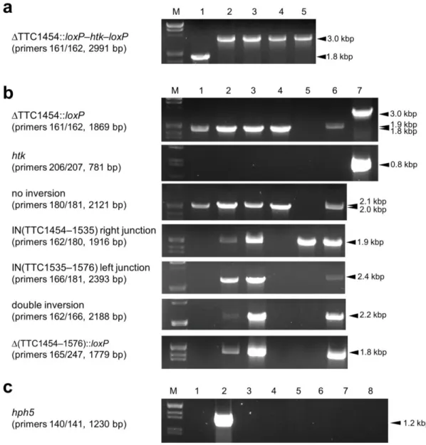
Fig. 10 PCR analyses of the triple gene disruptants. The genomic regions analyzed by PCR are indicated on the left. Each primer set and the expected lengths (bp) of the objective PCR products are also indicated in parentheses. The sizes (kbp) of the PCR products are indicated on the right. M indicates the lambda DNA/EcoRI+HindIII size marker. a Analysis of the gene disruption of TTC1454 using a loxP–htk–loxP cassette. The expected length of the PCR product for HB27 is 1785 bp. Lane 1 HB27, lanes 2–5 Km-resistant clones. b Analyses of the removal of htk and the junctions of the predicted chromosomal rearrangements inTTC1454, TTC1535, and TTC1576 triple disruptants. The predicted inversions and deletion are indicated in Fig. 8. Genomic DNA without any chromosomal rearrangements (no inversion) was examined by the amplification of
∆TTC1535KpnI::loxP. Double inversion indicates the co-occurrence of IN(TTC1454–1576) and IN(TTC1535–1576). Lane 1 HB27; lanes 2–6 Km-sensitive ST3 clones; lane 7 ST3. c Analysis for the absence of pSH-Cre from the ST3∆htk strain. Lane 1 HB27; lane 2 ST3/pSH-Cre; lanes 3–8 ST3∆htk candidate clones isolated from a third-passage culture of ST3∆htk/pSH-Cre.
37
Markerless gene disruption by irreversible Cre/lox recombination using a lox66–htk–lox71 cassette
In experiments for the markerless disruption of multiple genes, I found that each loxP site generated after the removal of htk was responsible for Cre-mediated chromosomal rearrangements during subsequent gene disruptions. In order to minimize this genetic instability, the mutant lox sites, lox66 and lox71, were used to suppress undesired chromosomal rearrangements in other organisms (Albert et al. 1995; Lambert et al. 2007; Kovács et al. 2010).
The lox72 site (Fig. 7b) produced by Cre-mediated recombination between lox66 and lox71 exhibited markedly reduced binding affinity for Cre, and thus allowed for repeated gene disruption. In order to evaluate its performance at 50 °C in T. thermophilus, I constructed another disruption cassette, lox66–htk–lox71 (Fig. 1a). Using the loxP–htk–loxP and lox66–htk–
lox71 cassettes, I constructed a set of gene disruption plasmids for TTC1535 (Fig. 3d) and TTC1537 (Fig. 3e), namely, pDELloxTTC1535 and pDELloxTTC1537 with a loxP–htk–loxP cassette, and pDELmloxTTC1535 and pDELmloxTTC1537 with a lox66–htk–lox71 cassette. As shown in Fig. 11a, all of the disruption cassettes in the four plasmids were in the same direction relative to the two genes on the chromosome, which, in principle, led to an undesired intergenic deletion between the lox sites via Cre-mediated recombination.
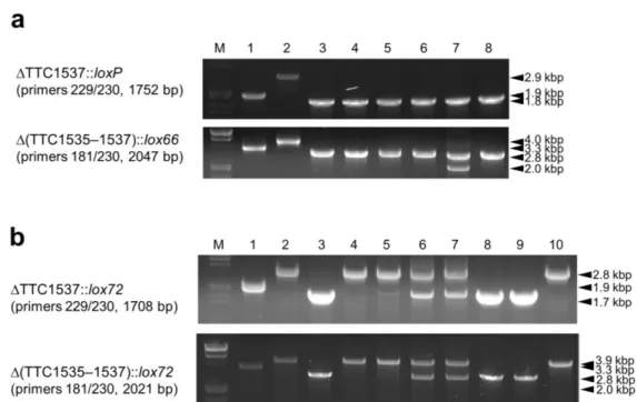
I attempted to generate the TTC1535 disruptant in the wild-type background using a
∆TTC1535::lox66–htk–lox71 construct derived from pDELmloxTTC1535. The transformation, identification of gene disruption (Fig. 12a, lanes 2–5), and introduction of pSH-Cre were successfully performed in the same manner as the methods using a loxP–htk–loxP cassette. In order to remove htk from the resultant ∆TTC1535::lox66–htk–lox71 strain designated as ST4, 16 colonies of ST4/pSH-Cre streaked on PY/Hm plates were incubated at 50 °C for five days.
Each single-colony isolate of the 16 clones was restreaked on PY and PY/Km plates and incubated at 70 °C overnight. However, in contrast to the high efficiency of the experiments
38
using the loxP–htk–loxP cassette, in which 100% of the Km-resistant clones examined were converted to Km-sensitive clones, all 16 clones grew on PY/Km plates. A PCR analysis was performed on seven clones that showed poorer growth than the others on PY/Km plates, and the results obtained indicated that a 2.9-kbp DNA fragment corresponding to the size of
∆TTC1535::lox66–htk–lox71 was amplified for all seven clones (Fig. 12b, lanes 3–9), while a putative 1.7-kbp DNA fragment for ∆TTC1535::lox72 was observed for one clone (Fig. 12b, lane 3). These results suggest that Cre-mediated recombination occurred between lox66 and lox71 in T. thermophilus under my experimental conditions, but with lower efficiency than that between two loxP sites. Since the mutant lox sites represented a marked reduction in the capacity for recombination, the 16 clones on the PY/Hm plate were subjected to an additional round of single colony isolation. In the second cycle on a fresh PY/Hm plate at 50 °C for five days, I found that four of the clones not only grew poorly, but exhibited distinct sensitivity to Km on the PY/Km plate. The four corresponding clones on the PY plate were subjected to a PCR analysis, and the results obtained suggested that each genomic DNA included
∆TTC1535::lox66–htk–lox71 and ∆TTC1535::lox72 (Fig. 12c, lanes 3–6). In order to obtain my objective ∆TTC1535::lox72 strain by chromosome segregation/isolation, 16 independent colonies derived from a frozen stock of clone 1 (Fig. 12c, lane 3) was subjected to a PCR analysis. The results obtained revealed that a 1.7-kbp DNA fragment derived from
∆TTC1535::lox72 was specifically amplified for two clones (Fig. 12d, lanes 5 and 14). I confirmed that the DNA sequence of the junction in the PCR fragment was completely identical to the lox72 sequence (Fig. 7b) produced by Cre-mediated recombination between the mutant lox sites on the lox66–htk–lox71 cassette. After the plasmid curing process for the two clones, I confirmed the absence of pSH-Cre and htk in their genomic DNAs by a series of PCR analyses (data not shown); therefore, the markerless gene disruptant, ∆TTC1535::lox72 designated as
39 ST4∆htk was established.
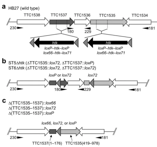
Fig. 11 Schematic representations of the gene disruption of TTC1535 and TTC1537 using loxP–htk–
loxP and lox66–htk–lox71 cassettes. a The chromosomal locus of TTC1534-TTC1538 in T.
thermophilus HB27. In order to insert the indicated cassettes in the ORF of TTC1535 and TTC1537, inverse PCR was performed to replace 300 bp and 280 bp of the respective coding regions by a BglII site. b ST5∆htk and ST6∆htk were constructed by the removal of the htk gene from ∆TTC1537::loxP–
htk–loxP and ∆TTC1537::lox66–htk–lox71 in the ∆TTC1535::lox72 background, respectively. c
∆(TTC1535–1537)::lox. Deletion of the chromosomal region between the two lox sites in TTC1535 and TTC1537. Cre-mediated recombination between lox72 and loxP and between two lox72 sites described in b generated lox66 and lox72, respectively. Numbers in parentheses are the base positions of each coding region. The positions of the primers used for PCR analyses are indicated by filled triangles. The 181 primer is located outside of the cloned region in both gene disruption constructs for TTC1535 and TTC1537. Shaded triangles show the orientation of the lox sites.
40
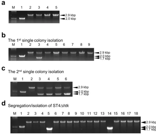
Fig. 12 PCR analyses of TTC1535 disruptants using the lox66–htk–lox71 cassette. The TTC1535 locus was analyzed by PCR using the primers 180/181. The expected lengths of the PCR products for the wild type, ∆TTC1535::lox66–htk–lox71, and ∆TTC1535::lox72 are 1995 bp, 2857 bp, and 1735 bp, respectively. a Analysis of the gene disruption of TTC1535 using a lox66–htk–lox71 cassette. Lane 1 HB27, lanes 2–6 ST4. b Analysis of the removal of htk from ST4 after the first cycle of single colony isolation at 50 °C. The seven ST4/pSH-Cre clones that exhibited moderate sensitivity to Km were analyzed. Lane 1 HB27; lane 2 ST4; lanes 3–9 Km-sensitive ST4/pSH-Cre clones. c Removal of htk after the second cycle of single colony isolation at 50 °C. The four ST4/pSH-Cre clones that exhibited distinct sensitivity to Km were analyzed. Lane 1 HB27; lane 2 ST4; lanes 3–6 Km-sensitive ST4/pSH-Cre clones. d Segregation/isolation of ST4∆htk. The 16 single-colony isolates derived from a frozen stock of one of the Km-sensitive ST4/pSH-Cre clones were analyzed for the identification of a single genotype of ∆TTC1535::lox72. Lane 1 HB27; lane 2 ST4; lanes 3–18 ST4∆htk/pSH-Cre candidate clones. The sizes (kbp) of the PCR products are indicated on the right. M indicates the lambda DNA/EcoRI+HindIII size marker.
41
Double gene disruption using a lox66–htk–lox71 cassette
In order to clarify whether lox72 is inactive during subsequent markerless gene disruption, ST4∆htk was subjected to the disruption of TTC1537. The transformation, identification of gene disruption using the loxP–htk–loxP cassette (Fig. 13a, top, lanes 1 and 2) and lox66–htk–lox71 cassette (Fig. 13b, top, lanes 1 and 2), and introduction of pSH-Cre were performed in the same manner. As a result, I obtained two strains (∆TTC1535::lox72, ∆TTC1537::loxP–htk–loxP) and (∆TTC1535::lox72, ∆TTC1537::lox66–htk–lox71) designated as ST5 and ST6, respectively, with pSH-Cre. All six independent ST5/pSH-Cre clones grown on PY/Hm plates at 50 °C were Km-sensitive, and the removal of htk from their chromosomes was identified by a PCR analysis (Fig. 13a, top, lanes 3–8). When I investigated the deletion of an intergenic region between loxP and lox72, designated as ∆(TTC1535–1537)::lox66 (Fig. 11c), in these clones, a 2.0-kbp DNA fragment derived from the deletion was detected for one clone by a PCR analysis (Fig. 13a, bottom, lane 7).
On the other hand, the removal of htk from the ∆TTC1537::lox66–htk–lox71 region of ST6/pSH-Cre required two cycles of single colony isolation at 50 °C. Among the resultant 16 independent clones examined, one exhibited significant and another seven had moderate sensitivity to Km. A PCR analysis to identify the removal of htk was performed for the eight Km-sensitive clones, and the results obtained suggested that a 1.7-kbp DNA fragment derived from ∆TTC1537::lox72 was specifically amplified for clones 1, 6, and 7 (Fig. 13b, top, lanes 3, 8, and 9). A PCR analysis resulted in no detectable band corresponding to the deletion of an intergenic region between two lox72 sites, designated as ∆(TTC1535–1537)::lox72 (Fig. 11c) for these 3 clones (Fig. 13b, bottom, lanes 3, 8, and 9), whereas the htk-derived DNA fragment was detectable for clone 1 (data not shown). Taken together, five clones of ST5∆htk/pSH-Cre and two clones of ST6∆htk/pSH-Cre were established in this experiment. Plasmid curing was