Introduction
Vascular endothelial growth factor (VEGF) is a major cytokine involved in angiogenesis. It is important to clarify the precise mechanisms by which VEGF expression is regulated. Expression of VEGF is regulated by oxygen concentrations,
VASCULAR ENDOTHELIAL GROWTH FACTOR(VEGF) EXPRESSION IS NEGATIVELY REGULATED BY BASIC-HELIX-LOOP-HELIX (BHLH)
TRANSCRIPTION FACTOR DEC2.
Hiroshi Kijima
1),Fuyuki Sato
1),Ujjal Kumar Bhawal
2),Takeshi Kawamoto
3), Katsumi Fujimoto
3),Tadaatsu Imaizumi
4),Tadanobu Imanaka
5),Jun Kondo
1),
Satoru Koyanagi
6),Mitsuhide Noshiro
3),Hidemi Yoshida
4)and Yukio Kato
3)Abstract The circadian rhythms in mammals are regulated by a pacemaker located in the suprachiasmatic nucleus of the hypothalamus. Five clock-gene families, i.e. Clock, Bmal, Per, Cry and Dec, have been found to be involved in a transcription-translation feedback loop that generates the circadian rhythm at the intracellular level. In this study, we examined functional analysis of the Dec gene. DEC1 and DEC2 are basic-helix-loop-helix (bHLH) transcription factors, involved in cellular diff erentiation, responses to hypoxia, and circadian rhythms. We recently showed that the expression of DEC1 and DEC2 was upregulated by hypoxia, however, the functions of these two factors under hypoxic conditions have not been elucidated in detail. It is well established that the expression of vascular endothelial growth factor (VEGF) is upregulated by hypoxia, and the expression of VEGF in response to hypoxia depends on transcriptional activation by a heterodimer comprising hypoxia-inducible factor 1 α (HIF-1α) and arylhydrocarbon receptor nuclear translocator 1 (ARNT1). In the present study, we showed that DEC2, but not DEC1, suppressed VEGF gene expression under hypoxic conditions. DEC2 protein was co-immunoprecipitated with HIF-1α but not with ARNT1. The binding of HIF-1α to the hypoxia response element (HRE) in the VEGF promoter was decreased by DEC2 overexpression, and increased by DEC2 knockdown. We also showed that the circadian expression of VEGF showed a reciprocal pattern to that of DEC2 in cartilage. DEC2 had a circadian oscillation in implanted Sarcoma 180 cells. We conclude that DEC2 negatively regulates VEGF expression and plays an important role in the pathological conditions in which VEGF is involved.
Hirosaki Med.J. 61, Supplement:S43―S52,2010
Key words: transcription factor; vascular endothelial growth factor (VEGF); hypoxia;
hypoxia-inducible factor-1
α(HIF-1
α)
1)Department of Pathology, Hirosaki University School of Medicine, Hirosaki, 036-8562 Japan; 2)
Department of Oral Maxillofacial Diagnostic Science, Division of Pathology & High-Tech Research Center, Kanagawa Dental College, Kanagawa, 238- 8580 Japan; 3)Department of Dental and Medical Biochemistry, Hiroshima University Graduate School of Biomedical Sciences, Hiroshima 734-8553 Japan; 4)Department of Vascular Biology, Institute of Brain Science, Hirosaki University School of Medicine, Hirosaki, 036-8562 Japan; 5)Department
of the Biology and Medicine of the Stem Cell, Hiroshima University Graduate School of Medical Science, Hiroshima, 734-8553 Japan; 6)Department of Medico-Pharmaceutical Sciences, Kyushu University, Fukuoka, 812-8582 Japan.
Corresponding author: Hiroshi Kijima, M.D.
Department of Pathology, Hirosaki University School of Medicine, Hirosaki 036-8562, Japan.
Tel +81 172 39 5029; Fax +81 172 39 5030 E-mail address: hkijima@cc.hirosaki-u.ac.jp
infl ammation, microbial infections, cytokines, and circadian rhythms
1-6).
Hypoxia is a physiological stress which
induces the expression of VEGF. Hypoxia-
inducible factor 1 (HIF-1) is a transcription
factor which plays a central role in the gene
expression induced by hypoxia, and also in the
development of cancer
7). HIF-1 is a heterodimer of HIF-1α and aryl hydrocarbon receptor nuclear translocator 1 (ARNT1; also known as HIF-1
β).
The expression of HIF-1
αis regulated by oxygen concentrations while ARNT1 is constitutively expressed. There is a hypoxia response element
(HRE) in the promoter region of the VEGF gene
7), and the binding of HIF-1
α/ARNT1 to the HRE initiates the transcription of VEGF mRNA. Mutation of the HRE abrogates the gene transcription induced by hypoxia
8).
It is reported that expression of the VEGF gene was regulated by circadian rhythms
6). Mammalian circadian rhythms are regulated by molecular clockwork systems based on a negative feedback loop in normal and tumor cells. Clock and Brain-muscle-arnt-like-protein
1/2(Bmal 1/2 ) are well known clock genes that positively regulate the expression of target genes.
Period 1/2/3(
Per 1/2/3),
Cryptochromes 1/2/3(
Cr y 1/2/3) and
Differentiated embr yo- chondrocyte 1(
Dec1; also named as Bhlhb2/Sharp2/Stra13) and Dec2
(Bhlhb3/Sharp1) are also designated as clock genes, but negatively regulate the expression of their targets. The heterodimer comprising CLOCK and BMAL 1/2
(CLOCK/BMAL 1/2) enhances transcription of
Dec, Per,and
Cryvia CACGTG E-boxes, and the products of these genes, DEC, PER, and CRY, suppress the transactivation by CLOCK/BMAL 1/2
9,10). It was also reported that PER, but not CRY, regulated the expression of the VEGF gene induced by HIF-1
α/ARNT1
6). However, there have been no reports about the regulation of VEGF by other clock genes.
The basic helix-loop-helix (bHLH) transcrip- tion factors are involved in the expression of various genes. We reported that DEC1 and DEC2 are bHLH transcription factors, and play an important role in the circadian rhythm of the suprachiasmatic nucleus (SCN)
11)or peripheral tissue
10,12,13)as a negative regulator. These two DECs were shown to regulate the diff erentiation
of chondrocytes, skeletal muscles, and nervous systems
14 -16). Recently, we reported that the expression of DEC1 and DEC2 is induced by hypoxia
17). This suggests that DEC1 and DEC2 are involved in the expression of other hypoxia- inducible genes.
In the present study, we investigated the role of DEC1 and DEC2 in hypoxia-induced VEGF expression. Our results showed that DEC2 regulated the expression of the VEGF gene induced by HIF-1
α/ARNT1 transactivation by interacting with HIF-1
αand interfered with the binding of HIF-1
αto the VEGF promoter under hypoxic conditions, and that DEC2 showed the opposite phase to VEGF in circadian rhythm.
Materials and Methods
Cell culture
NIH3T3, Sarcoma 180, and COS-7 cells were obtained from the American Type Culture Collection (ATCC, Manassas, VA, USA). HSC- 3 cells were obtained from the Japanese Cancer Research Resources Bank. Cells were cultured in Dulbeccoʼs Modified Eagleʼs Medium-high glucose (Sigma Chemical Co., St. Louis, MO, USA) supplemented with 10% fetal bovine serum. The cells were cultured at 37°C in a humidified atmosphere of 95% air and 5% CO
2(20% O
2). Hypoxic exposure was performed in a hypoxic chamber (1% O
2for NIH3T3 or HSC- 3 cells, and 1% O
2for 4 h or 3% O
2for 24 h for Sarcoma 180 cells; Sarcoma 180 cells died in the presence of 1% O
2for 24 h).
Construction of reporter and expression plasmids
The luciferase reporter plasmid,
Veg f-luc,
containing a 1289-bp fragment of the mouse
Vegfgene
in pGL3-basic (Fig. 1A), was previously
described
6). A 34-bp fragment containing the
hypoxia- response element (HRE), as well
as mutated HRE (HREm), of
Veg fwith the
flanking sequence was made by annealing the
oligonucleotides as described previously
18). These fragments were ligated into the
NheIand
XhoIsites of pTK-Luc upstream of the TK promoter
(Fig. 1A; pVEGF-HRE-TK-Luc and pVEGF- HREm-TK-Luc). The promoter constructs of HRE and HREm of DEC1 (Fig. 1A; pDEC1- HRE-TK-Luc and pDEC1-HREm-TK-Luc), and the expression plasmids for FLAG -HIF-1
α, FLAG-DEC2, FLAG-DEC1, DEC2 pcDNA, DEC1 pcDNA, and PER2 pcDNA were previously described
11,15,17,19). Expression constructs encoding full-length mouse ARNT1 were obtained by subcloning PCR-generated fragments into the vector pcDNA3.1.
Transient Transfection and luciferase assay NIH3T3 or Sarcoma 180 cells were seeded at 1×10
4cells per 16 -mm well 24 h before transfection. The transfection of plasmid DNA was performed using Lipofectamine 2000 reagent
(Invitrogen Corp., Carlsbads, CA, USA) as previously described
12). After the transfection, t he cells were incubated under normoxic conditions for 16 h. Then, these cells were incubated for an additional 24 h under hypoxic or normoxic conditions. The cells were harvested and the luciferase activity in the cell lysate was determined using a Dual Luciferase Reporter Assay System (Promega, Madison, WI, USA) . Luciferase activity was normalized using pTK-luc activity as a control. Values are mean ± SD for three wells.
Reverse transcription-polymerase chain reaction
(RT-PCR)
Total RNA was isolated using an RNEasy RNA isolation kit (QIAGEN, Hilden, Germany).
First-strand cDNA was synthesized from 1
μg of total RNA, using ReverTra Ace (TOYOBO, Osaka, Japan). PCR was performed using an aliquot of fi rst-strand cDNA as a template under standard conditions with
ExTaqDNA polymerase
(Takara, Siga, Japan). The cDNA for mouse
V E GF, A R NT1, DE C1, DE C 2 , PE R 2 , a nd GAPDH were amplified up to 30 cycles. The cDNA for mouse HIF-1
αwas amplifi ed 32 cycles.
The primers used were previously described
18).
Enzyme-linked immunosorbent assay (ELISA)
The concentration of mouse VEGF in the conditioned medium was measured using an ELISA kit (R&D systems, Minneapolis, MN, USA). This assay recognizes the soluble forms of VEGF (mouse VEGF
120and VEGF
164). The values were represented as the mean ± SD for the three wells.
Western blot analysis
The cells were seeded at 1×10
5cells per 3 5 -mm well and lysed using M-PER lysis buffer (PIERCE, Rockford, IL, USA). Protein concentrations were determined by BCA assay.
The lysates (30
μg protein) were subjected to SDS/PAGE, and proteins were transferred to PVDF membranes. The membranes were incubated with antibodies specific for DEC2
(1:2000)
12), DEC1 (1:1000)
19), HIF-1
α(1:1000, Abcam, Cambridge, England), ARNT1 (1:3000, Abca m), PE R 2 ( 1 : 5 0 0 , Tra ns G en ic I nc , Kobe, Japan) or actin (1:30000) followed by a horseradish peroxidase-conjugated secondary antibody (1:5000). An ECL Advance Western Blotting Detection kit (Amersham, Uppsala, Sweden) was used for detection.
Immunoprecipitation
COS-7 cells were seeded at 1×10
6cells per 10 0 -mm well 24 h before transfection, and transfected with expression vectors using Lipofectamine 2000 reagent. After 40 h, cells were lysed in lysis buff er containing 50 mM Tris- HCl (pH 7.4), 150 mM NaCl, 1 mM EDTA, and 1% Triton X-100. The lysates were incubated with anti-FLAG M2 agarose affi nity gel (Sigma)
overnight at 4°C. Immunoprecipitates were
washed three times with buffer containing
0.5 M Tris-HCl (pH7.4) and 1.5 M NaCl, and then were eluted in SDS sample buffer. The immunoprecipitated proteins were analyzed by Western blotting.
Animal models
Six-week- old male C57/ BL6 mice (Crea Japan, Tokyo, Japan) were housed under 12:12- h LD conditions for 19 days, and killed at the indicated zeitgeber (ZT-an environmental agent or event that provides the setting or resetting of the biological clock of an organism: ZT-0 corresponds to 8:00 AM lights on) time on day 20, and mouse rib cartilage samples were placed within phosphate-buffered saline (PBS)
and stored in a deep freezer. Mice were housed under LD conditions. Sarcoma 180 cells were implanted into the mice and RNA was prepared as previously described
6). The cDNA of HIF-1α, DEC2, VEGF, and GAPDH was amplifi ed by RT- PCR. To evaluate the quantitative reliability of RT-PCR, we performed a kinetic analysis of amplified products to ensure that signals were derived only from the exponential phase of amplification. The density of each band was analyzed with the use of NIH image software.
Results
Effects of DEC1 and DEC2 on the promoter activity of
Vegf.
We examined whether DE C1 or DE C2 aff ects the promoter activity of
Vegf. Cells were transiently transfected with a full-length
Veg fpromoter containing a HRE (Fig. 1A) together with the expression vector for DEC1 or DEC2.
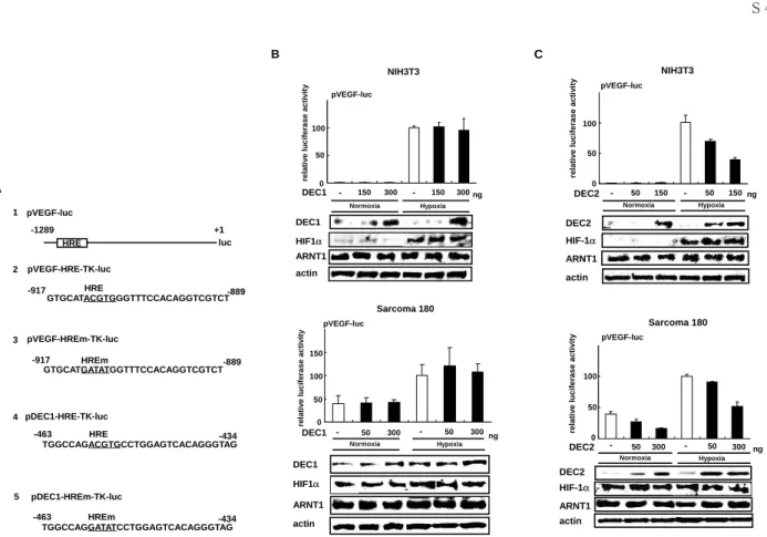
The promoter activity of
Veg fwas increased by hypoxia about 100 fold and the increase was not aff ected by DEC1 in NIH3T3 cells (Fig. 1B, upper panel). In Sarcoma 180 cells, the promoter activity of
Vegfwas strong even under normoxic conditions, and was not altered by DEC1 (Fig.
1B, lower panel). The promoter activity of Vegf under hypoxia was not suppressed by DEC1.
On the other hand, DEC2 suppressed, in a dose- dependent manner, the promoter activity of
Vegfinduced by hypoxia in NIH3T3 cells (Fig.
1C, upper panel) or the activity in Sarcoma 180 cells under both normoxia and hypoxia (Fig. 1C, lower panel). We confirmed, by Western blot analysis, that overexpression of DEC1 or DEC2 does not alter the endogenous levels of HIF-1
αand ARNT1 proteins in cells.
Eff ect of DEC2 on the VEGF promoter activity induced by HIF-1
α/ARNT1
transactivation.
The HIF-1
α/ARNT1 heterodimer plays a critical role in the transcription of
Vegfinduced by hypoxia
7). We next examined if DEC2 aff ects the promoter activity of Vegf induced by HIF-1α/
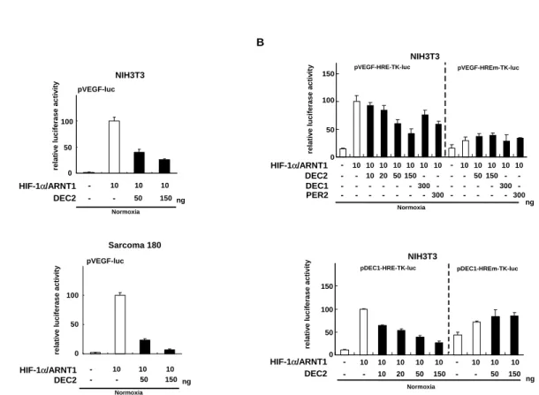
ARNT1 transactivation. When the constructs for HIF-1α and ARNT1 were co-transfected into NIH3T3 or Sarcoma 180 cells, the promoter activity of Vegf increased about 100 fold even under normoxic conditions (Fig. 2A upper and lower panel), and the increase was suppressed by DEC2 in a dose-dependent manner.
We also examined if DEC2 affects the promoter activity of truncated constructs containing only HRE (pVEGF-HRE-TK-luc)
or HREm (pVEGF-HREm-TK-luc) (Fig. 1A).
When the constructs for HIF-1
αand ARNT1
were co-transfected into NIH3T3 cells, the
promoter activity of VEGF-HRE increased about
5 fold under normoxic conditions (Fig. 2B, upper
panel). Co-transfection of the PER2 construct
slightly suppressed the promoter activity of
VEGF-HRE as previously described
6). When
the DEC2 construct was co-transfected, the
promoter activity of VEGF-HRE was suppressed
in a dose-dependent manner (Fig. 2B, upper
panel). The inhibitory effect of DEC2 on the
promoter activity was more signifi cant than that
of PER2. The eff ect of the DEC1 construct was
weak. Co-expression of HIF-1α and ARNT1
did not induce the promoter activity of VEGF-
HREm. The activity of VEGF-HREm was not changed by the co-transfection of DEC2, DEC1, or PER2.
Dec1
is a target of HIF-1
αlike
Vegf17)
. We next examined whether DEC2 suppresses the activity of the DEC1-HRE promoter (Fig. 1A)
induced by HIF-1
α/ARNT1 transactivation. The promoter activity of DEC1-HRE induced by the transactivation was also suppressed by the co- transfection of DEC2 (Fig. 2B, lower panel). On the other hand, the promoter activity of DEC1-
HREm was not induced by HIF-1
α/ARNT1 transactivation, similar to VEGF-HREm, and the activity of DEC1-HREm was not changed by co- transfection of DEC2.
DEC2 protein interacts with HIF-1
αprotein in cells.
We performed an immunoprecipitation assay in order to examine the interaction between DEC2 and HIF-1
αor ARNT1 (Fig. 3). COS-7 cells were transfected with expression vectors
Figure 1 Eff ect of DEC1 or DEC2 on the promoter activity of the VEGF gene. (A) The diagram shows the structure of luciferase reporter constructs containing the hypoxia response element (HRE) or HRE mutant (HREm) of the 5ʼ-fl anking regions of Veg f and Dec1. 1. pVEGF-luc; full-length mouse Veg f containing 1289 bp upstream of the gene. 2. pVEGF-HRE-TK-luc; HRE with 29 bp of fl anking sequence in Veg f was subcloned into pGL3-TK. 3.
pVEGF-HREm-TK-luc; HREm with 29 bp of fl anking sequence in Veg f was subcloned into pGL3-TK. 4. pDEC1- HRE-TK-luc; HRE with 30 bp of fl anking sequence in Dec1 was subcloned into pGL3-TK. 5. pDEC1-HREm-TK- luc; HREm with 30 bp of fl anking sequence in Dec1 was subcloned into pGL3-TK. (B) The reporter construct pVEGF-luc was co-transfected with the expression vector for DEC1 into NIH3T3 or Sarcoma 180 cells. After 16 h of transfection, the cells were incubated under normoxic or hypoxic conditions for an additional 24 h. Each value represents the mean ± SE (bars). The expression of DEC1, HIF-1α, ARNT1, and actin protein was confi rmed by Western blot analysis. (C) The reporter construct pVEGF-luc was co-transfected with the expression vector for DEC2 into NIH3T3 or Sarcoma 180 cells. The cells were cultured under normoxic or hypoxic conditions and subjected to a luciferase assay or Western blot analysis as in (B). Each value represents the mean ± SE (bars). The expression of DEC2, HIF-1α, ARNT1 and actin protein was confirmed by Western blotting. (modified fi gures of ref. #18)
A
HRE luc
pVEGF-luc
pVEGF-HRE-TK-luc
pVEGF-HREm-TK-luc
pDEC1-HRE-TK-luc
pDEC1-HREm-TK-luc
GTGCATACGTGGGTTTCCACAGGTCGTCT
-917 -889
GTGCATGATATGGTTTCCACAGGTCGTCT
-917 HREm -889
TGGCCAGACGTGCCTGGAGTCACAGGGTAG
-463 HRE -434
TGGCCAGGATATCCTGGAGTCACAGGGTAG
-463 HREm -434
1
-1289 +1
2
3
4
5
HRE
B
NIH3T3
DEC1 - 50 300 ng
0 100 50
50 100
relativeluciferaseactivityrelativeluciferaseactivity 0
Sarcoma 180
Normoxia Hypoxia
DEC1 - 150 300 - 150 300ng
DEC1
actin DEC1
pVEGF-luc
actin HIF1α
HIF1α 150
pVEGF-luc
- 50 300
Normoxia Hypoxia
ARNT1
ARNT1
C
NIH3T3
DEC2 50 ng
Normoxia
- 300
0 100
50
relativeluciferaseactivityrelativeluciferaseactivity
Sarcoma 180 Normoxia Hypoxia DEC2 - 50 150 - 50 150ng
DEC2
actin
actin DEC2
pVEGF-luc
pVEGF-luc
- 50 300 Hypoxia HIF-1α
HIF-1α ARNT1
ARNT1 0 100
50
for ARNT1, FLAG, FLAG-HIF-1
α, FLAG-DEC1, or FLAG-DEC2. After 40 h of transfection, the cells were lysed and the expression of ARNT1, DEC2, or DEC1 protein was confi rmed by immunoblotting using aliquots of total cell lysate. FLAG-tagged proteins in the lysates were immunoprecipitated with anti-FLAG antibody, and immunoblotted with antibodies against ARNT1 (Fig. 3A), DEC2, or DEC1
(Fig. 3B). HIF-1α was co-immunoprecipitated with ARNT1 as previously described
20). DEC2 was co-precipitated with HIF-1
αbut not with ARNT1. On the other hand, DEC1 was not bound to HIF-1
αor ARNT1. These results demonstrated that DEC2 protein could physically bind to HIF-1
αin cells.
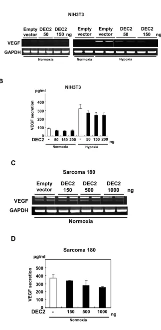
DEC2 suppressed the endogenous expression of VEGF.
We further examined if DEC2 suppresses the endogenous VEGF expression induced by hypoxia. NIH3T3 cells were transfected with the expression vector for DEC2. After 16 h of transfection, the cells were incubated for an additional 24 h under hypoxic conditions. The expression level of VEGF mRNA was examined by RT-PCR (Fig. 4A), and the amount of VEGF protein in the conditioned medium was measured by an ELISA (Fig. 4B). DEC2 suppressed, in a dose-dependent manner, the endogenous expression of VEGF mRNA and protein induced by hypoxia, but had no eff ect on the expression under normoxic conditions. In Sarcoma 180 cells,
Figure 2 DEC2 suppressed the VEGF promoter activity induced by HIF-1α/ARNT1 transactivation in NIH3T3 and Sarcoma 180 cells. (A) The reporter construct pVEGF-luc was co-transfected with the expression vectors for HIF-1α and ARNT1 together with DEC2 into NIH3T3 or Sarcoma 180 cells. After incubation under normoxic conditions for 40 h, a Luciferase assay was performed. Each value represents the mean ± SE (bars). (B) The reporter construct pVEGF-HRE-TK-luc or pVEGF-HREm-TK-luc was co-transfected with expression vectors for HIF-1α and ARNT1 together with DEC2, DEC1, or PER2 into NIH3T3 cells (upper panel). The reporter construct pDEC1-HRE-TK-luc or pDEC1-HREm-TK-luc was co-transfected with expression vectors for HIF-1α and ARNT1 together with DEC2 into NIH3T3 cells (lower panel). After 40 h of incubation, the cells were lysed and subjected to a luciferase assay. Each value represents the mean ± SE (bars). (modifi ed fi gures of ref. #18)
A
NIH3T3
0 100
50
relativeluciferaseactivity
DEC2 - 50 150ng
pVEGF-luc
HIF-1α/ARNT1 - -
10 10 10
pVEGF-luc
DEC2 - 50 150ng
- -
10 10 10
relativeluciferaseactivity
Sarcoma 180
Normoxia
Normoxia
HIF-1α/ARNT1 0 100
50
B
NIH3T3
relativeluciferaseactivity 0
DEC2
ng
- 50 150
- - 10 10 10
relativeluciferaseactivity
Normoxia 50
100
10 10 10 10 - 10 10 10 DEC1
PER2
- - - - 50 150 - -
- - - - 300
- -
- 300
-
- - - 300
- - 300
DEC2 - 50 150 ng
- -
10 10 10 - 10 10 10
- - 50 150 Normoxia
0 50 HIF-1α/ARNT1
HIF-1α/ARNT1
NIH3T3
pDEC1-HREm-TK-luc pDEC1-HRE-TK-luc
100 150
10 10 10 20
- - - - - - -
- - - - 150
pVEGF-HRE-TK-luc pVEGF-HREm-TK-luc
20 10 10 10
DEC2 suppressed the endogenous expression of VEGF during normoxia in a dose-dependent manner (Fig. 4C and 4D).
Day-night difference of VEGF and DEC2 mRNA in implanted tumor cells.
As shown in Fig. 5, mice were housed under a LD cycle and a 50
μl volume containing 1.5×
10
6viable Sarcoma 180 cells was injected into the right hind footpad of each mouse. The tumor volume was estimated as previously described
6). The mRNA levels for DEC2 and VEGF in implanted Sarcoma 180 cells exhibited an opposite circadian phase at ZT2 and ZT14
(
P< 0.01), but HIF-1
αdid not show a circadian rhythm.
Figure 3 DEC2 interacts with HIF-1α. (A, B) COS- 7 cells were transfected with the expression vector for ARNT1, FLAG, FLAG -HIF-1α, FLAG-DEC1 or FLAG-DEC2. After 40 h of transfection, cells were lysed and the lysates were immunoprecipitated (IP) with anti- FLAG antibody, and immunoblotted (IB) with antibodies against ARNT1 (A), DEC2 or DEC1
(B). To confirm the expression of ARNT1, DEC2, and DEC1 proteins, aliquots of total cell lysate were immunoblotted with each antibody.
(modifi ed fi gures of ref. #18)
Figure 4 DEC2 suppressed the endogenous expression of VEGF. NIH3T3 cells were transfected with the expression vector for DEC2. After 16 h of transfection, the cells were exposed to hypoxia and incubated for an additional 24 h. (A) RT-PCR analysis for VEGF mRNA was performed as shown in Fig.1. (B) The conditioned medium was collected and the secretion of VEGF protein into the medium was measured by an ELISA. Each value represents the mean ± SE (bars). Sarcoma 180 cells were transfected with the expression vector for DEC2. After 40 h of transfection, RT-PCR (C) and ELISA (D) were performed as above. Each value represents the mean ± SE (bars). (modifi ed fi gures of ref. #18)
ARNT1
IP:FLAG IB:ARNT1 Cell lysate IB:ARNT1 FLAG FLAG-HIF-1α FLAG-DEC2 FLAG-DEC1
A
+
+ + + + +
+
-
-
+- - -
+-
+
- -
- - - - -
+- - -
+- -
COS-7
Cell lysate IB:DEC2 IP:FLAG IB:DEC2
Cell lysate IB:DEC1 IP:FLAG IB:DEC1
B
DEC2 FLAG FLAG-HIF-1α
+ ++
- -
+DEC1 FLAG FLAG-HIF-1α
+ ++
- -
+COS-7
NIH3T3
DEC2 50 150 ng
Normoxia Hypoxia
- 200 - 50 150 200 0
200 300 400
VEGFsecretion 100
A
VEGF GAPDH
NIH3T3
Empty vectorDEC2
50 DEC2150
ng DEC2
50 150
DEC2
Normoxia Normoxia Hypoxia
ng
B
Empty vector Empty
vector
pg/ml
C
VEGF GAPDH
Empty vector DEC2
150 500 1000 DEC2 DEC2
ng
Normoxia Sarcoma 180
DEC2 - 150 500 1000 ng Sarcoma 180
Normoxia 0
100 500
200 300 400
VEGFsecretion
D
pg/ml
Discussion
The hypoxia-inducible factor HIF-1
αis the key regulator in cellular adaptation to hypoxia.
Hypoxia stabilizes HIF-1
αallowing it to form a heterodimer with ARNT1
7): The heterodimer HIF-1α/ARNT1 binds to the HRE, and causes an increase in the expression of target genes such as the VEGF and erythropoietin genes
21). In the present study, we demonstrated that DEC2 interacts with HIF-1
α, and interferes w it h t he bind ing of H I F-1
αto t he H R E , resulting in downregulation of the expression of HIF-1
α-inducible genes such as the VEGF gene during hypoxia. Recent studies have shown that both DEC1 and DEC2 were induced to express by hypoxic conditions
17,22-24). Here we showed that the expression of DEC1 and DEC2 was also induced by hypoxia in mouse fibroblasts, Sarcoma 180 cells, and human squamous cell carcinoma cells. Although HIF-1
αwas activated by hypoxia in these cells, the induction of DEC2 expression by hypoxia was slower than that of HIF-1
αand DEC1 expression.
This is a new molecular mechanism in which the DEC2-mediated feedback loop participates in the regulation of gene expression induced by hypoxia. We also found that DEC1 did not bind to HIF-1
α, and had little eff ect on the promoter activity of the V EGF gene. This finding indicates that DEC1 and DEC2, which diff er in structure of the C-terminal, have distinct roles in adaptation to hypoxia: DEC2 has the alanine and glycine-rich region in its C-terminal half, but DEC1 does not.
In contrast to DEC1 and DEC2 expression, PER2 expression was downregulated by hypoxia in mouse Sarcoma 180 and human squamous cell carcinoma cells. It was reported that PER2 also inactivates HIF-1
αin a similar manner to DEC2
6): PER2 inhibited HIF-1
α/ARNT1-induced VEGF promoter activity in Sarcoma 180 cells by interacting with HIF-1α protein. The different expression profiles of DEC2 and PER2 under hypoxic conditions suggest distinct functions in the transcriptional regulation of target genes of HIF-1
α.
Both DEC1 and DEC2 show a circadian expression in many tissues
11,13 , 2 5). We also showed, in the present study, that DEC2 and VEGF protein expression had a circadian rhythm in cartilage. The expression of VEGF showed a reciprocal pattern to that of DEC2 at ZT2 and ZT14. Furthermore, both DEC2 and VEGF had circadian oscillations in implanted Sarcoma 180 cells, and the expression of VEGF mRNA showed the opposite circadian phase to that of DEC2 mRNA. These findings indicate that DEC2 regulates VEGF expression in both normal and tumorous tissues.
Several recent reports showed that clock genes play important roles in cancer biology:
(i) The mRNA expression of DEC1 was sup- pressed by the overexpression of VHL in renal cell carcinoma
26), (ii) Knockdown of BMAL2 enhanced cell proliferation in hepatocellular carcinoma
27), and (iii) PER1 plays an important
Figure 5 Circadian expression of VEGF and DEC2 mRNA in implanted tumor cells. Temporal profi les of mRNA expression of HIF-1α, DEC2, VEGF164, and VEGF120 in tumor masses. For plots of RNA, the mean peak values for HIF-1α, DEC2, VEGF164, and VEGF120, are set at 100. Each point represents the mean ± SE
(bars; n=4-6). The mRNA levels for DEC2, VEGF164, and VEGF120 exhibit significant circadian variations. (modifi ed fi gures of ref.
#18)
2 6 10 14 18 22
Zeitgeber time (ZT) 120
100 80 60 40 20 0
RelativeRNAlevels
DEC2 VEGF164
VEGF120
HIF-1α
role in cell growth and DNA damage control in human cancer cells
28). DEC2 may contribute to the regulatory mechanism in tumor cell biology.
In summary, we revealed that DEC2 nega- tively regulates VEGF expression by interacting with HIF-1
α, and interferes with the binding of HIF-1
αto the HRE in the VEGF promoter under hypoxic conditions. Therefore, DEC2 may regulate the other genes targeted by HIF-1
α. In addition, DEC2 regulates the transcription of its targets by interacting with E-box in a circadian fashion
15). DEC2 may be a key transcription factor which plays an important role in the crosstalk between the hypoxia-response system and circadian regulation
18).
Acknowledgements
This work was supported by Grants-in-Aid for Science from the Ministry of Education, Culture, Sports, Science and Technology of Japan; Grant for Hirosaki University Institutional Research;
and The Karoji Memorial Fund for Medical Research.
References
1)Shweiki D, Itin A, Soffer D, Keshet E. Vascular endothelial growth factor induced by hypoxia may mediate hypoxia-initiated angiogenesis.
Nature 1992;359:843-5.
2)Hlatky L, Tsionou C, Hahnfeldt P, Coleman CN.
Mammary fibroblasts may influence breast tumor angiogenesis via hypoxia-induced vascular endothelial growth factor up-regulation and protein expression. Cancer Res 1994;54:6083-6.
3)Ben-Av P, Crofford LJ, Wilder RL, Hla T. In- duction of vascular endothelial growth factor expression in synovial fi broblasts by prostaglandin E and interleukin-1: a potential mechanism for infl ammatory angiogenesis. FEBS Lett 1995;372:83- 7.
4)Li J, Perrella MA, Tsai JC, Yet SF, Hsieh CM, Yoshizumi M, Patterson C, Endege WO, Zhou F, Lee ME. Induction of vascular endothelial growth factor gene expression by interleukin-1 beta in rat
aortic smooth muscle cells. J Biol Chem 1995;270:
308-12.
5)Sato F, Imaizumi T, Sashinami H, Yoshida H, Kusumi T, Mori F, Wakabayashi K, Nakane A, Satoh K, Kijima H. Upregulation of vascular endothelial growth factor by heat-killed Listeria monocytogenes in macrophages. Biochem Biophys Res Commun 2007;354:608-12.
6)Koyanagi S, Kuramoto Y, Nakagawa H, Aramaki H, Ohdo S, Soeda S, Shimeno H. A molecular mechanism regulating circadian expression of vascular endothelial growth factor in tumor cells.
Cancer Res 2003;63:7277-83.
7)Forsythe JA, Jiang BH, Iyer NV, Agani F, Leung SW, Koos RD, Semenza GL. Activation of vascular endothelial growth factor gene transcription by hypoxia-inducible factor 1. Mol Cell Biol 1996;16:
4604-13.
8)Kimura H, Weisz A, Kurashima Y, Hashimoto K, Ogura T, DʼAcqisto F, Addeo R, Makuuchi M, Esumi H. Hypoxia response element of the human vascular endothelial growth factor gene mediates transcriptional regulation by nitric oxide: control of hypoxia-inducible factor-1 activity by nitric oxide.
Blood 2000;95:189-97.
9)Sangoram AM, Saez L, Antoch MP, Gekakis N, Staknis D, Whiteley A, Fruechte EM, Vitaterna MH, Shimomura K, King DP, Young MW, Weitz CJ, Takahashi JS. Mammalian circadian autoregulatory loop: a timeless ortholog and mPer1 interact and negatively regulate CLOCK-BMAL1- induced transcription. Neuron 1998;21:1101-13.
10)Hamaguchi H, Fujimoto K, Kawamoto T, Noshiro M, Maemura K, Takeda N, Nagai R, Furukawa M, Honma S, Honma K, Kurihara H, Kato Y.
Expression of the gene for Dec2, a basic helix- loop-helix transcription factor, is regulated by a molecular clock system. Biochem J 2004;382:43-50.
11)Honma S, Kawamoto T, Takagi Y, Fujimoto K, Sato F, Noshiro M, Kato Y, Honma K. Dec1 and Dec2 are regulators of the mammalian molecular clock. Nature 2002;419:841-4.
12)Kondo J, Sato F, Fujimoto K, Kusumi T, Imanaka T, Kawamoto T, UK Bhawal, Noshiro M, Kato Y, Sato T, Kijima H. 57Arg in the bHLH transcription factor DEC2 is essential for the suppression of
CLOCK/BMAL2-mediated transactivation. Int J Mol Med 2006;17:1053-6.
13)Noshiro M, Kawamoto T, Furukawa M, Fujimoto K, Yoshida Y, Sasabe E, Tsutsumi S, Hamada T, Honma S, Honma K, Kato Y. Rhythmic expression of DEC1 and DEC2 in peripheral tissues: DEC2 is a potent suppressor for hepatic cytochrome P450s opposing DBP. Genes Cells 2004;9:317-29.
14)Shen M, Kawamoto T, Yan W, Nakamasu K, Tamagami M, Koyano Y, Noshiro M, Kato Y.
Molecular characterization of the novel basic helix-loop-helix protein DEC1 expressed in differentiated human embryo chondrocytes.
Biochem Biophys Res Commun 1997;236:294-8.
15)Fujimoto K, Hamaguchi H, Hashiba T, Nakamura T, Kawamoto T, Sato F, Noshiro M, UK Bhawal, K suardita, Kato Y. Transcriptional repression by the basic helix-loop-helix protein Dec2: multiple mechanisms through E-box elements. Int J Mol Med 2007;19:925-32.
16)Boudjelal M, Taneja R, Matsubara S, Bouillet P, Dolle P, Chambon P. Overexpression of stra13, a novel retinoic acid-inducible gene of the basic- helix-loop-helix family, inhibits mesodermal and promotes neuronal differentiation of P19 cells.
Genes Dev 1997;11:2052-65.
17)Miyazaki K, Kawamoto T, Tanimoto K, Nishiyama M, Honda H, Kato Y. Identification of functional hypoxia response elements in the promoter region of the DEC1 and DEC2 genes. J Biol Chem 2002;277:47014-21.
18)Sato F, Bhawal UK, Kawamoto T, Fujimoto K, Imaizumi T, Imanaka T, Kondo J, Koyanagi S, Noshiro M, Yoshida H, Kusumi T, Kato Y, Kijima H. Basic-helix- loop-helix (bHLH) transcription factor DEC2 negatively regulates vascular endothelial growth factor expression. Genes Cells 2008;13:131-44.
19)Sato F, Kawamoto T, Fujimoto K, Noshiro M, Honda KK, Honma S, Honma K, Kato Y.
Functional analysis of the basic helix-loop-helix transcription factor DEC1 in circadian regulation.
Interaction with BMAL1. Eur J Biochem 2004;
271:4409-19.
20)Jiang BH, Rue E, Wang GL, Roe R, Semenza GL.
Dimerization, DNA binding, and transactivation properties of hypoxia-inducible factor 1. J Biol Chem 1996;271:17771-8.
21)Grimm C, Wenzel A, Groszer M, Mayser H, Seeliger M, Samardzija M, Bauer C, Gassmann M, Reme CE. HIF-1-induced erythropoietin in the hypoxic retina protects a gainst light- induced retinal degeneration. Nat Med 2002;8:718-24.
22)Yun Z, Maecker HL, Johnson RS, Giaccia AJ.
Inhibition of PPAR2. Oncogene 2002;22:5306-14.
23)Chakrabarti J, Turley H, Campo L, Han C, Harris AL, Gatter KC, Fox SB. The transcriptional factor DEC1 (stra13, Sharp2) is associated with the hypoxic response and high tumor grade in human breast cancers. Br J Cancer 2004;91:954-8.
24)Ivanov SV, Salnikow K, Ivanova AV, Bai L, Lerman MI. Hypoxic repression of STAT1 and its downstream genes by a pVHL/HIF-1 target DEC1/STRA13. Oncogene 2007;26:802-12.
25)Furukawa M, Kawamoto T, Noshiro M, Honda KK, Sakai M, Fujimoto K, Honma S, Honma K, Hamada T, Kato Y. Clock gene expression in the submandibular glands. J Dent Res 2005;84:1193-7.
26)Ivanova AV, Ivanov SV, Danilkovitch-Miagkova A, Lerman MI. Regulation of stra13 by the von Hippel-Lindau tumor suppressor protein, hypoxia, and the UBC9/ubiquitin proteasome degradation pathway. J Biol Chem 2001;276:15306-15.
27)Yeh CT, Lu SC, Tseng IC, Lai HY, Tsao ML, Huang SF, Liaw YF. Antisense overexpression of BMAL2 enhances cell proliferation. Oncogene 2003;22:5306-14.
28)Gery S, Komatsu N, Baldjyan L, Yu A, Koo D, Koeffler HP. The circadian gene per1 plays an important role in cell growth and DNA damage control in human cancer cells. Mol Cell 2006;
22:375-82.