「
UDP/P2Y6 receptor signaling regulates IgE-dependent degranulation in human basophils
」(
UDP/P2Y6
受容体によるヒト好塩基球IgE
依存性脱顆粒反応の調節)
弘前大学大学院保健学研究科保健学専攻
提出者氏名: 中 野 学
所 属: 医療生命科学領域 病態解析学分野
指導教員: 高 見 秀 樹
Contents
Abbreviations ... 2
Introduction ... 3
Materials and Methods ... 5
Results ... 9
Discussion ... 16
Acknowledgments ... 19
References... 20
和文要旨 ... 25
Abbreviations
AC : adenylate cyclase
ENPP : ecto-nucleotide pyrophosphatase/phosphodiesterase ENTPDase : ecto-nucleoside triphosphate diphosphohydrolase GPCR : G-protein coupled receptor(Gタンパク共役型受容体)
MFI : mean fluorescence intensity(平均蛍光強度)
PKC : protein kinase C(プロテインキナーゼC) PLC : phospholipase C(ホスホリパーゼC) P2YR : P2Y purinergic receptors
7-AAD : 7-Amino-Actinomycin D
Introduction
Nucleotides are released from damaged cells, secreted by activated cells, or spontaneously secreted1,2). Extracellular nucleotides regulate a variety of biological functions through P2 purinergic receptors expressed on cell surfaces3,4). P2 purinergic receptors are divided into two subfamilies: P2X receptors (P2X1–7), which function as ion channels, and P2Y receptors (P2Y1, P2Y2, P2Y4, P2Y6, P2Y11, P2Y12, P2Y13, P2Y14), which function as G protein-coupled-receptors (GPCRs). The ligands of P2Y receptors vary according to subtype. ATP is the ligand for P2Y2 and P2Y11, ADP for P2Y1, P2Y12, and P2Y13, UTP for P2Y2 and P2Y4, UDP for P2Y6, and UDP-glucose for P2Y14. The effects of P2Y receptor activation vary according to the coupled G protein. The Gαs-coupled receptor subtype (P2Y11) activates adenylate cyclase (AC).
Conversely, Gαi-coupled receptor subtypes (P2Y12, P2Y13, and P2Y14) inhibit AC. The Gαq-coupled receptor subtypes (P2Y1, P2Y2, P2Y4, and P2Y6) activate phospholipase C (PLC) to increase intracellular Ca2+ and activate protein kinase C (PKC)5-7) .
The P2Y6 receptor is distributed in immune cells and non-immune cells and is selectively stimulated by UDP and selectively inhibited by MRS2578. Mast cells are activated by P2Y6 receptor stimulation to release MIP-1β8). The P2Y6 receptor inhibits the activation of CD4+ T cells and protects against allergic pulmonary inflammation9). During allergic airway inflammation and allergic bronchospasm, increased P2Y6 receptor expression in epithelial cells is involved in the control of inflammation and remodeling10). In addition, microglial phagocytosis is accelerated by UDP stimulation via the P2Y6 receptor11). By activating PKC, UDP stimulation of the P2Y6 receptor also protects against apoptosis induced by TNF secretion by astrocytoma cells12). However, to date, there have been no reports regarding the effects of P2Y6 on basophil function during allergic inflammation.
Activated basophils release histamine and leukotriene and produce cytokines such as IL-4 and IL-1313,14); this has an important role in allergic reactions and biological
defense against parasitic infections15,16). The signal transduction associated with IgE-dependent and IgE-independent signaling in basophils is similar to intracellular signaling of the Gαq protein through the P2Y receptor. IgE-dependent activation induced by an allergen results in PKC activation and intracellular Ca2+ increase17). The MCP-1 receptor, C5a receptor, and fMLP receptor induce IgE-independent activation of basophils via the GPCR, which induces an increase in intracellular Ca2+ and activation of PKC18-21).
It was reported that Gα12/13, which is induced through the regulation of RhoGEF, as well as increased cAMP and PKA activation, are associated with the P2Y6 receptor22). cAMP and PKA act on ion channels to induce extracellular Ca2+ flow into the cytoplasm23,24). If the G protein associated with the P2Y6 receptor is Gαq, stimulation of this receptor results in degranulation of basophils via PKC activation and intracellular Ca2+ increase, thus enhancing IgE-dependent and IgE-independent basophil degranulation. If the associated G protein is Gα12/13, stimulation increases intracellular Ca2+ and enhances IgE-dependent and IgE-independent basophil degranulation.
Therefore, it was expected that stimulation of UDP/P2Y6 receptor signaling would induce the degranulation of basophils. However, to date, there have been no reports regarding the effects of the P2Y6 receptor on basophil function.
In this study, we demonstrated that stimulation of the UDP/P2Y6 receptor affected IgE-dependent degranulation of basophils. These cells produced UDP spontaneously, possibly via constant P2Y6 stimulation. Furthermore, degranulation of basophils was inhibited by MRS2578. Taken together, our findings showed that the P2Y6 receptor might be a potential target for the regulation of IgE-dependent degranulation in basophils.
Materials and Methods
1. Basophil isolation
Peripheral blood samples were taken from 10 healthy volunteers after obtaining informed consent. Subjects ranged in age from 20 to 38 years. Peripheral blood (10–
20 ml) was collected and basophils were isolated using Hetasep™ and Easysep™
Human Basophil Enrichment kits (StemCell Technologies, Vancouver, Canada), according to the manufacturer’s instructions25). The purity of enriched basophils was determined by flow cytometry using a Cytomics FC500 (Beckman Coulter, Fullerton, CA, USA) with APC-conjugated anti-CD123 (Miltenyi Biotec, Bergisch Gladbach, Germany) and FITC-conjugated anti-FcεRIα (BioLegend, San Diego, CA, USA). The resulting purity was determined to be > 94%. This study was approved by The Committee for Medical Ethics of Hirosaki University Graduate School of Medicine, Hirosaki, Japan.
2. RNA extraction and reverse transcription PCR analysis
Total RNA was extracted from 1 × 10⁵ cells using a total RNA isolation Nucleospin® RNA XS kit (MACHEREY-NAGEL, Düren, Germany). Total RNA was reverse transcribed using the PrimeScript™II 1st strand cDNA Synthesis Kit (Takara Bio, Siga, Japan). P2Y receptor amplifications were performed with 1 µl of cDNA in GoTaq® Green Master Mix solution (Promega, Madison, WI, USA) using a thermal cycler (Bio-Rad Laboratories, Hercules, CA, USA). The cycle conditions were as follows: 3 min at 94 °C and 35 cycles of 1 min at 94 °C, 1 min at the appropriate annealing temperature (P2Y1, 2, 12, 13 = 55 °C, P2Y4, 6, 11 = 62.5 °C, P2Y14 = 60 °C, GAPDH = 58 °C), and 1 min at 72 °C, followed by 30 cycles of 1 min at 94 °C, 1 min at 60 °C, 1 min at 72 °C and a final 7 min step at 72 °C.
Ecto-nucleoside triphosphate diphosphohydrolase (ENTPDase) amplifications were
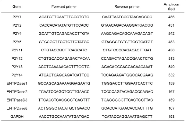
performed with 1 µl of cDNA in GoTaq® Green Master Mix solution using a Thermal cycler (Bio-Rad Laboratories). The cycle conditions were as follows: 3 min at 94 °C and 20 cycles of 1 min at 94 °C, 1 min at 75 °C (and then decreasing by 1 °C/cycle), and 1 min at 72 °C, followed by 30 cycles of 1 min at 94 °C, 1 min at 60 °C, 1 min at 72 °C and a final 7 min step at 72 °C. The primer sequences and expected sizes of the PCR products are shown in Table 1. The primer sequence for P2Y receptor and GAPDH were described by Kudo et al26), and the primer sequence of ENTPDase was described by Bahrami et al27). PCR products were analyzed after electrophoretic separation using 1.5% agarose gels containing ethidium bromide and were visualized using a ChemiDoc XRS and Quantity One software (Bio-Rad Laboratories).
Table 1. Sequence specific primers for P2Y receptor, ENTPDase, and GAPDH
3. Measurement of intracellular Ca2+
Cellular concentrations of Ca2+ were measured using the Calcium Kit II - iCellux (Dojindo, Kumamoto, Japan). Isolated basophils were incubated at 1 × 10⁶
cells/ml in HEPES-HBSS supplemented with a Ca2+ probe and 1.25 mM probenecid for 60 min at 37 °C and 5% CO2. After washing twice with HEPES-HBSS, cells were suspended at 1 × 10⁵ cells/ml in HEPES-HBSS or HEPES-HBSS with 10 μM MRS2578 (Sigma Aldrich, St. Louis, MO, USA). Subsequently, 100 pM of UDP and 0.1 µg/ml anti-IgE were added to calcium probe-labeled cells and fluorescence measurements of Ca2+ were performed using a TriStar LB941 (Berthold Technologies, Germany).
4. Basophil degranulation
Isolated basophils were suspended at 1 × 10⁵ cells/ml in HEPES-HBSS.
Basophils were stimulated with 0.1 µg/ml of goat monoclonal anti-IgE (Beckman Coulter) and 1 pM to 10 nM of UDP at 37 °C for 15 min. Degranulation was stopped by chilling the samples on ice, after which FITC-conjugated anti-CD63 (BioLegend) antibodies were added with subsequent incubation for 30 min on ice. Mean fluorescence intensity (MFI) of CD63 in basophils after stimulation was measured by flow cytometry. All basophils were acquired using a Cytomics FC500 and analysis was performed using FlowJo software (Tree Star, Ashland, OR, USA).
5. MRS2578 treatment of basophils
Isolated basophils were cultured at 1 × 10⁵ cells/ml in RPMI1640 containing 5% serum from the blood sample donor, 2 mM glutamine, 100 U/ml penicillin, 100 µg/ml streptomycin, and 1 ng/ml recombinant human IL-3 (Wako Pure Chemical, Osaka, Japan). Basophils were treated in 24-well plates with 10 µM MRS2578 for 0.5, 18, and 42 h. Controls were treated with 0.1% DMSO. Cultured basophils were suspended at 1 × 10⁵ cells/ml in HEPES-HBSS with 10 μM MRS2578 or 0.1%
DMSO. Cultured basophils were stimulated using 0.1 μg/ml anti-IgE, and the MFI of CD63 in basophils was measured by flow cytometry. All basophils were acquired using a Cytomics FC500 and analysis was performed using FlowJo software.
6. Viability assay
Apoptosis and necrosis were detected using APC-conjugated annexin V and 7-amino-actinomycin D (7-AAD) assays. Briefly, basophils that were cultured with 0.1% DMSO or 10 μM MRS2578 for 18 or 42 h were suspended in Annexin V Binding Buffer and stained with APC-conjugated Annexin V and 7-AAD (BioLegend) for 30 min on ice. All basophils were acquired using a Cytomics FC500 and analysis was performed using FlowJo software.
7. Detection of spontaneous nucleotide secretion from basophils by HPLC analysis Isolated basophils were cultured at 1 × 10⁵ cells/ml in RPMI1640 medium containing 2 mM glutamine, 100 U/ml penicillin, and 100 µg/ml streptomycin at 37 °C in 5% CO2 for 18 h. The supernatant was separated from the basophil culture medium by centrifugation at 14,000 × g for 5 min, and maintained at -20 °C until use.
Reversed-phase HPLC was performed using a Hydrosphere C18 column (150 mm × 4.6 mm, particle size, 5 µm; YMC Co., Kyoto, Japan). The column was eluted with 100 mM KH2PO4-K2HPO4 (pH 5.5) at a flow rate of 1.0 ml/min and nucleotides were detected at 260 nm using a Hitachi L-7100 (Hitachi High-Technologies Corporation, Tokyo, Japan).
8. Statistical analysis
Data are presented as the means ± SEM. Statistical significance was determined using a paired Student’s t-test. P < 0.05 was considered statistically significant.
Results
1. UDP stimulates the P2Y6 receptor in basophils
The expression of P2Y receptors on basophils had not previously been confirmed. RNA was extracted from isolated human basophils, and P2Y receptor mRNA expression was confirmed by RT-PCR. In five independent experiments, bands were detected by electrophoresis for P2Y2, P2Y4, P2Y6, P2Y12, P2Y13, and P2Y14 (Fig. 1A). Therefore, it was hypothesized that the basophils would be responsive to UDP, which is a ligand of the P2Y6 receptor.
UDP stimulation through the P2Y6 receptor increased intracellular Ca2+. We confirmed the change in intracellular Ca2+ in basophils following UDP stimulation (three independent experiments). Intracellular Ca2+ in basophils was immediately increased by UDP stimulation and was significantly inhibited by MRS2578 (Fig. 1B).
The fluorescence intensity was increased to 1305.5 ± 35.6 with UDP stimulation, and reduced to 251.7 ± 81.3 through the addition of MRS2578. The increased fluorescence intensity after UDP stimulation was decreased by 80.7% with MRS2578 treatment. The increased fluorescence intensity with anti-IgE stimulation was similar to that after UDP stimulation (1026.5 ± 270.0). Furthermore, UDP and stimulation with anti-IgE elevated the fluorescence intensity (3285.9 ± 223.7). The increase in fluorescence intensity after UDP stimulation was increased by 220.3% with anti-IgE treatment. The increase in fluorescence intensity with UDP, anti-IgE, and MRS2578 stimulation was similar to that induced by anti-IgE stimulation (1207.6 ± 344.6).
Therefore, UDP has the potential to induce degranulation and enhance IgE-dependent degranulation in basophils.
Figure 1 UDP stimulates the P2Y6 receptor in basophils.
(A) mRNA expression of P2Y receptors and GAPDH in human basophils was confirmed by RT-PCR (representative of five experiments). (B) Basophils were loaded with a calcium fluorescent probe in HBSS. Fluorescence intensities were determined over 1,200 s. Peaking of UDP induced Ca2+ increase during pretreatment. Data represent the mean ± SEM (n = 3); statistical significance was assessed by performing a Student’s t-test. *P < 0.05; **P < 0.01, significantly different.
2. UDP enhances IgE-dependent degranulation of basophils
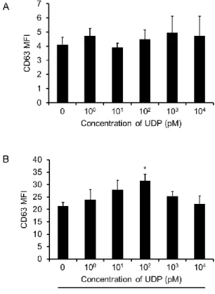
Based on previous results, it was suggested that UDP might induce degranulation and enhance IgE-dependent degranulation in human basophils. We confirmed the effect of UDP stimulation on degranulation by measuring CD63 expression (three independent experiments). CD63 is known as a granular-associated molecule28,29,30). Basophils incubated with UDP for 15 min exhibited expression of CD63. There was no significant difference in the MFI of CD63 after UDP stimulation, regardless of concentration, when compared to that in the unstimulated control (Fig. 2A).
Next, we confirmed the effect of UDP/P2Y6 receptor signaling on IgE-dependent degranulation in basophils. Basophils incubated with UDP and anti-IgE for 15 min exhibited CD63 expression. IgE-dependent degranulation reached its maximum with 100 pM UDP (Fig. 2B). There was a statistically significant difference in the MFI of CD63 in basophils stimulated with anti-IgE (21.4
± 1.4) compared to that in cells treated with 100 pM UDP and anti-IgE (31.6 ± 2.7).
Figure 2 Effects of UDP on IgE-dependent basophil degranulation.
After stimulating basophils, the mean fluorescent intensity of CD63 was measured by flow cytometry. (A) Basophils were stimulated for 15 min with 1 pM to 10 nM UDP (n = 3). Data represent the mean ± SEM (n = 3); statistical significance was assessed by a Student's t-test. A significant difference compared to medium alone was not observed. (B) Basophils were stimulated with 1 pM to 10 nM UDP and 0.1 µg/ml anti-IgE (n = 3). Data represent the mean ± SEM (n = 3);
statistical significance was assessed by performing a Student’s t-test. *P < 0.05, significantly different from the value of the anti-IgE group.
3. Basophils secrete UTP
Some immune cells secrete UDP spontaneously, and activated basophils secrete ATP31,32). We confirmed the presence of nucleotides in the culture supernatant of basophils by HPLC, even when the basophils had not been activated by secreted nucleotides (three independent experiments). In the culture supernatant of basophils, a peak was confirmed at a retention time of 2.33 min (Fig. 3A). The retention time of ATP was 6.30 min, that of ADP was 7.75 min, that of UTP was 2.32 min, that of UDP was 2.53 min, and that of UDP-glucose was 2.87 min (Fig. 3B). Nucleotides were not detected in the RPMI1640 medium (Fig. 3C). These findings confirmed that basophils secrete UTP.
Figure 3 Spontaneous secretion of nucleotides.
Nucleotides in the culture supernatant of basophils were detected by reversed-phase HPLC.
Each peak was identified by comparison with the retention times of a standard mixture. (A) Culture supernatant; (B) nucleotide standard mixture; (C) RPMI1640 medium only (representative of three experiments).
4. Expression of ENTPDase in basophils
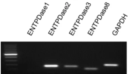
ENTPDase and ecto-nucleotide pyrophosphatase/phosphodiesterase (ENPP) are known as extracellular nucleotidic hydrolases. CD203c, a marker of basophils, is an ENPP3 that hydrolyzes nucleotides33,34) such as ATP. We confirmed the mRNA expression of ENTPDase1, ENTPDase2, ENTPDase3, and ENTPDase8, which are extracellular nucleotide hydrolases, in human basophils by RT-PCR (Fig. 4). In three independent experiments, bands were detected by electrophoresis for ENTPDase2, ENTPDase3, and ENTPDase8; these enzymes hydrolyze nucleotide triphosphate to nucleotide diphosphoric acid. However, the expression of ENTPDase1, which hydrolyzes nucleotide triphosphate and nucleotide diphosphoric acid to nucleotide monophosphate, was not confirmed35). Therefore, UTP secreted by basophils might be hydrolyzed to UDP by ENTPDase2, ENTPDase3, and ENTPDase8.
Figure 4 Expression of ENTPDase mRNA.
The mRNA expression of ENTPDase family members and GAPDH in human basophils was evaluated by RT-PCR (representative of three experiments).
5. MRS2578 inhibits IgE-dependent degranulation in basophils
Our findings suggested that basophil P2Y6 receptors might be stimulated by the spontaneous production of UDP. Even when UDP was not added exogenously, basophils continuously produced UDP, which could enhance IgE-dependent
degranulation. We confirmed the IgE-dependent degranulation of basophils by inhibiting the P2Y6 receptor and measuring the expression of CD63. Basophils were cultured with MRS2578, a specific antagonist of P2Y6 receptor, for 0.5, 18, and 42 h.
Controls consisted of treatment with the MRS2578 vehicle solution, DMSO (three independent experiments). Basophils cultured with MRS2578 showed decreased expression of CD63 following anti-IgE stimulation compared to that in basophils cultured with DMSO (Fig. 5). The MFI of CD63 on basophils following anti-IgE antibody stimulation was 18.6 ± 0.8 at 0.5 h, 16.4 ± 0.3 at 18 h, and 1.7 ± 0.2 at 42 h, with MRS2578 treatment. The MFI of CD63 on basophils following anti-IgE antibody stimulation was 20.0 ± 0.4 at 0.5 h, 22.4 ± 0.3 at 18 h, and 21.5 ± 0.1 at 42 h, with DMSO treatment. Thus, MFI decreased by 7.0% at 0.5 h, 26.8% at 18 h, and by 92.2% at 42 h with MRS2578 treatment. This confirmed that MRS2578 inhibits the IgE-dependent degranulation of basophils, indicating that the IgE-dependent degranulation of basophils is regulated by UDP via the P2Y6 receptor.
Figure 5 Effects of MRS2578 on IgE-dependent basophil degranulation.
Basophils were cultured in suspension at 1 × 10⁵ cells/ml in RPMI1640 with 1 ng/ml rIL-3 and 10 μM MRS2578. After culture for 0.5, 18, or 42 h, cells were stimulated with 0.1 μg/ml anti-IgE for 15 min. Degranulation of basophils was measured by CD63 expression (n = 3). Data represent the mean ± SEM (n = 3); statistical significance was assessed by a Student’s t-test. **P <
0.01, significantly different from the value of the DMSO condition.
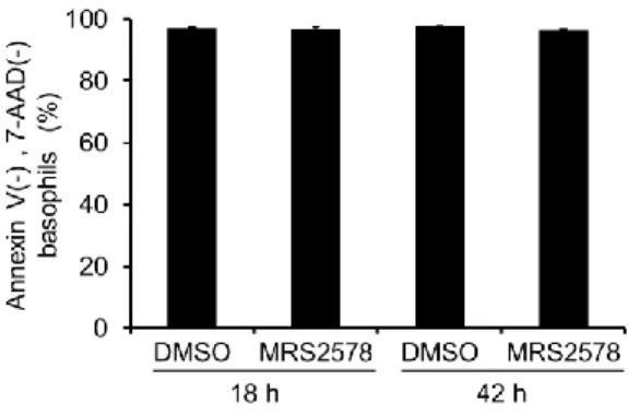
6. MRS2578 does not alter the survival of basophils
We then examined the cytotoxicity of MRS2578 using Annexin-V and 7-AAD (three independent experiments). The number of cells negative for Annexin-V and 7-AAD was 96.9 ± 0.7% following the addition of MRS2578 at 18 h and 97.3 ± 0.4%
following the addition of DMSO at the same time point. The number of cells negative for Annexin-V and 7-AAD was 96.3 ± 0.5% following the addition of MRS2578 at 42 h and 97.6 ± 0.2% following the addition of DMSO (Fig. 6). No significant difference in cytotoxicity between the MRS2578 and DMSO groups was observed at either time point.
Figure 6 MRS2578 does not induce cytotoxicity in basophils.
The cytotoxicity of 0.1% DMSO and 10 μM MRS2578 was confirmed using in Annexin V and 7-AAD assays (n = 3). Data represent the mean ± SEM (n = 3); through performing a Student's t-test, a significant difference, compared to the DMSO control, was not observed.
Discussion
The P2Y6 receptor is involved in the biological activity of various cells, and the aggravation of allergic symptoms is associated with allergic reactions8-10). However, the effect of the P2Y6 receptor on the function of basophils, which have an important role in allergic reactions, has not been reported. In this study, we showed that stimulation of UDP/P2Y6 receptor signaling enhanced the IgE-dependent degranulation of basophils.
Furthermore, we showed that MRS2578, a specific antagonist of the P2Y6 receptor, inhibited IgE-dependent degranulation. Therefore, inhibition of P2Y6 receptor stimulation inhibits the degranulation of basophils, and might be effective for allergy treatment.
Expression patterns of the P2Y receptor in basophils have not been reported to date. In this study, we detected the expression of P2Y receptor mRNA in basophils, and determined that the selective agonistic effect of UDP on P2Y6 receptors increased intracellular Ca2+ in basophils, which was inhibited by incubation with MRS2578. These results indicate that expression of the P2Y6 receptor is present in basophils. However, the P2Y6 receptor protein has not been detected to date and this should be assessed in future studies.
Inhibition of IgE-dependent basophil degranulation by MRS2578 indicates that basophils are constantly stimulated by UDP and that stimulation via the P2Y6 receptor is necessary for IgE-dependent basophil degranulation. IgE-dependent degranulation was enhanced after 15 min of UDP stimulation. In contrast, inhibition of IgE-dependent degranulation by MRS2578 required a reaction time of 18 h or longer. The possibility that the basophils had been constantly stimulated by UDP was considered. One inhibitory mechanism of MRS2578 is to block the P2Y6 receptor and its response to UDP.
Therefore, the lack of an effect is due to the fact that UDP stimulation is thought to require 18 h or longer.
Expression of ENTPDase mRNA and the spontaneous secretion of UTP
suggested the possibility that the P2Y6 receptor on basophils is stimulated by an autocrine mechanism. The P2Y6 receptor constantly stimulates basophils, and therefore, should have an important role in their degranulation. Furthermore, this receptor reacts minimally with UTP37). UDP was not detected by HPLC analysis.It was considered that UDP reacted with the P2Y6 receptor or ENTPDase on the cell surface such that UDP might be undetectable in the culture supernatant. Therefore, hydrolysis of UTP by basophils, and detection of UDP and ENTPDase proteins on the cell surface is necessary in further studies to confirm autocrine activation of P2Y6.
Intracellular signaling induces many cell functions. PKC and PLCγ signal transduction molecules are Ca2+-dependent37,38). Furthermore, molecules released during the degranulation of basophils and mast cells that fuse to cell membranes are also Ca2+-dependent39,40). Therefore, intracellular Ca2+ might have an important role in the activation and degranulation of basophils. Indeed, the IgE-dependent and IgE-independent activation of basophils is induced by the activation of PKC, which increases intracellular Ca2+21,41).
Gαq protein-coupled GPCRs induce the activation of PKC, increase intracellular Ca2+, and induce the degranulation of basophils. Furthermore, the C5a receptor, MCP-1 receptor, and fMLP receptor (a GPCR) induce the IgE-independent degranulation of basophils18-21). It has been suggested that when the G protein coupled with the P2Y6 receptor in basophils is Gαq, degranulation is induced by UDP stimulation.
However, UDP stimulation did not induce the degranulation of basophils, herein. It was shown that Gα12/13 increased the intracellular Ca2+ necessary for degranulation, but did not activate PKC21). If Gα12/13 is coupled with the P2Y6 receptor in basophils, this might explain why degranulation was not induced by UDP stimulation. Furthermore, these results indicate that Gα12/13 promotes IgE-dependent degranulation because an increase in intracellular Ca2+ strengthens FcεRI signaling42). However, further studies will be necessary to confirm the activation of Rho as well as increases in cAMP via UDP stimulation, as proving that Gα12/13 is coupled with the P2Y6 receptor was beyond the
scope of this study.
IL-4, which is produced by basophils, is associated with IgE production43) and Th2 differentiation44). Since this study did not examine the effect of UDP/P2Y6 receptor signaling on cytokine production in basophils, future investigation into the effect of this pathway on cytokine production is necessary.
Basophils have an important role in allergic disease15) and in biophylaxis against parasitic infections16). Inhibiting basophil function prevents allergic reactions, but consequently immunity to parasitic infection decreases. Furthermore, UDP/P2Y6 receptor signaling has a role in biophylaxis against viruses, bacteria, and parasites31,45,46). Inhibition of UDP/P2Y6 receptor signaling might decrease immunity against infections.
Therefore, it is important to determine the effect of inhibition of the P2Y6 receptor, a target for the treatment of allergic disease, on infection. Because this study investigated cellular responses in vitro, investigating the effect of UDP/P2Y6 receptor signaling using in vivo models of allergic reaction and immunity against infections is required in future studies.
Acknowledgments
The authors would like to thank Mai Kumeta, Hidemi Numasawa, Rei Sawaki, and Kyoko Ito for helpful assistance. This work was supported by a grant from the Hirosaki University Grant for Exploratory Research by Young Scientists and Newly-appointed Scientists. We would like to thank Editage (www.editage.jp) for English language editing.
References
1) Sakaki H, Tsukimoto M, Harada H, Moriyama Y, Kojima S. Autocrine regulation of macrophage activation via exocytosis of ATP and activation of P2Y11 receptor.
PLoS One, 8:e59778, 2013.
2) Bours MJ, Swennen EL, Di Virgilio F, Cronstein BN, Dagnelie PC. Adenosine 5'-triphosphate and adenosine as endogenous signaling molecules in immunity and inflammation. Pharmacol Ther, 112:358–404, 2016.
3) Burnstock G, Williams M. P2 purinergic receptors: modulation of cell function and therapeutic potential. J Pharmacol Exp Ther, 295:862–869, 2000.
4) Burnstock G, Boeynaems JM. Purinergic signalling and immune cells. Purinergic signal, 10:529–564, 2014.
5) Surprenant A, North RA. Signaling at purinergic P2X receptors. Annu Rev Physiol, 71:333–359, 2009.
6) Erlinge D. P2Y receptors in health and disease. Adv Pharmacol, 61:417–439, 2011.
7) Communi D, Janssens R, Suarez-Huerta N, Robaye B, Boeynaems JM. Advances in signalling by extracellular nucleotides. the role and transduction mechanisms of P2Y receptors. Cell Signal, 12:351–360, 2000.
8) Jiang Y, Borrelli L, Bacskai BJ, Kanaoka Y, Boyce JA. P2Y6 receptors require an intact cysteinyl leukotriene synthetic and signaling system to induce survival and activation of mast cells. J Immunol, 182:1129–1137, 2009.
9) Giannattasio G, Ohta S, Boyce JR, Xing W, Balestrieri B, Boyce JA. The purinergic G protein-coupled receptor 6 inhibits effector T cell activation in allergic pulmonary inflammation. J immunol, 187:1486–1495, 2011.
10) Vieira RP, Muller T, Grimm M, von Gernler V, Vetter B, Durk T, et al. Purinergic receptor type 6 contributes to airway inflammation and remodeling in experimental allergic airway inflammation. Am J Respir Crit Care Med, 184:215–223, 2011.
11) Neher JJ, Neniskyte U, Hornik T, Brown GC. Inhibition of UDP/P2Y6 purinergic
signaling prevents phagocytosis of viable neurons by activated microglia in vitro and in vivo. Glia, 62:1463–1475, 2014.
12) Kim SG, Gao ZG, Soltysiak KA, Chang TS, Brodie C, Jacobson KA. P2Y6 nucleotide receptor activates PKC to protect 1321N1 astrocytoma cells against tumor necrosis factor-induced apoptosis. Cell Mol Neurobiol, 23:401–418, 2003.
13) Schroeder JT, MacGlashan DW Jr, Kagey-Sobotka A, White JM, Lichtenstein LM.
IgE-dependent IL-4 secretion by human basophils. The relationship between cytokine production and histamine release in mixed leukocyte cultures. J Immunol, 153:1808–1817, 1994.
14) Ochensberger B, Daepp GC, Rihs S, Dahinden CA. Human blood basophils produce interleukin-13 in response to IgE-receptor-dependent and -independent activation.
Blood, 88:3028–3037, 1996.
15) Schroeder JT. Basophils: emerging roles in the pathogenesis of allergic disease.
Immunol Rev, 242:144–160, 2011.
16) Schwartz C, Turqueti-Neves A, Hartmann S, Yu P, Nimmerjahn F, Voehringer D.
Basophil-mediated protection against gastrointestinal helminths requires IgE-induced cytokine secretion. Proc Natl Acad Sci USA, 111:E5169–5177, 2014.
17) Kempuraj D, Madhappan B, Christodoulou S, Boucher W, Cao J, Papadopoulou N, et al. Flavonols inhibit proinflammatory mediator release, intracellular calcium ion levels and protein kinase C theta phosphorylation in human mast cells. Br J Pharmacol, 145:934–944, 2005.
18) Bischoff SC, Krieger M, Brunner T, Dahinden CA. Monocyte chemotactic protein 1 is a potent activator of human basophils. J Exp Med, 175:1271–1275, 1992.
19) Uguccioni M, Mackay CR, Ochensberger B, Loetscher P, Rhis S, LaRosa GJ, et al.
High expression of the chemokine receptor CCR3 in human blood basophils. Role in activation by eotaxin, MCP-4, and other chemokines. J Clin Invest, 100:1137–1143, 1997.
20) Chirumbolo S, Vella A, Ortolani R, De Gironcoli M, Solero P, Tridente G, et al.
Differential response of human basophil activation markers: a multi-parameter flow cytometry approach. Clin Mol Allergy, 6:12, 2008.
21) Jimenez-Sainz MC, Fast B, Mayor F Jr, Aragay AM. Signaling pathways for monocyte chemoattractant protein 1-mediated extracellular signal-regulated kinase activation. Mol Pharmacol, 64:773–782, 2003.
22) Nishida M, Sato Y, Uemura A, Narita Y, Tozaki-Saitoh H, Nakaya M, et al. P2Y6 receptor-Galpha12/13 signalling in cardiomyocytes triggers pressure overload-induced cardiac fibrosis. EMBO J, 27:3104–3115, 2008.
23) Pifferi S, Boccaccio A, Menini A. Cyclic nucleotide-gated ion channels in sensory transduction. FEBS Lett, 580:2853–2859, 2006.
24) Ismailov II, Benos DJ. Effects of phosphorylation on ion channel function. Kidney Int, 48:1167–1179, 1995.
25) Gibbs BF, Papenfuss K, Falcone FH. A rapid two-step procedure for the purification of human peripheral blood basophils to near homogeneity. Clin Exp Allergy, 38:480–485, 2008
26) Kudo F, Nishiguchi N, Mizuike R, Sato H, Ito K, Nakano M, et al. Neutrophil phagocytosis is down-regulated by nucleotides until encounter with pathogens.
Immunology Lett. 144:24–32, 2012.
27) Bahrami F, Kukulski F, Lecka J, Tremblay A, Pelletier J, Rockenbach L, et al.
Purine-metabolizing ectoenzymes control IL-8 production in human colon HT-29 cells. Mediators Inflamm, 2014:879895, 2014.
28) Knol EF, Mul FP, Jansen H, Calafat J, Roos D. Monitoring human basophil activation via CD63 monoclonal antibody 435. J Allergy Clin Immunol. 88:
328-338, 1991.
29) MacGlashan D Jr. Expression of CD203c and CD63 in human basophils:
relationship to differential regulation of piecemeal and anaphylactic degranulation processes. Clin Exp Allergy. 40: 1365-1377, 2010.
30) Hoffmann HJ, Santos AF, Mayorga C, Nopp A, Eberlein B, Ferrer M, et al. The
clinical utility of basophil activation testing in diagnosis and monitoring of allergic disease. Allergy. 70: 1393-1405, 2015.
31) Li R, Tan B, Yan Y, Ma X, Zhang N, Zhang Z, et al. Extracellular UDP and P2Y6 function as a danger signal to protect mice from vesicular stomatitis virus infection through an increase in IFN-β production. J Immunol, 193:4515–4526, 2014.
32) Tsai SH, Kinoshita M, Kusu T, Kayama H, Okumura R, Ikeda K, et al. The ectoenzyme E-NPP3 negatively regulates ATP-dependent chronic allergic responses by basophils and mast cells. Immunity, 42:279–293, 2015.
33) Tsai SH, Takeda K. Regulation of allergic inflammation by the ectoenzyme E-NPP3 (CD203c) on basophils and mast cells. Semin Immunopathol, 38:571–579, 2016.
34) Zimmermann H, Zebisch M, Sträter N. Cellular function and molecular structure of ecto-nucleotidases. Purinergic Signal, 8:437–502, 2012.
35) Kukulski F, Levesque SA, Lavoie EG, Lecka J, Bigonnesse F, Knowles AF, et al.
Comparative hydrolysis of P2 receptor agonists by NTPDases 1, 2, 3 and 8.
Purinergic Signal, 1:193–204, 2005.
36) Communi D, Parmentier M, Boeynaems JM. Cloning, functional expression and tissue distribution of the human P2Y6 receptor. Biochem Biophys Res Commun, 222:303–308, 1996.
37) Ryan MJ, Gross KW, Hajduczok G. Calcium-dependent activation of phospholipase C by mechanical distension in renin-expressing As4.1 cells. Am J Physiol Endocrinol Metab, 279:E823–829, 2000.
38) Luo JH, Weinstein IB. Calcium-dependent activation of protein kinase C. The role of the C2 domain in divalent cation selectivity. J Biol Chem, 268:23580–23584, 1993.
39) Tadokoro S, Nakanishi M, Hirashima N. Complexin II regulates degranulation in RBL-2H3 cells by interacting with SNARE complex containing syntaxin-3. Cell Immunol, 261:51–56, 2010.
40) Gaisano HY. Here come the newcomer granules, better late than never. Trends Endocrinol Metab, 25:381–388, 2014.
41) Kempuraj D, Madhappan B, Christodoulou S, Boucher W, Cao J, Papadopoulou N, et al. Flavonols inhibit proinflammatory mediator release, intracellular calcium ion levels and protein kinase C theta phosphorylation in human mast cells. Br J Pharmacol, 145:934–944, 2005.
42) He SH, Xie H, He YS. Induction of tryptase and histamine release from human colon mast cells by IgE dependent or independent mechanisms. World J Gastroenterol, 10:319–322, 2004.
43) Yanagihara Y, Kajiwara K, Basaki Y, Ikizawa K, Ebisawa M, Ra C, et al. Cultured basophils but not cultured mast cells induce human IgE synthesis in B cells after immunologic stimulation. Clin Exp Immunol, 111:136–143, 1998.
44) Yoshimoto T, Yasuda K, Tanaka H, Nakahira M, Imai Y, Fujimori Y, et al. Basophils contribute to T(H)2-IgE responses in vivo via IL-4 production and presentation of peptide-MHC class II complexes to CD4+ T cells. Nat Immunol, 10:706–712, 2009.
45) Zhang Z, Wang Z, Ren H, Yue M, Huang K, Gu H, et al. P2Y(6) agonist uridine 5'-diphosphate promotes host defense against bacterial infection via monocyte chemoattractant protein-1-mediated monocytes/macrophages recruitment. J Immunol, 186:5376–5387, 2011.
46) Moreira-Souza AC, Marinho Y, Correa G, Santoro GF, Coutinho CM, Vommaro RC, et al. Pyrimidinergic receptor activation controls Toxoplasma gondii infection in macrophages. PLoS One, 10:e0133502, 2015.
要 旨
UDP/P2Y6受容体によるヒト好塩基球IgE依存性脱顆粒反応の調節
中野 学
医療生命科学領域 病態解析科学分野
細胞外ヌクレオチド受容体である P2Y受容体は、Gタンパク共役型受容体で ある。細胞外ヌクレオチドの刺激は、P2Y受容体を介し cAMP産生、PKC活性 化や細胞内 Ca2+濃度上昇を誘導することで、細胞機能を調節する。P2Y 受容体 ファミリーである P2Y6受容体は、UDPを特異的アゴニスト、MRS2578を特異 的アンタゴニストとする。好塩基球の脱顆粒は、アレルギー反応を誘導するこ とで知られている。本研究では、好塩基球の脱顆粒反応に対するP2Y6受容体の 作用について検討した。ヒト好塩基球は、P2Y6受容体 mRNAを発現しており、
UDP刺激により細胞内Ca2+濃度の上昇が誘導された。また、UDP刺激による細 胞内Ca2+濃度の上昇はMRS2578により抑制され、好塩基球に発現しているP2Y6 受容体が、細胞内にシグナル伝達していることが示唆された。UDP は、単独刺 激では好塩基球の脱顆粒を誘導しないが、IgE依存性脱顆粒を促進した。この結 果から、好塩基球のIgE依存性脱顆粒の調節にP2Y6受容体が関与していること が考えられた。好塩基球の培養上清からUTPが検出され、好塩基球でENTPDase mRNA が発現していることを確認した。これらの結果から、好塩基球がオート クラインにより、P2Y6受容体を刺激していることが考えられた。実際、MRS2578 存在下で培養した好塩基球は、抗IgE抗体の刺激による脱顆粒が誘導されず、こ の抑制効果は経時的に増強された。本研究の結果、好塩基球はオートクライン によりP2Y6受容体を刺激し、IgE依存性脱顆粒を促進している可能性が示唆さ れた。また、MRS2578による IgE依存性脱顆粒を抑制したことから、アレルギ ー症状の緩和治療のターゲットとしてP2Y6受容体は有用であると考える。