Pediatric Cardiology and Cardiac Surgery 32(3): 223‒229 (2016) 症例報告
川崎病冠動脈病変の血管壁評価における
MR coronary vessel wall imaging と光干渉断層法の比較
真田 和哉1),田原 昌博1),新田 哲也1),下薗 彩子1), 佐藤 友保2),野中 春輝2),石川 優子2)
1)あかね会土谷総合病院小児科
2)あかね会土谷総合病院放射線科
Vessel Wall Evaluations of Coronary Arterial Lesions in Kawasaki Disease: Comparison of Magnetic Resonance Imaging and Optical Coherence Tomography Findings
Kazuya Sanada
1), Masahiro Tahara
1), Tetsuya Nitta
1), Saiko Shimozono
1), Tomoyasu Sato
2), Haruki Nonaka
2), and Yuko Ishikawa
2)1) Department of Pediatrics, Tsuchiya General Hospital, Hiroshima, Japan
2) Department of Radiology, Tsuchiya General Hospital, Hiroshima, Japan
Evaluation of the coronary arterial intima is important in the follow-up of coronary arterial lesions complicated by Kawasaki disease (KD). We evaluated the lesions in four cases of KD using optical coherence tomography (OCT), which can show vessel wall thickness on magnetic resonance imaging (MRI) of the coronary vessel wall using the black blood method (BB). All cases showed intimal thickening on OCT at areas of thickened vessel wall using BB. Although vessel wall thickening was detected as iso-intensity with BB, OCT showed various histological features of the intima at these lesions. We revealed that intimal thickening is evaluable using BB compared with OCT findings. MRI of coronary vessel walls with BB may be useful in screening for intimal thickening of coronary arterial lesions complicated with KD.
Keywords: Kawasaki disease, magnetic resonance coronary angiography, black blood method, coro- nary arterial wall thickness, optical coherence tomography
川崎病冠動脈病変後遺症における血管内膜の評価は重要である.Black blood(BB)法を用いたmag- netic resonance coronary vessel wall imagingで血管壁肥厚を指摘された川崎病冠動脈病変合併症例 4例に対し,光干渉断層法(optical coherence tomography: OCT)を用いて,血管内膜病変の観察を行っ た.全症例においてBB法で血管壁肥厚が存在した部位に一致して,OCTで内膜肥厚を認めた.BB法 で均一に描出された血管壁にはOCTで様々な病変を認めた.BB法とOCTの比較で,BB法で血管内 膜肥厚の存在が評価可能なことが示唆された.BB法は川崎病冠動脈病変における血管内膜病変のスク リーニングに有用であると考えた.
はじめに
川崎病冠動脈病変後遺症の遠隔期において血管内膜 病変が存在し,その評価に
black blood
(BB
)法を用いた
magnetic resonance
(MR
)coronary vessel wall imaging
が有用であると言われている1‒3).しかし,BB
法で血管壁肥厚を指摘された病変に対し,光干渉 断層法(optical coherence tomography: OCT
)で内2015年11月2日受付,2016年3月15日受理
著者連絡先:〒730‒8655 広島県広島市中区中島町3‒30 あかね会士谷総合病院 真田和哉 doi: 10.9794/jspccs.32.223
224
膜病変の詳細な評価を行った報告はない.我々は川崎 病冠動脈瘤合併症例
4
例の冠動脈の血管壁をBB
法とOCT
で評価し,比較検討を行った.画像検査の撮像方法
Magnetic resonance imaging
(MRI
) 装 置 はPhil- lips
社製Intera 1.5T Achieva
を使用し,MR coronary angiography
(MRCA
) とBB
法 を 用 い たMR coro- nary vessel wall imaging
を撮像した.BB
法の撮像 方法はT1 BB Turbo field echo
(TFE
)に脂肪抑制(
spectral presaturation with inversion recovery: SPIR
法)を併用し,収集法はspiral sequence
を使用でき ないため,low high
法を使用した.OCT
はST. JUDE MEDICAL
社製ILUMIEN OCT Imaging System
を使 用し,6Fr
シースを挿入し,ガイディングカテーテル は同社製Dragonfly JP Imaging Catheter
を使用した.症 例 症例1
症例:
11
歳男性現病歴:
6
歳時に川崎病を発症し初回治療のガンマグ ロブリン大量療法(IVIg
)2 g/kg
に反応なく,IVIg
1 g/kg
の追加投与を行った.発症1
か月後に施行した
MRCA
で右冠動脈segment 2
に最大径4.5 mm
の 冠動脈瘤を認めた.7
歳時,9
歳時に施行したMRCA
では冠動脈瘤は不変だった.画像検査所見:
11
歳時に施行したMRCA
でsegment 2
の瘤内に隔壁様の構造物を認め,BB
法では瘤部に全 周性の血管壁肥厚を認めた.OCT
では全周性に内膜 の線維性肥厚を認めた.石灰化や血栓は認めなかっ た.MRCA
で認めた隔壁様構造物の部分には内膜の 剥離を認めた(Fig. 1
).症例2
症例:
8
歳男性現病歴:生後
3
か月時に川崎病を発症し,初回治療Fig. 1 Image findings for Case 1
Cross-section imaging with the number correspond with the dotted line of the same number . On coronary angiography, the maximum diameter of the aneurysm (segment 2) is 4.5 mm (a-1), and the aneurysm is in the form of a slit on mag- netic resonance coronary angiography (b-1). Vessel wall imaging shows thickening at the aneurysm (c-1, arrows). On optical coherence tomography imaging (d-1, e-1), the slit-like aneurysm has separation of the intima (closed triangles), and the intima of this aneurysm is hyperplastic with fibrosis (dashed arrows).
の
IVIg 2 g/kg
に反応なく,追加治療でIVIg 2 g/kg
×2
回,methylprednisolone pulse
療法,ulinastatin
投 与を行った.右冠動脈segment 2
に最大径5.6 mm
, 左冠動脈前下行枝segment 6
に最大径5.6 mm
の冠動 脈瘤が残存した.その後segment 6
の瘤は退縮した.3
歳時に施行した冠動脈造影(coronary angiography:
CAG
)でsegment 2
の瘤入口部に28
%狭窄を認めた.8
歳時に労作時胸痛を認め,運動負荷心電図で一過性 にII
,III
,aVf
のST
低下を認めた.画像検査所見:
8
歳時に施行したMRCA
でsegment 2
に最大径5.1 mm
の冠動脈瘤を認め,入口部は2.2 mm
と狭小化していた.BB
法ではsegment 2
の近位部か ら遠位部にかけて全周性に血管壁の肥厚を認めた.CAG
でsegment 2
の瘤入口部に40
%狭窄を認め,OCT
では瘤全体に内膜の線維性肥厚を認めた.瘤の 中央には一部,脂質に富んだ内膜が存在し,遠位部に は半周性に内膜の石灰化を認めた.冠血流予備量比(
fractional flow reserve: FFR
)は0.84
であり,イン ターベンションは行わなかった(Fig. 2
).症例3
症例:
17
歳女性現病歴:
5
歳時に川崎病を発症し初回治療でIVIg 2 g/kg
の投与を行った.発症2
か月後に施行したCAG
で 左前下行枝segment 6
に最大径5.3 mm
,segment 7
に最大径3.0 mm
の冠動脈瘤を認めた.9
歳時に施行 したCAG
でsegment 6
に最大径6.2 mm
の冠動脈瘤 と瘤入口部に25
%狭窄を認め,segment 7
の瘤は退縮 していた.その後MRCA
で経過観察し,病変の変化 はなかった.画像検査所見:
17
歳時に施行したMRCA
でsegment 6
の瘤だった部分が狭窄し内腔不整となっており,BB
法では全周性に血管壁の肥厚を認めた.CAG
ではsegment 6
に50
%狭窄を認め,OCT
ではsegment 6
の狭窄部に内膜の線維性肥厚を認め,石灰化や中膜の 剥離を伴っていた.狭窄部より近位側にも内膜の線維 性肥厚と新生血管を認めた.FFR
は0.85
〜0.94
であ り,インターベンションは施行しなかった.症例4
症例:
21
歳女性 Fig. 2 Image findings for Case 2On coronary angiography and magnetic resonance coronary angiography, the maximum diameter of the aneurysm (segment 2) is 5.4 mm, and the proximal part of the aneurysm shows localized stenosis (a-2, b-2). Vessel wall imaging shows thickening at all parts of the aneurysm (c-2, arrows). On optical coherence tomography imaging (d-2), intimal thickening with fibrosis (dashed arrows), a lipid-laden intima (open triangles), and calcified intima (asterisks) are evident.
226
現病歴:
1
歳時に川崎病を発症し初回治療のIVIg 200 mg/kg
×5
日の投与に反応なく,追加治療でIVIg 500 mg/kg
×3
日の投与を行った.右冠動脈segment 1
, 左冠動脈主幹部segment 5
,前下行枝segment 6
に冠 動脈瘤を形成した.発症3
か月後に施行したCAG
でsegment 5
に冠動脈瘤を認め,segment 1
,6
の瘤退縮 を認めた.15
歳時に施行したMRCA
でsegment 5
に 最大径4 mm
の拡張を認め,BB
法で血管壁の肥厚を 認めた.画像検査所見:
21
歳時に施行したCAG
で右冠動脈,左冠動脈ともに瘤や狭窄はなかった.
OCT
ではseg- ment 1
,segment 5
,segment 6
に内膜の線維性肥厚 を認め,segment 6
に内膜の血管新生を豊富に認めた(
Fig. 3
).CAG
上退縮した瘤やその周囲ではOCT
で 内膜肥厚を認めたが,急性期に正常だった冠動脈にはOCT
を施行した範囲で,内膜肥厚は認めなかった.一方,
BB
法では解像度が低いため血管壁は均一に描 出され,性状の評価はできなかった.考 察
川崎病に対する急性期治療の進歩により,冠動脈後 遺症の発生頻度は
2.6
%と減少傾向であるが,新規患 者数は増加傾向で冠動脈後遺症のある患者数は2013
年〜2014
年の2
年間で887
人と2007
年から横ばい である4).Tsuda
らは発症100
日以内の初回CAG
で 冠動脈瘤の径が4 mm
以上だと,血管内超音波(intra- vascular ultrasound: IVUS
)で内膜肥厚が認められた と報告している5).また,6 mm
以上だとリモデリン グが強くなり,遠隔期に狭窄病変や石灰化を起こす とも報告している6).冠動脈後遺症の経過観察におい て,血管内膜の評価は重要である.近年,dual source computed tomography
(DSCT
)やBB
法を用いた内 膜評価の報告が散見される1‒3, 7).BB
法は血流を無信号にし,プラークや内膜を描出 する撮像法で,脂肪抑制を併用することで内膜肥厚 の検討が可能と言われている2).OCT
はカテーテル ベースの冠動脈内画像診断法で近赤外線光の干渉によ Fig. 3 Image findings for Cases 3 and 4(Case 3) Localized stenosis is seen at segment 6. In vessel wall imaging, thickening is seen at the lesion (b-3, c-3). On the optical coherence tomography (OCT) image (d-3), intimal hyperplasia with fibrosis (arrows), calcification (asterisks), neovascularization (double arrows), and separation of the media (white arrows) are apparent. (Case 4) The aneurysm at the bifurcation of the left coronary artery has regressed. Upon vessel wall imaging, thickening is apparent at the lesion.
This lesion shows intimal hyperplasia with fibrosis (d-4, arrows) and neovascularization (double arrows) on OCT. Side branches originate from the coronary artery (hash marks).
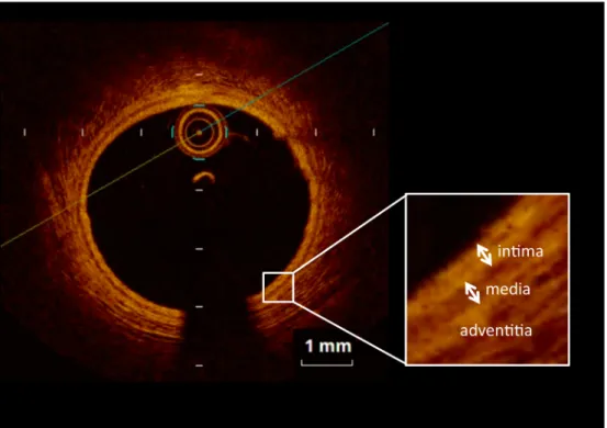
Fig. 4 Optical coherence tomography imaging of normal coronary artery (Case 3, segment 11)
Table 1 Summary of four cases
Case Sex Age (years)
Age at KD diagnosis
years)
Initial therapy
Additional therapy
CAG MRI (BB) OCT
Lesions Wall thick- ness (mm)
Intima thick-
ness (mm) Findings
1 M 11 6 IVIg IVIg RCA #1 N N/A 0.14‒0.26 N
2 g/kg 1 g/kg #2 AN 2.20‒2.79 0.24‒0.67 IH, Fib, SI
LCA N N/A N/A N/A
2 M 8 0 IVIg IVIg RCA #1 N N/A 0.41‒0.52 IH, Fib, NV
2 g/kg 2 g/kg*2, mPSL #2 AN, LS 2.74‒6.47 0.18‒0.75 IH, Fib, Cal, LI, NV
pulse, UTI LCA #6 RA N/A N/A N/A
3 F 17 5 IVIg none RCA N N/A N/A N/A
2 g/kg LCA #5 N N/A 0.23‒0.38 IH, Fib
#6 LS 2.72‒6.74 0.52‒1.71 IH, Fib, SM, Cal, NV
#7 RA N/A 0.19‒1.05 IH, Fib, Cal
#11 N N/A 0.16‒0.23 N
4 F 21 1 IVIg IVIg RCA #1 RA N/A 0.47‒0.55 IH, Fib
200 mg/kg 500 mg/kg #2 N N/A 0.07‒0.10 N
*5 day *3 day LCA #5 AN 2.44 0.57‒0.66 IH, Fib
#6 RA N/A 0.46‒0.58 IH, Fib, NV
#9 N N/A 0.11‒0.19 N
Vessel wall thickness of coronary artery lesions on black-blood magnetic resonance imaging was measured using the half- width of the signal level. M; male, F; female, KD; Kawasaki disease, IVIg; intravenous immunoglobulin, mPSL; methylpredonis- olone, UTI; ulinastatin, RCA; right coronary artery, LCA; left coronary artery, CAG; coronary angiography, MRI; magnetic reso- nance imaging, BB; black blood method, OCT; optical coherence tomography, AN; aneurysm, N; normal coronary artery, LS;
localized stenosis, RA; regressed aneurysm, N/A; not available, IH; intimal hyperplasia, Fib; fibrosis, SI; separation of the intima, Cal; calcification, LI; lipidOladen intima, NV; neovascularization, SM; separation of the media.
228
り組織を画像化するため,軸解像度が約
15 nm
と極 めて高く,正常な冠動脈は三層構造として描出され,プラークや血栓の性状評価,組織診断に有用である8)
(
Fig. 4
).当院では2009
年からMRCA
とBB
法を用 いたMR vessel wall imaging
を併用し,冠動脈後遺症 における血管壁肥厚の観察を行っている.今回,BB
法で壁肥厚を指摘された部位の内膜病変評価のためにOCT
を行った.患者のまとめを
Table 1
に示した.4
症例,4
か所 の病変でBB
法を撮像し,すべての病変に血管壁肥厚 を認め,OCT
でも内膜肥厚を認めた.内膜の多くは 石灰化や,線維性肥厚などの川崎病遠隔期に認めら れる病変9)だったが,症例2
では瘤の内膜に脂質に 富んだ病変を認めた.成人冠動脈の粥状硬化病変は マクロファージの集簇により同様の画像を示す8).Dionne
らは川崎病冠動脈瘤を有する小児18
例に対し,川崎病罹患後平均
9
年後にOCT
を施行し,8
例に内膜へマクロファージの浸潤を認めたと報告し た10).Suzuki
らは小児死亡例の病理所見で,川崎病 冠動脈病変にマクロファージの浸潤はなかったと報告 しており11),長期経過の冠動脈病変における病態解 明が期待される.BB
法の最大信号の半分の範囲(半値幅)で測定し た血管壁肥厚とOCT
での内膜肥厚を比較したとこ ろ,BB
法 の 血 管 壁 肥 厚 は2.20
〜6.74 mm
,OCT
の 内膜肥厚は0.18
〜1.71 mm
とかなりの差があった.Greil
らはBB
法を用いて川崎病冠動脈瘤の血管壁肥厚を測定し,平均
2.5
±0.5 mm
であったと報告して おり12),当院の検討とほぼ同等であった.一方,Dionne
らはOCT
における冠動脈瘤の内膜肥厚は右冠動脈で平均
347.1
±173.4
µm,左前下行枝で435.0
±158.1
µm,左回旋枝で360.0
±165.5
µmであったと報 告している10).このように,過去の報告の比較でもBB
法での血管壁肥厚とOCT
での内膜肥厚に差があっ た.BB
法で血管壁が厚く見える原因は体動,心拍の 影響やスライス厚が5 mm
と厚いことが原因と言われ ている2).さらに,BB
法で評価した血管壁肥厚は内 膜だけでなく,内膜を含めた血管壁全体,または血管 壁と血管周囲組織の変化を合わせて評価している可能 性があり,今後の検討が必要である.BB
法の問題点の一つ目は前述のごとく,解像度が 低い点である.二つ目は,当院で用いたBB
法では血 管の全長を描出できず,撮像部位以外の内膜肥厚の存 在が否定できないことである.鈴木らは1
回の撮像で 冠動脈全長の3D
画像を作成可能なvolume isotropic turbo-spin-echo acquisition
(VISTA
)-BB
法の有用性を報告している3).一方,
OCT
における問題点の一 つ目は患者への侵襲が大きいことである.デバイスを 使用するのに最低でも6Fr
のシースが必要で,体格の 小さい小児には出血や動脈閉塞のリスクが高くなる.また画像を得るには,病変に直接デバイスを挿入し,
赤血球を排除するために,血管内を透明な液体で置換 する必要がある.二つ目は光の浸達度が
1
〜2 mm
と 浅く8),大きな冠動脈瘤では血管壁の全周を評価でき ないことである.本検討により
BB
法で血管壁肥厚が存在する部位 にはOCT
で内膜肥厚が存在することが示された.近 年,経過観察目的のCAG
は侵襲が大きいため減少傾 向にあり13),OCT
も侵襲が大きいため施行頻度は少 ない.本検討では正常冠動脈壁でBB
法とOCT
の比 較を行えなかったが,北爪らは14
歳未満の冠動脈に おいて,BB
法で瘤の退縮部位は47.4
%で血管壁が描 出され,正常冠動脈壁は1
か所も描出されなかった と報告している14).BB
法を併用したMRCA
は,非 侵襲的に血管内腔と血管壁を評価できるため内膜肥 厚のスクリーニングに有用であると思われる.しか し現状では,BB
法の解像度が低いため,内膜肥厚の 定量やプラーク性状の評価は困難である.そのためイ ンターベンションの適応が考慮される場合はIVUS
,OCT
による内膜病変の詳細な観察が必要となる13). 今後,MR
装置の高速化,3T
装置による高解像度化,3D
収集による冠動脈全長の評価など,MRCA
の更な る進歩で,川崎病冠動脈後遺症の経過観察における患 者の負担軽減が期待される.結 語
BB
法を用いたvessel wall imaging
とOCT
で血管 壁肥厚を比較した川崎病冠動脈瘤合併の4
例を経験し た.いずれの症例もBB
法で血管壁肥厚を指摘された 部位に一致してOCT
で内膜肥厚が存在した.BB
法を 用いたvessel wall imaging
による血管壁の評価は冠動 脈内膜病変のスクリーニングに有用であると考えた.付 記
この論文の電子版にて動画を配信している.
引用文献
1) Suzuki A, Takemura A, Inaba R, et al: Magnetic reso- nance coronary angiography to evaluate coronary arterial lesions in patient with Kawasaki disease. Cardiol Young
2006; 16: 563‒571
2) 武村 濃,鈴木淳子,稲葉利佳子,ほか:川崎病冠動脈 障害に対するMR coronary vessel wall imagingの検討.
Prog Med 2005; 25: 1833‒1836
3) 鈴木淳子,林 慈明,飯山利健,ほか:MRIによる川崎 病冠動脈障害の経過観察̶特に瘤内血栓の経過̶.Prog Med 2014; 34: 1250‒1256
4) 第23回川崎病全国調査成績,2015年9月,http://www.
jichi.ac.jp/dph/kawasakibyou/20150924/mcls23report1013.
5) Tsuda E, Kamiya T, Kimura K, et al: Coronary artery dil-pdf atation exceeding 4.0 mm during acute Kawasaki disease predicts a high probability of subsequent late intima- medial thickening. Pediatr Cardiol 2002; 23: 9‒14 6) Tsuda E, Kamiya T, Ono Y, et al: Incidence of stenotic
lesions predicted by acute phase changes in coronary arterial diameter during Kawasaki disease. Pediatr Car- diol 2005; 26: 73‒79
7) 辻 井 信 之, 津 田 悦 子, 神 崎 歩, ほ か: 川 崎 病 冠 動 脈 障 害 に お け るDual-Source Computed Tomography
(DSCT)の有用性.Prog Med 2015; 35: 1187‒1192 8) 高野雅充:OCTによってなにがみえるか.Heart View
2011; 15: 635‒643
9) 横内 幸,大原関利章,勝碕譲児,ほか:川崎病冠動脈 病変の病理.日本臨床2014; 72: 1518‒1521
10) Dionne A, Ibrahim R, Gebhard C, et al: Coronary wall structural change in patients with Kawasaki disease: New insight from optical coherence tomography (OCT). J Am Heart Assoc 2015; 4: e001939
11) Suzuki A, Miyagawa-Tomita S, Komatsu K, et al: Active remodeling of the coronary arterial lesions in late phase of Kawasaki disease immunohistochemical study. Circu- lation 2000; 101: 2935‒2941
12) Greil GF, Seeger A, Miller S, et al: Coronary magnetic res- onance angiography and vessel wall imaging in children with Kawasaki disease. Pediatr Radiol 2007; 37: 666‒673 13) 日本循環器学会学術委員会合同研究班:川崎病心臓血管
後遺症の診断と治療に関するガイドライン(2013年改 訂版),http://www.j-circ.or.jp/guideline/pdf/JCS2013_
ogawas_h.pdf
14) 北爪 勉,鈴木淳子,武村 濃,ほか:MRIによる川 崎病の冠動脈壁肥厚の検討.日小児循環器会誌2011; 27:
35‒42