異時性に結核後膿胸関連リンパ腫を発症した 1 例
神宮 大輔 矢島 剛洋 生方 智 庄司 淳
小西 一央 高橋 洋
緒 言
膿胸関連リンパ腫(pyothorax-associated lymphoma: PAL) は慢性結核性膿胸,結核性胸膜炎,人工気胸術後 20 年以 上を経た患者の胸腔内に発生するびまん性大細胞型 B 細 胞リンパ腫(diffuse large B-cell lymphoma: DLBCL)で, 組織学的には DLBCL associated chronic infl ammation とさ れる。局所制御例も報告されているが1) ∼ 4),長期観察例 の報告は少ない。今回,われわれは初回手術後から 10 年以上を経て異時性に PAL を発症した 1 例を経験した ので報告する。 症 例 患 者:78 歳,男性。 主 訴:右胸痛,血痰。 既往歴:18 歳で結核性胸膜炎に罹患し,内服で加療 された。61 歳時( 1994 年)に右側胸部から背部にかけて の痛みが出現し,右胸壁腫瘍が認められた。術前の経皮 生検で脂肪肉腫が疑われ,右第 4 ∼第 9 肋骨切除を含む 胸壁腫瘍切除および広背筋充塡術が実施された。肋骨を 切離し,腫瘍を観察したところ,腫瘍に癒着した被包化 腔が認められた。被包化腔は石灰化し,陳旧性胸膜炎後 の変化と判断された。腫瘍との一塊切除が試みられた が,被包化腔と胸壁の癒着が強固であり,被包化腔を全 て摘出することは困難であった。被包化腔は脂肪肉腫と 無関係で,腫瘍切除を優先すべきと判断され,被包化腔 の一部は胸腔内に遺残することとなった。腫瘍および被 包化腔との癒着剝離時に被包化腔から異臭を伴う膿汁が 確認され,陳旧性胸膜炎後の膿胸腔と判断されたが,膿 汁は抗酸菌および好気性菌・嫌気性菌いずれも培養され なかった。また,手術検体の病理診断でも脂肪肉腫と診 断され,摘出病変の断端は脂肪肉腫としては陰性であっ たことから,完全切除と判断された。術後経過も良好 で,陳旧性胸膜炎後の膿胸腔の一部は遺残した状態でか かりつけ医にて慎重経過観察となった。血液検査および CT 検査が定期的に実施されていたが,炎症反応は正常 であった。 喫煙歴:ex-smoker,40 本 ⁄日 ×50 年(20∼75 歳)。 現病歴:2011 年 10 月初旬より感冒様症状,11 月下旬よ り右胸痛,血痰を呈し当院を紹介受診し,精査加療目的 宮城厚生協会坂総合病院呼吸器科 連 絡 先 : 神 宮 大 輔,宮 城 厚 生 協 会 坂 総 合 病 院 呼 吸 器 科,〒 985 _ 8506 宮城県塩釜市錦町 16 _ 5 (E-mail: d.jinguuu@gmail.com)
(Received 23 Feb. 2019 / Accepted 28 May 2019)
要旨:〔背景〕膿胸関連リンパ腫(pyothorax-associated lymphoma: PAL)は慢性結核性膿胸,結核性胸
膜炎,人工気胸術後20年以上を経た患者の胸腔内に発生するびまん性大細胞型B細胞リンパ腫(diffuse large B-cell lymphoma: DLBCL)であるが,詳細な発症経過を確認した報告はない。〔症例〕78 歳,男性。 18 歳で結核性胸膜炎に罹患し,61 歳で右胸壁腫瘍に対して胸壁腫瘍切除術が施行された。その後,他 院で画像検査も含め定期経過観察となっていた。78 歳時に右胸痛,血痰のため当科を受診した。右胸 壁腫瘍を認め,DLBCL の組織像を認め PAL と診断し,61 歳時の病理標本を再検討し,DLBCL の組織 像を確認した。〔結語〕異時性に PAL を発症し,画像および病理学的検討も含め詳細な臨床経過を把 握できた貴重な 1 例と考え,報告した。 キーワーズ:膿胸関連リンパ腫(PAL),結核,びまん性大細胞型 B 細胞性リンパ腫(DLBCL),EB ウ イルス(EBV),悪性リンパ腫,異時性
Table Laboratory data on admission Hematology WBC Neut Lymph Mono Eo RBC Hb Ht Plt Biochemistry CRP AST ALT T-Bil 2900 /μμL 58.6 % 22.5 % 14.1 % 4.8 % 391 ×104/μμL 12.5 g/dL 35.8 % 17.3 ×10⁴/μμL 2.05 mg/dL 35 IU/L 21 IU/L 0.9 mg/dL ALP LDH CK Na K Cl BUN Cre TP Alb Serology sIL-2R
EBV EA-DR IgG EBV VCA IgM EBV VCA IgG EBNA IgG 296 IU/L 366 IU/L 77 IU/L 141 mEq/L 3.8 mEq/L 105 mEq/L 11.3 mg/dL 0.7 mg/dL 6.9 g/dL 3.9 g/dL 1790 U/mL <×10 <×10 ×320 ×160
Fig. 1 Chest X-ray on admission shows the
post-operative change and the low density area in the right lung fi eld. (a) Chest computed tomography (CT) on admission shows a tumor originating from the posterior empyema wall, involving the chest wall. (b) CT image at the level of the carina. (c) Coronal reformatted CT image. に当科入院となった。 入院時身体所見:血圧 117/71 mmHg,脈拍 88 回 ⁄ 分・ 整,体温 36.6 度,呼吸数 16 回 ⁄分,SpO2 95%(室内気)。 心音・呼吸音に異常なし。右側胸部から上腹部にかけて 広範な手術痕あり(61 歳の手術創)。その他特記なし。 入院時検査所見(Table):CRP 2.05 mg/dL,LDH 366 IU/L,可 溶 性 IL-2 受 容 体 1790 U/mL と 軽 度 上 昇 あ り。 EBV 抗体価は既感染パターンを呈した。 入院時画像所見(Fig. 1):胸部 X 線では右肺野の透過 性低下を認め,胸部 CT では右胸腔内外に突出し,不均 一に造影される腫瘤影を認めた。腫瘤の肺内浸潤および 右腋窩リンパ節転移も示唆された。 入院後経過:右胸壁腫瘤に対し,エコーガイド下経皮 針生検を実施した。病理組織所見(Fig. 2a)では CD20 陽性の大型異型リンパ球のびまん性増殖像がみられたこ とから,DLBCL cStage ⅡA 期と診断された。EBER-ISH 法で EBER 陽性細胞が確認され,臨床経過と併せ,PAL と判断した。腫瘍径は大きく,手術および放射線での局
Fig. 2 Pathological fi ndings. (a) Histology of echo-guided needle biopsy from the tumor in 2011 shows diffuse proliferation
of large atypical lymphocytes, stained positively by CD20 and EBER in situ hybridization. (b, c) The specimen of pathological autopsy in 2012 and the surgical specimen in 1994 show the same pathological fi ndings as echo-guided biopsy. The histology of surgical specimen in 1994 stained negatively by S-100. (※The staining method of EBER in situ hybridization in 2012 was different from the other methods.)
a: Echo-guided needle biopsy from the tumor in 2011
b: Pathological autopsy in 2012 c: Surgical specimen in 1994
所治療は困難であることから,化学療法の方針となった。 同 年 12 月 初 旬 よ り R-CHOP 療 法(Rituximab, Cyclo-phosphamide, Doxorubicin, Vincristine, Prednisone)を開始 した。治療後,一時的に腫瘍は縮小したが,3 コース終 了時点で再増大した。2012 年 3 月中旬よりサルベージ 療 法 と し て PEP-C 療 法(Prednisone, Cyclophosphamide, Etoposide, Procarbazine)にレジメンを変更したが,7 月 上旬に脳転移,8 月下旬に Grade 4 の血液毒性が出現し, 治療継続困難となった。以後,Best Supportive Care の方 針となり,11 月下旬に死亡した。ご家族にご了承いただ き,病理解剖を行った。 病理解剖所見:右胸腔内から側胸壁,右腋窩,肺内, 縦隔に広がる白色均質で柔軟な腫瘤を認めた。腫瘤の壁 は石灰化し,内部は壊死組織で充満しており,被包化さ れた膿胸で矛盾しないと判断した。肉眼所見でも腫瘍と 膿胸腔は近接し,顕微鏡所見でも膿胸腔内には壊死組織 と好中球が認められ,腫瘍細胞が膿胸腔に接して存在し ていることが確認された(Fig. 3)。その他,左前頭葉, 肝,両側副腎,胸椎,右鎖骨下リンパ節および縦隔リン パ節に生検時と同様の DLBCL を確認した(Fig. 2b)。 今回の検討において,前回手術後の経過観察の CT を 後方視的に検討した(Fig. 4)。61 歳(1994 年)の初回手 術前の画像は残存していなかったが,術後の 1995 年の CT から保管されており,1995 年の CT で遺残膿胸腔が確 認された(Fig. 4a)。膿胸腔は経過観察の CT では著変な く経過していたが,2009 年に遺残膿胸腔内部の低吸収領 域が消失し,膿胸腔の境界が不明瞭となっていた。2010 年以降,膿胸腔に接する腫瘤影の存在が疑われ,膿胸腔 も徐々に増大した。2011 年 7 月から 11 月にかけて腫瘤 影は急速に増大し,胸腔内外に浸潤したことが判明した。 さらに,1994 年の手術時の組織標本の再検討を実施し たところ,CD20 および EBER-ISH 陽性の大型腫瘍細胞 のびまん性増殖像を示す部分も認められ(Fig. 2c),以 前の腫瘍も PAL であったことが判明した。胸壁浸潤を 呈していたが,肺浸潤および他臓器転移は認められない ことから,pStage Ⅰ期であったと考えられた。 考 察 膿胸関連リンパ腫は慢性膿胸の約 2 % に合併する悪性 リンパ腫として 1987 年に報告された6)。Nakatsuka らの 106 例の検討では平均発症年齢 64 歳,男女比 12.3:1 で, 患者の 80% で結核に対する人工気胸術既往,17% で結核
Fig. 4 Chest CT scan for follow-up after surgery, at the level of the carina. (a) The empyema cavity (arrow head) remained
in 1995. (b, c) No remarkable change was detected until 2009, but the empyema cavity disappeared at that time. (d, e) The tumor in contact with the empyema cavity had been growing gradually from 2010 to July 2011. (f) The tumor grew rapidly within four months.
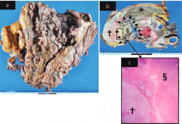
Fig. 3 Autopsy fi ndings. (a) The right lung, thoracic wall, and mediastinum were resected en bloc. (b) There were necrotic
tissues in the empyema cavity. Horizontal slice showed the tumor (†) in contact with the empyema cavity (arrow and §) and the right lung (*). (c) Microscopic fi ndings also identifi ed the tumor (†) in contact with the empyema cavity (§).
性慢性膿胸を認め,人工気胸術後または結核性膿胸治療 後から 20 年以上を経て発症していた1)。
組織学的には 2008 年の WHO 分類で DLBCL associated chronic infl ammation に位置付けられている。PAL 患者に おいて腫瘍細胞内の EBER 陽性率は 70% 程度1)であり, EBV に感染した B リンパ球が外因性の慢性炎症を背景に 腫瘍化するとも考えられている7)。本症例でも,1994 年 の手術検体,今回のエコーガイド下生検検体および病理 解剖検体のすべてにおいて EBER 陽性であり,EBV 感染 が示唆された。 自験例では 1994 年の組織標本を用いた検討で,パラ フィンブロックの保存状態も良好とはいえず,病理学的 検討の問題も懸念されたが,1994 年の手術検体でもエコ ー下穿刺および病理解剖時と同様の組織像がhematoxylin-eosin 染色で確認され,免疫組織化学染色においても同 様の染色パターンを呈しており,長期保存後ではあった が,病理学的な診断に問題はないと判断した。 PAL の治療法は確立されておらず,Stage I _Ⅱ期の中 には化学療法のみの長期寛解例や外科的治療による根 治例も報告されている2) 3)が,一般的には 5 年生存率は Stage Ⅰ期 47%, Ⅱ 期 35%,Ⅲ_Ⅳ 期 15%4)で, 他 の 非 Hodgkin リンパ腫と比較して予後不良である。また,放 射線治療の有効性も示唆されている1) 7)が,自験例では 78 歳の腫瘍診断時および R-CHOP 2 コース終了時いずれ も腫瘍径が大きく,放射線療法は困難であった。 本症例は 17 年の期間をおいて同一の腫瘍組織が確認 された。われわれが医中誌および PubMed で検索したか ぎりでは過去に PAL が異時性に発症した例は確認でき ず,自験例も生前に膿胸の確定診断には至らなかった。し かし,18 歳で結核性膿胸に罹患し,1994 年の手術時に内 部に膿汁を伴う石灰化胸膜腔が確認され,術後の CT で も被包化腔が確認され,死後の病理解剖時にも腫瘍に接 して膿胸腔が確認されたことから,臨床的に結核性胸膜 炎後の慢性膿胸腔が長期間遺残していたと判断した。 自験例において 2011 年の DLBCL が 1994 年の DLBCL の遺残として長期経過で増大して顕在化したか,1994 年 の時点で完全切除され遺残膿胸腔を発生母地として新規 に 出 現 し た か の 判 断 は 難 し い。1994 年 の 手 術 検 体 で DLBCL が完全切除されているかどうかの検討を試みた が,残存検体が一部のみであるため,DLBCL の完全切 除が達成できていたかは分からなかった。また,2011 年 6 月以前も 1995 年からわずかではあるが画像上も腫瘤 影に変化が認められてはいた。しかし,1994 年の腫瘍切 除術の際,DLBCL が残存していれば,10 年以上無症候 で経過するとは考えにくく,今回の入院時の DLBCL は 遺残膿胸腔を発生母地として,異時性に新たに DLBCL が発症したと判断した。また,1994 年の手術時に腫瘍が 完全切除され,遺残膿胸腔から新規に DLBCL が発症し たと考えた場合の発症時期も検討した 。しかし,2011 年 7 月(Fig. 4e)では腫瘍増大は明らかであるが,2009 年 (Fig. 4c)も腫瘍辺縁は不明瞭となり,周囲への増大傾 向が認められた。従って,2009 年頃に新規に DLBCL が 出現した可能性はあるが,残存情報で発症時期を断定す ることは困難であった。 腫瘍摘出術と膿胸腔全摘術による長期生存例が報告さ れている2) 3) 5)。自験例においても 1994 年の手術時に膿胸 腔が全摘できていれば,PAL の異時性発症を防げたかも しれない。また,1994 年の術後の経過観察中は自覚症状 および血液検査における炎症反応上昇も認められず,遺 残した膿胸は被包化されたまま制御されていると考えら れた。従って,膿胸の制御のみでは不十分であり,PAL に対し手術療法を行う場合,発生母地となる膿胸腔を含 め全て摘出することの重要性が示唆された。 謝 辞 本例の治療および病態検討に際し多大なご尽力をいた だきました当院呼吸器科 渡辺洋先生,東北大学血液内 科 藤原亨先生,病理学的見地からご指導いただきまし た当院病理部 伊東干城先生に深謝致します。
著者の COI(confl icts of interest)開示:本論文発表内 容に関して特になし。
文 献
1 ) Nakatsuka S, Yao M, Hoshida Y, et al.: Pyothorax-Associated Lymphoma: A Review of 106 Cases. J Clin Oncol. 2002 ; 20 : 4255 4260. 2 ) 中島由槻, 輪久宋明, 小島 玲, 他:慢性結核性膿胸由 来の悪性リンパ腫に対する胸膜肺全摘術の11例の治療 成績. 日胸外会誌. 1996 ; 44 : 484 492. 3 ) 伊藤博通, 遠藤勝幸:膿胸関連リンパ腫の 1 手術例. 日呼外会誌. 2002 ; 16 : 645 649.
4 ) Narimatsu H, Ota Y, Kami M, et al.: Clinicopathological features of pyothorax-associated lymphoma; a retrospective survey involving 98 patients. Annals of Oncology. 2007 ; 15 : 122 128.
5 ) 河岡 徹, 松井洋人, 長島 淳, 他:放射線治療ならび に 外 科 切 除 で 局 所 制 御 し 得 た 膿 胸 関 連 リ ン パ 腫 の 1 例. 癌と化学療法. 2009 ; 36 : 2127 2129.
6 ) Aozasa K: Pyothorax-associated Lymphoma. J Clin Exp Hematopathol. 2006 ; 46 : 5 10.
7 ) 黒崎敦子:胸膜, 胸壁のリンパ腫. 日本胸部臨床. 2011 ; 20 : 560 569.
Abstract [Background] This is a report of
pyothorax-associated lymphoma (PAL), a diffuse large B-cell lymphoma (DLBCL) that developed in the pleural cavity after more than a 20-year history of pyothorax resulting from artifi cial pneumothorax treatment for pulmonary tuberculosis or tuber-culous pleuritis. The clinical course of PAL is not clear. [Case] A 78-year-old man with right chest pain and bloody sputum entered our hospital. He had past history of tubercu-losis pleuritis at age 18, and resection of a right chest wall tumor at age 61. Chest computerized tomography (CT) on admission revealed a tumor of the right chest wall. A biopsy was performed, and was diagnosed as DLBCL, which we diagnosed as PAL. In addition, we reexamined the specimen collected at age 61, and identifi ed the same histopatholog-ical features in the new specimen.
[Conclusions] This is the fi rst case report of heterochronic PAL, from which we could observe the process of PAL development for a 17-year period.
Key words: Pyothorax-associated lymphoma: PAL,
Tuber-culosis, Diffuse large B-cell lymphoma: DLBCL, Epstein-Barr virus: EBV, Malignant lymphoma, Heterochronic
Department of Respiratory Medicine, Saka General Hospital Correspondence to: Daisuke Jingu, Department of Respira-tory Medicine, Saka General Hospital, 16_5, Nishiki-cho, Shiogama-shi, Miyagi 985_8506 Japan.
(E-mail: d.jinguuu@gmail.com) −−−−−−−−Case Report−−−−−−−−
A CASE OF HETEROCHRONIC PYOTHORAX-ASSOCIATED LYMPHOMA
WITH PAST HISTORY OF TUBERCULOUS PYOTHORAX
Daisuke JINGU, Takehiro YAJIMA, Satoshi UBUKATA, Makoto SHOJI, Kazuhisa KONISHI, and Hiroshi TAKAHASHI