Abstract The occurrence of pulmonary tuberculosis (PTB) and lung cancer as comorbidities has been extensively discussed in many studies. In the past, it was well known that lung cancer is a specific epidemi-ological successor of PTB and that lung cancer often develops in scars caused by PTB. In recent years, the relevance of the two diseases has drawn attention in terms of the close epidemiological connection and chronic inflammation-associated carcinogenesis. In Japanese case series studies, most lung cancer patients with tuberculous sequelae received supportive care alone in the past, but more recently, the use of aggressive lung cancer treatment is increasing. Many studies on PTB and lung cancer as comorbidities have revealed that active PTB is noted in 2_5% of lung cancer cases, whereas lung cancer is noted in 1_ 2% of active PTB cases. In such instances of comorbidity, many active PTB cases showed Type II (non-extensively cavitary disease) and Spread 2_ 3 (intermediate_extensive diseases) on chest X-rays, but standard anti-tuberculosis treatment easily eradicates negative conversion of sputum culture for M.tuberculosis; lung cancer cases were often stage III_ IV and squamous cell carcinoma predominant, and the administration of aggressive treatment for lung cancer is increasing. The major clinical problems associated with PTB and lung cancer as comor-bidities include delay in diagnosis (doctor’s delay) and therapeutic limitations. The former involves two factors of radiographic interpretation: the principles of parsimony (Occam’s razor) and visual search; the latter involves three factors of lung cancer treatment: infectivity of M.tuberculosis, anatomical limitation due to lung damage by tuberculosis, and drug-drug interactions between rifampicin and anti-cancer drugs, especially molecularly targeted drugs. The comorbidity of these two diseases is an important health-related issue in Japan. In the treatment of PTB, the possibility of concurrent lung cancer should be kept in mind, while in the treatment of lung cancer, the possibility of concurrent PTB should also be considered.
Key words : Tuberculosis, Lung cancer, Comorbidity, Epidemiology, Scar cancer, Doctor’s delay, Therapeutic limitation
Pathology and Epidemiology Division, Center for Pulmonary Diseases and Clinical Research Center, National Hospital Organization Tokyo National Hospital
Correspondence to : Atsuhisa Tamura, Pathology and Epidemiology Division, Center for Pulmonary Diseases and Clinical Research Center, National Hospital Organization Tokyo National Hospital, 3_1_1, Takeoka, Kiyose-shi, Tokyo 204_8585 Japan.
(E-mail: tamura-in@tokyo-hosp.jp)
(Received 19 Sep. 2015 / Accepted 11 Dec. 2015)
−−−−−−−−Review Article−−−−−−−−
TUBERCULOSIS AND LUNG CANCER
Atsuhisa TAMURA
1. Introduction
The occurrence of pulmonary tuberculosis (PTB) and lung cancer as comorbidities was first described in an autopsy record by Bayle in 18101), followed by the first case report
by Penard in 18462), and has subsequently been discussed in
many studies3)_7). In Japan, the comorbidity of these two
diseases has drawn attention since the mid-20th century8)_12).
Japan is a middle-ranked country for PTB prevalence, and lung cancer is the leading cause of cancer death in Japan. Furthermore, the proportion of elderly patients affected by both diseases is high. Therefore, the comorbidity of these two diseases is an important issue in Japan.
In this review, we introduce epidemiological studies focus-ing on the relationship between previous tuberculosis and lung cancer development, as well as etiological studies
investiga-ting the relationship between tuberculosis-related lesions and lung carcinogenesis. We then document case series studies in terms of various pathological conditions related to the comor-bidity of PTB and lung cancer.
2. Epidemiological studies
In epidemiological studies of lung cancer in patients with previous tuberculosis, long-term surveys in England/Wales and Australia since the early 1900s have shown that the overall mortality rate for tuberculosis+lung cancer was constant, at approximately 20%. As this close relationship was not seen between other diseases, lung cancer appears to be a specific epidemiological successor of PTB. It was assumed that the reduced number of tuberculosis deaths in young individuals may be associated with the increased incidence of lung cancer in elderly patients13)_15). It is unclear
80 *65 87 80 53 73 60 27 53 95 *74 90 15 85 45 65 55 25 0 50 100 Mean age
(*years) surgical Post-cases
Performance
status 0_2 Squamous cell carcinoma Stages III_IV Chemo-therapy cases Supportive care cases 1984_1995 (n=15) 2003_2011 (n=20) % Male Smokers
Fig. 1 Lung cancer in patients with tuberculosis sequelae
Smokers: current and previous smokers
Post-surgical cases: patients who had a history of surgical therapy for pulmonary tuberculosis
indicated that scar cancer of the lung is rare in actual clinical practice. In recent years, it has been found that in various types of cancer, chronic inflammation and cancer development are closely related32). Tuberculosis-induced chronic
inflam-mation and the scarring process involves various cytokines, chemokines, and DNA damage of epithelial cells; therefore, this may lead to cancer development33)34).
4. Comorbidity of tuberculosis and lung cancer (1) Previous tuberculosis and lung cancer
a) Tuberculosis sequela/state-after thoracoplasty
Tuberculosis sequela is the term for conditions that develop with various anatomical changes during the treatment process of PTB, such as respiratory dysfunction, cor pulmonale, and secondary infection35). Patients with tuberculosis sequelae
occasionally develop lung cancer, and in our earlier report36),
we found that 15 out of 294 patients (5.1%) who died from tuberculosis sequelae between 1984 and 1995 had concurrent lung cancer. Most patients with lung cancer were male, smokers, and underwent surgical treatment for tuberculosis, whereas more than half of the patients ended up receiving supportive care alone. However, in patients with tubercu-losis sequelae from 2003 to 201137), performance status was
good, and there were more patients receiving aggressive cancer therapy despite the increasing age of patients (Fig. 1). These changes result from not only advances in cancer treat-ments but also changes in the concept of tuberculosis sequelae; there are few post-surgical cases today, and the majority of tuberculosis sequelae were based on widespread lung injury due to severe PTB.
In the 1950s, prior to the development of anti-tuberculosis drug therapy, surgical treatment with thoracoplasty was widely performed in Japan, and many patients successfully recovered that whether the association between these two diseases
described in the dawn (mid-20th century) of anti-tuberculosis treatment is still established, but in recent years, it has been demonstrated that mostly in Asian countries, having a history of tuberculosis irrespective of smoking is an independent risk factor for lung cancer occurrence16)_20). Furthermore, with
regard to prognosis, a prospective population-based cohort study has shown that having a history of tuberculosis predicts poor prognosis in lung cancer patients21).
In Japan, it has been demonstrated that the risk of lung cancer among patients with active PTB is 20 times greater than that of the general population22), and patients with active
PTB are more likely to die of lung cancer compared with other malignancies23).
3. Etiological studies
Various theories have been proposed with regard to the pathogenetic relationship between tuberculosis and lung cancer, including the antagonist theory of Rokitansky report-ed in 185524), scar cancer theory25), and coincident theory26).
Among these, the scar cancer theory, which states that lung cancer is likely to develop in scars caused by PTB and other causes, has been widely accepted27)_29). However, Shimosato
et al.30) demonstrated that scar formation in peripheral lung
adenocarcinoma is the result of cancer growth and not a pre-existing lesion, and Noguchi et al.31) revealed a close
relationship between the degree of scarring within pulmonary adenocarcinoma and lung cancer prognosis. In an earlier report, Kageyama et al.10) described that tuberculosis lesions,
compared with healthy lung tissue, can be an origin of lung cancer development, but this carcinogenesis has no specific relationship with the tuberculosis lesion. Aoki23) also reviewed
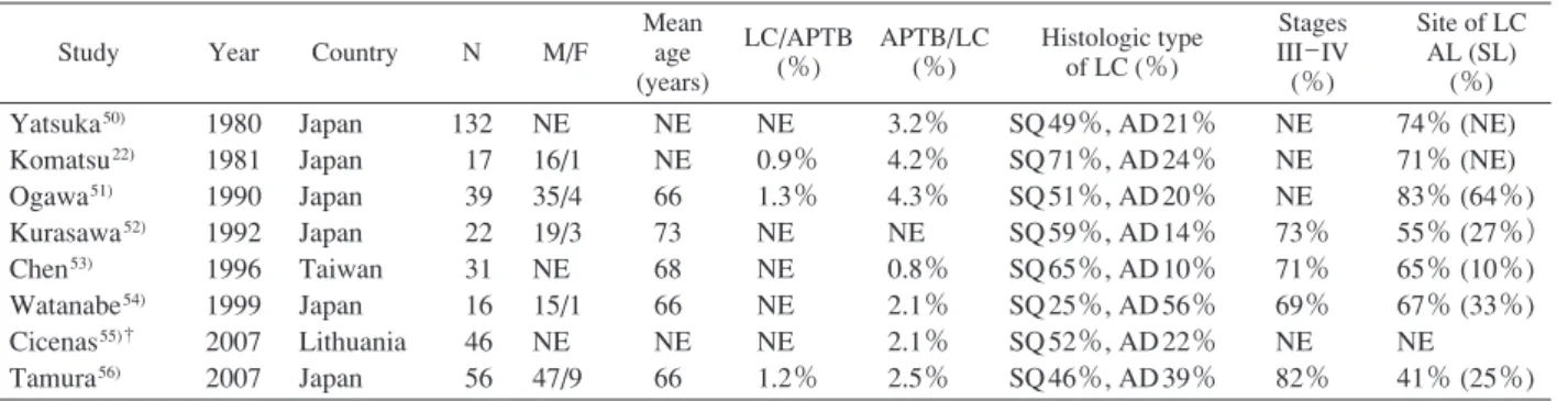
Table 1 Previous studies on the comorbidity of active pulmonary tuberculosis and lung cancer
Study Year Country N M/F
Mean age (years) LC/APTB (%) APTB/LC (%) Histologic type of LC (%) Stages III_IV (%) Site of LC AL (SL) (%) Yatsuka50) Komatsu22) Ogawa51) Kurasawa52) Chen53) Watanabe54) Cicenas55)† Tamura56) 1980 1981 1990 1992 1996 1999 2007 2007 Japan Japan Japan Japan Taiwan Japan Lithuania Japan 132 17 39 22 31 16 46 56 NE 16/1 35/4 19/3 NE 15/1 NE 47/9 NE NE 66 73 68 66 NE 66 NE 0.9% 1.3% NE NE NE NE 1.2% 3.2% 4.2% 4.3% NE 0.8% 2.1% 2.1% 2.5% SQ 49%, AD 21% SQ 71%, AD 24% SQ 51%, AD 20% SQ 59%, AD 14% SQ 65%, AD 10% SQ 25%, AD 56% SQ 52%, AD 22% SQ 46%, AD 39% NE NE NE 73% 71% 69% NE 82% 74% (NE) 71% (NE) 83% (64%) 55% (27%) 65% (10%) 67% (33%) NE 41% (25%) APTB : active pulmonary tuberculosis, LC : lung cancer, AL : affected-side lung, SL : same lobe, NE : not evaluated,
SQ : squamous cell carcinoma, AD : adenocarcinoma, †: surgical case study from tuberculosis with this treatment. However, some of these patients developed tuberculosis sequelae over 20_ 30 years after surgery, and during management of sequelae, lung cancer was sometimes detected. During the period from 1982 to 1998, 20 of the 1,635 lung cancer patients (1.2%) seen in our hospital had a history of thoracoplasty38). Patients with
tuberculosis sequelae and patients who had received thoraco-plasty in our studies developed lung cancer in all lung lobes, irrespective of the site of the tuberculosis lesion. Yoneyama et al.39) reported that in patients with pre-existing unilateral
lung disease including tuberculosis, lung cancer predomi-nantly developed in the healthy side of the lung. They assumed that as a result of ventilator impairment of the affected side, the healthy side may then have had increased exposure to carcinogens. From the perspective of these clinical data, it appears that most lung cancers in patients with previous tuberculosis are not scar cancers. There are few reports on such pathologies outside Japan, and only a few case reports can be found with regard to lung cancer development following surgery for tuberculosis40)_ 43).
b) Lung cancer in chronic pyothorax
The most well-known malignancy seen in patients with chronic pyothorax due to tuberculosis is pyothorax-associated lymphoma, a disease specific to Japan44); its onset primarily
involves Epstein-Barr virus infection caused by artificial pneumothorax45)46). It has long been known that patients
with chronic pyothorax have lung cancer47). At our hospital,
we found that in the 15 patients with chronic pyothorax and concurrent thoracic malignancies between 1977 and 2002, lung cancer and pyothorax-associated lymphoma occurred in four and nine patients, respectively48). In a study of 12
patients with chronic pyothorax and concurrent lung cancer examined by the Japan National Hospital Organization Study Group for Lung Cancer (JNHOSGLC) between 1980 and 200549), unlike lung cancer in tuberculosis sequelae and
state-after thoracoplasty, lung cancer existed around the chronic pyothorax in nine patients. Lung cancer might commonly develop around chronic pyothorax because persistent chronic inflammation around the pyothorax contributes to cancer development; thus, lung cancer around chronic pyothorax
can be considered to be analogous to scar cancer. (2) Active PTB and lung cancer
Concurrent active PTB and lung cancer is a condition most commonly addressed in case series studies. Previously, Nuessle26) summarized past autopsy reports in which lung
cancer was observed in 1.4% of active PTB cases, and active PTB was observed in 6.4% of lung cancer cases. There have been many case series studies on comorbidity of the two diseases, especially in Japan (Table 1)22)50)_ 56). In summary,
these studies indicated that lung cancer was comorbid with 1_ 2% of active PTB cases, while active PTB was comorbid with 2_ 5% of lung cancer cases. Clinical features associated with the comorbidity of these two diseases include the follow-ing: i) the comorbidity is male predominant, ii) squamous cell carcinoma is more common than adenocarcinoma, iii) the site of lung cancer often lies on the same lung side as the PTB, and iv) lung cancer is often detected at an advanced stage (stage III_ IV). Furthermore, reports from the past three decades were mostly from Asian countries, whereas there have been no reports from the US and Europe, which have a low prevalence of tuberculosis.
At our hospital from 1991 to 2004, there were 56 patients with comorbid active PTB and lung cancer. Similar to patients with previous tuberculosis, there was a high rate of men, elderly individuals, and smokers among these cases; comorbid lung cancer was found in 1.2% of cases with active PTB and comorbid active PTB was found in 2.5% of cases with lung cancer56). Active PTB was often sputum-smear positive, Type
II (non-extensively cavitary disease), and Spread 2_ 3 (inter-mediate_ extensive diseases), whereas lung cancer was fre-quently squamous cell carcinoma, stage III_ IV, and occurred on the same lobe as PTB. This trend was comparable with a study of comorbid cases at our hospital between 2005 and 201257) (Fig. 2). The comorbid cases with the two diseases may
be divided into three groups according to the time of detect-ion: the PTB preceding group, concurrent detection group, and lung cancer preceding group. In the PTB preceding group (cases with lung cancer detected during treatment for PTB), a small lung cancer shadow may be identified in many cases upon review of chest X-rays at the time of PTB diagnosis;
%
Pulmonary tuberculosis Lung cancer
84 50 86 48 55 68 83 66 25 46 82 82 64 79 57 43 82 89 57 32 50 89 0 50 100 2005_2012 (n=28)
Concur-rent Squamouscell
carcinoma 1991_2004 (n=56)
Male Over 70
years Smokers Perfor-mance status 0_1
Type II Spreads
2_3 smear forPositive AFB
Coexis-tence III _ IVStages
Concurrent group and tuberculosis preceding group
Lung cancer preceding group 19 25 32 56 14 35 19 5 5 42 5 85 58 0 50 100 0 50 100 Resection Chemotherapy
Radiation therapy Supportive care alone 1991_2004 (n=37) 2005_2012 (n=16) 1991_2004 (n=19) 2005_2012 (n=12) % %
Fig. 2 Relationship between active pulmonary tuberculosis and lung cancer
Smokers : current and previous smokers, Type II : non-extensively cavitary shadow on chest X-ray, Spreads 2_3 : intermediate_extensive diseases on chest X-ray, AFB : acid fast bacilli,
Concurrent : lung cancer concurrently detected with pulmonary tuberculosis, Coexistence : coexisting lung cancer and pulmonary tuberculosis in the same lobe
Fig. 3 Selection of lung cancer treatment in patients with active pulmonary tuberculosis and lung cancer
thus, this group may essentially be considered to be similar to the concurrent detection group. In contrast, PTB in the lung cancer preceding group is often found when lung cancer is advanced and patients are receiving anti-cancer chemotherapy, corticosteroids for brain metastasis, or supportive care alone. Compared with other groups, patients in this group are often in poor general condition, with severe illness resulting from both diseases. With advanced age, an immunosuppressive state, and corticosteroid administration, it is commonly known that there is an increased risk of PTB arising from endogenous reactivation58)_60). During the treatment of elderly patients
with lung cancer in whom incidence rates of tuberculosis are
high, appropriate care should be taken regarding the onset of pulmonary tuberculosis due to reactivation.
With the treatment of the two diseases in our study56), we
found that the negative conversion rate of sputum culture with PTB treatment was good at 56% after 1 month and at 94% after 2 months in the concurrent detection group and the tuberculosis preceding group combined. In other case series studies52)58)61) and a case-control study62), it has been shown
that if PTB is adequately treated, it can easily yield negative conversion of sputum culture, and anti-cancer chemotherapy is not an obstacle in the treatment of PTB. In contrast, it has been reported that in lung cancer treatment, the clinical course of each disease runs independently61); however, it has also been
reported that patients with concurrent tuberculosis have a worse prognosis than patients without tuberculosis53). In our
hospital, the lung cancer preceding group included many patients with poor performance status who were receiving supportive care alone, while in the concurrent detection and tuberculosis preceding groups, there were many patients undergoing aggressive cancer therapy, such as resection and chemotherapy. Furthermore, as shown in Fig.2, although there has been no change in patient background, aggressive cancer therapy has been increasingly performed in all groups; there has been a remarkable increase particularly in the num-ber of cases treated with chemotherapy, increasing from 32% to 56% in the concurrent detection group and tuberculosis preceding group, and increasing from 5% to 42% in the lung cancer preceding group (Fig.3). Data on the detailed out-comes of patients with both active PTB and lung cancer who receive more recently developed anti-cancer chemotherapy
drugs are greatly anticipated.
Interestingly, with comorbid active PTB and lung cancer, both diseases commonly co-occur in the same lung lobe. At our hospital, we have sometimes experienced lung cancer and pulmonary mycobacteriosis coexisting in the same lobe of the lung63); the tuberculosis lesions were within or lied
adjacent to lung cancer. Pathological findings of resected cases have revealed disruption of the fibrocaseous tubercu-losis lesion wall due to lung cancer invasion and associated tuberculosis reactivation, while there were no cases of lung cancer developing within the tuberculosis lesion. Kageyama et al.10) noted that tuberculosis lesions are prone to
reactiva-tion when there are coexisting tuberculosis lesions and lung cancer. Karnak et al.64) also reported that bronchoscopy
re-vealed that malignant tumors, particularly lung cancer, led to the reactivation of pulmonary tuberculosis. It is thought that the two diseases coexist in the same lobe often due to the onset of tuberculosis secondary to the progression of lung cancer. Furthermore, the coexistence of the two diseases is a condition that requires careful clinical attention, because many investigators have indicated that radiographic diagnosis of this coexistence is difficult65)_68).
(3) Active PTB after resection of lung cancer
Gastrectomy for gastric cancer has been found to be a risk factor for the onset of pulmonary tuberculosis69). However,
there have been few reports focused on PTB following resec-tion of lung cancer. Of the 4,393 patients hospitalized at our hospital for active PTB between 1996 and 2007, 14 patients (0.3%) developed PTB following lung cancer resection70).
Patients who developed PTB were often men, elderly, within 5 years after lobectomy, with previous tuberculosis or recurrent lung cancer, and receiving corticosteroids. In the treatment of PTB, as is the case when two diseases are concurrently detected, if standard anti-tuberculosis treatment is possible then the negative conversion of sputum culture may easily be achieved.
5. Clinical problems (1) Delay in diagnosis (doctor’s delay)
When clinically analyzing diagnosis of tuberculosis, there is a period from the time of symptom onset to the time of diagnosis, called total delay, which is further categorized into patient’s delay (from the time of symptom onset to the time of first visit to a medical institution) and doctor’s delay (from the time of first visit to a medical institution to the time of diagnosis [delay in diagnosis])71). Total delay is a
major problem for PTB in clinical practice72). Since patients
with active PTB, previous tuberculosis, and lung cancer are typically under the management of medical institutions, patient’s delay is rare in these cases; doctor’s delay more than patient’s delay is considered to be the primary factor contributing to total delay with regard to comorbid PTB and lung cancer.
There are two main factors that cause doctor’s delay. First,
in the interpretation of chest X-rays, pulmonologists attempt to classify chest shadows with various patterns as a single disease or as simply as possible ― i.e., they often apply the principle of parsimony (Occam’s razor) when making a diag-nosis. In general, when faced with various clinical data, parsimony is useful for making a differential diagnosis and determining treatment. However, it is also well known that this tendency to simplify can result in erroneous diagnoses73)74).
It is thought that in concurrent PTB and lung cancer, espe-cially in cases in which the two diseases coexist in the same lobe, this principle of parsimony can easily result in a doctor’s delay63). Another factor is that the effectiveness of a visual
search75) depends on the number and nature of interfering
objects. For example, it is difficult to detect a small lung cancer shadow (target object) among widespread previous tuberculosis shadows (interfering objects) in patients with tuberculosis sequelae36).
To eliminate any doctor’s delay caused by these factors, it is important to keep in mind the possibility of coexisting PTB and lung cancer. More specifically, the following should be noted: i) a shadow on peripheral consolidations of chest X-ray of hilar-type lung cancer should not simply be considered obstructive pneumonia, but the possibility of a tuberculosis lesion such as caseous pneumonia should be taken into consideration; ii) one should perform sputum examination for M.tuberculosis in patients with lung cancer, even when they are receiving supportive care; iii) it is important to carefully consider whether or not there are any growing shadows on chest X-ray during PTB treatment; and iv) pulmonologists should compare prior X-rays and assess any changes when examining patients with previous tuberculosis. (2) Therapeutic limitations
As mentioned above, if the PTB treatment proceeds uneventfully, negative conversion of sputum culture can be easily achieved; therefore, any limitations in the treatment of comorbid PTB and lung cancer are primarily the limitations of lung cancer treatment. These include1) postponing the timing of surgery for lung cancer due to tuberculosis infectivity; 2) limitations to treatment methods for anatomical injury due to tuberculosis, especially avoidance of surgical treatment; and 3) interference of cancer chemotherapy due to anti-tuberculosis therapy, particularly the induction of CYP3A4 by rifampicin (RFP).
With regards to JNHOSGLC questionnaire survey by Hayashi et al.76) regarding the time until lung cancer surgery
for cases in which cancer and PTB were concurrently detected, it was found that the majority of respondents were of the opinion that the surgical period for lung cancer was more than 4 weeks after the start of anti-tuberculosis treatment, and upon negative conversion of sputum smear with or without confirmation of drug-sensitivity . Although there has been no evidence regarding the appropriate timing of surgery, the opinion that takes into account tuberculosis infectivity for others before and after surgery is presumed to be valid77). With
Table 2 Pharmacokinetic drug-drug interactions of molecular targeted drugs with rifampicin
Drug Metabolic mediator Decrease† of AUC
∞ Decrease†of Cmax Gefitinib78) Erlotinib79) Afatinib80) Crizotinib81) Alectinib82) CYP3A4 CYP3A4 P-glycoprotein CYP3A4 CYP3A4 83% 69% 34% 82% 73% 65% 39% 22% 69% 51% AUC∞: area under the plasma concentration-time curve from time zero to infinity,
Cmax: peak plasma concentration, †: Percent decrease by co-administration of rifampicin
regards to the second limitation mentioned above, when the tuberculosis lesions extensively exist in one side of the lung, it is often difficult to perform resection or radiotherapy for lung cancer in the contralateral lung. In our experience, lung cancers in the side of the lung contralateral to the thoracoplasty were unable to undergo resection, even if the lung cancer was stage I or II. This anatomical problem is difficult to solve, but early detection of lung cancer may increase treatment options, such as stereotactic irradiation. With regards to the third limitation listed above, it has been found that the induction of CYP3A4 by RFP affects the drug metabolism of anti-cancer agents. This mostly attenuates the decrease of blood concentration by increasing anti-cancer drug metabolism upon CYP3A4 induc-tion. In particular, excluding bevacizumab, molecularly tar-geted drugs combined with RFP reduce the area under the plasma concentration-time curve from time zero to infinity (AUC∞) and peak plasma concentration (Cmax) to a high
degree; thus, caution is required (Table 2)78)_82). On the other
hand, with cytotoxic drugs, there is a case report in which the use of RFP lowered blood levels of irinotecan83). In
addition, it is possible that blood concentration levels of docetaxel, paclitaxel, and vinorelbine are also lowered by CYP3A induction84)85); however, there is no evidence
indicat-ing that the degree of such attenuations reach significant levels in clinical practice. To avoid lowering blood concentrations of molecularly targeted drugs, it is thought that when choosing an anti-tuberculosis agent, RFP should be changed to rifabutin, which induces CYP3A4 to a lesser degree86); however,
ad-verse effects associated with rifabutin should be taken into consideration87).
6. Conclusion and Future Perspective
The close relationship between PTB and lung cancer has again come to light as a result of large scale epidemiological studies recently conducted in Asia. At the same time, it is thought that investigations into scar cancer will be acceler-ated from the perspective of chronic inflammation and cancer development. In studying the mechanisms of carcinogenesis, instead of studies regarding PTB with regional differences in prevalence rates, studies of the relationship between pul-monary nontuberculous mycobacteriosis, the prevalence of which is increasing worldwide, and lung cancer may be significant88)_91).
Doctor’s delay is a major clinical problem related to PTB and lung cancer. To eliminate this delay, it is necessary to consider concurrent lung cancer in the treatment of active PTB and to consider concurrent active PTB in the treatment of lung cancer; it is also important to perform X-rays and sputum examination with bronchoscopy if necessary. Further-more, when treating active PTB, it is thought that the ade-quate introduction of standard anti-tuberculosis treatment92)
will be useful for early detection of lung cancer and verifica-tion of Interferon-Gamma Release Assays93) for elderly lung
cancer patients in Japan will be useful for early diagnosis of active PTB. Anti-cancer chemotherapy, which has made remarkable developments, should be managed after fully understanding drug-drug interactions, especially the attenua-tion of the effects of molecularly targeted drugs as a result of CYP3A4 induction by RFP.
Confl ict of interest
The author declares that there is no conflict of interest related to this article.
References
1 ) Bayle GL: Recherches sur la Phthisie Pulmonaire. Gabon, Paris, 1810, 310.
2 ) Penard M: Cancer et tubercule du poumon. Bull Soc Anat De Paris. 1846 ; 21 : 260.
3 ) Fried BM: Bronchiogenic cancer combined with tuberculosis of the lungs. Am J Cancer. 1935 ; 23 : 247 266.
4 ) Robbins E, Silverman G: Coexistent bronchogenic carcinoma and active pulmonary tuberculosis. Cancer. 1949 ; 2 : 65 97. 5 ) Sakula A: Carcinoma of bronchus in association with active
pulmonary tuberculosis. Br Med J. 1955 ; 1 : 759 762. 6 ) Sherman PH, Conant JS, Peereboom G: Carcinoma of the
lung in a tuberculosis hospital population. Am Rev Respir Dis. 1967 ; 96 : 451 459.
7 ) Berroya RB, Polk JW, Raju P, et al.: Concurrent pulmonary tuberculosis and primary carcinoma. Thorax. 1971 ; 26 : 384 387.
8 ) Miyaji T, Kitamura H, Senoo T, et al.: Morphological study of 406 cases of bronchogenic carcinoma in Japan. Gan. 1955 ; 46 : 523 547.
9 ) Kawai N, Katsuki H: Relationship between pulmonary tuberculosis and lung cancer. Kekkaku. 1956 ; 31 (supple) :
1 28.
10) Kageyama K, Hanaoka K: Pulmonary tuberculosis and lung cancer. Kekkaku. 1975 ; 50 : 607 611.
11) Matsushima T: Statistical study on association of tubercu-losis and carcinoma of the lung in Chugoku and Shikoku area. Kekkaku. 1978 ; 53 : 377 383.
12) Aoki K: Epidemiological study on pulmonary tuberculosis and lung cancer. Kekkaku. 1985 ; 60 : 629 642.
13) Cherry T: Cancer and acquired resistance to tuberculosis, part 1. Med J Austral. 1924 ; 2 : 372 378.
14) Campbell AH: The relationship between cancer and tuber-culosis mortality rates. Br J Cancer. 1961 ; 15 : 10 18. 15) Haybittle JL: Study of cancer mortality in England and
Wales using birth-standardized populations. Br J Prev Soc Med. 1962 ; 16 : 93 104.
16) Zheng W, Blot WJ, Liao ML, et al.: Lung cancer and prior tuberculosis infection in Shanghai. Br J Cancer. 1987 ; 56 : 501 504.
17) Brenner AV, Wang Z, Kleinerman RA, et al.: Previous pulmonary diseases and risk of lung cancer in Gansu Province, China. Int J Epidemiol. 2001 ; 30 : 118 124. 18) Wu CY, Hu HY, Pu CY, et al.: Pulmonary tuberculosis
increases the risk of lung cancer: a population-based cohort study. Cancer. 2011 ; 117 : 618 624.
19) Brenner DR, Boffetta P, Duell EJ, et al.: Previous lung diseases and lung cancer risk: a pooled analysis from the International Lung Cancer Consortium. Am J Epidemiol. 2012 ; 176 : 573 585.
20) Simonsen DF, Farkas DK, Søgaard M, et al.: Tuberculosis and risk of cancer: a Danish nationwide cohort study. Int J Tuberc Lung Dis. 2014 ; 18 (10) : 1211 1219.
21) Heuvers ME, Aerts JG, Hegmans JP, et al.: History of tuberculosis as an independent prognostic factor for lung cancer survival. Lung Cancer. 2012 ; 76 : 452 456. 22) Komatsu H, Ishizuka Y, Yoneda R: A study of coexistence
of bronchogenic carcinoma and active pulmonary tubercu-losis. Kekkaku. 1981 ; 56 : 49 55.
23) Aoki K: Excess incidence of lung cancer among pulmonary tuberculosis patients. Jpn J Clin Oncol. 1993 ; 23 : 205 220. 24) Rokitansky C: A Manual of Pathological Anatomy. vol 1.
Blanchard and Lea, Philadelphia, 1855 : 237 238.
25) Friedrich G : Periphere Lungenkrebse auf dem Boden Pleuranaher Narben. Virchows Arch (Pathol Anat). 1939 ; 304 : 230 247.
26) Nuessle WF: Association of bronchogenic carcinoma and active pulmonary tuberculosis: with report of four cases. Dis Chest. 1953 ; 23 : 207 216.
27) Raeburn G, Spencer H: Lung scar cancers. Br J Tuberc Dis Chest. 1957 ; 51 : 237 245.
28) Yokoo H, Suckow EE: Peripheral lung cancers arising in scars. Cancer. 1961 ; 14 : 1205 1215.
29) Auerbach O, Garfinkel L, Parks VR: Scar cancer of the lung: increase over a 21 year period. Cancer. 1979 ; 43 : 636 642.
30) Shimosato Y, Suzuki A, Hashimoto T, et al.: Prognostic implications of fibrotic focus (scar) in small peripheral lung cancer. Am J Surg Pathol. 1980 ; 4 : 365 373.
31) Noguchi M, Morikawa A, Kawasaki M, et al.: Small adenocarcinoma of the lung; histologic characteristics and prognosis. Cancer. 1995 ; 75 : 2844 2852.
32) Moss SF, Blaser MJ: Mechanisms of disease: Inflammation and the origins of cancer. Nat Clin Pract Oncol. 2005 ; 2 : 90 97.
33) Bobba RK, Holly JS, Loy T, et al.: Scar carcinoma of the lung: a historical perspective. Clin Lung Cancer. 2011 ; 12 : 148 154.
34) Nalbandian A, Yan BS, Pichugin A, et al.: Lung carcino-genesis induced by chronic tuberculosis infection: the exper-imental model and genetic control. Oncogene. 2009 ; 28 : 1928 1938.
35) The Japanese Society for Tuberculosis. New Terminology of Tuberculosis, Nankodo Co., Tokyo, 2008, 44.
36) Tamura A, Nagai H, Sagara Y, et al.: Lung cancer in patients with sequelae of tuberculosis. Kekkaku. 1998 ; 73 : 619 624. 37) Mori A, Kusaka K, Kawashima M, et al.: Lung cancer in
patients with sequelae of tuberculosis. Haigan. 2013 ; 53 : 546 (abstract).
38) Tamura A, Hebisawa A, Hayashi K, et al.: Lung cancer in patients who had received thoracoplasty for pulmonary tuberculosis. Jpn J Clin Oncol. 1999 ; 29 : 541 545. 39) Yoneyama T, Naruke T, Suemasu K, et al.: Bronchial
carcinoma in patients with pre-existing unilateral lung disease. Thorax. 1976 ; 31 : 650 651.
40) Fulton D, Rolleston C: Bronchial carcinoma in a case of pulmonary tuberculosis undergoing artificial pneumothorax. Br J Tuberc Dis Chest. 1946 ; 40 : 129 133.
41) Bruce T, Dahlstorm G, Uggla LG: Squamous epithelial cancer of the pleura following extrapleural pneumothorax for pulmonary tuberculosis. Acta Tuberc Scand. 1960 ; 38 : 261 266.
42) Harland RW, Sharma M, Rosenzweig DY: Lung carcinoma in a patient with Lucite sphere plombage thoracoplasty. Chest. 1993 ; 103 : 1295 1297.
43) Rena O, Casadio C, Maggi G : Primitive squamous-cell carcinoma after extrapleural pneumothorax for active tuber-culosis. Eur J Cardiothorac Surg. 2001 ; 19 : 92 95. 44) Iuchi K, Ichimiya A, Akashi A, et al. : Non-Hodgkin’s
lymphoma of the pleural cavity developing from long-standing pyothorax. Cancer. 1987 ; 60 : 1771 1775. 45) Fukayama M, Ibuka T, Hayashi Y, et al.: Epstein-Barr virus
in pyothorax-associated pleural lymphoma. Am J Pathol. 1993 ; 143 : 1044 1049.
46) Nakatsuka S, Yao M, Hoshida Y, et al.: Pyothorax-associated lymphoma: a review of 106 cases. J Clin Oncol. 2002 ; 20 : 4255 4260.
47) Deaton WR Jr.: Carcinoma arising in chronic empyema cavity. Case report with review of the literature. Dis Chest 1962 ; 42 : 563 566.
48) Tamura A, Hebisawa A, Sagara Y, et al.: Thoracic malig-nancies in patients with chronic tuberculous empyema. Kekkaku. 2004 ; 79 : 301 307.
49) Tamura A, Hebisawa A, Iuchi K, et al.: Lung cancer in patients with chronic pyothorax. Respirology. 2008 ; 13 : 585 589.
50) Yatsuka Y, Matuyama T, Sawamura K, et al.: Pulmonary tuberculosis and pulmonary carcinoma from a clinical point of view-based on 4000 cases recorded by the National Sanatorium Lung Cancer Research Group. Haigan. 1980 ; 20 Suppl : 21 32.
51) Ogawa N, Arai T, Inagaki K, et al.: A study on active pulmonary tuberculosis with coexistent lung carcinoma. Nihon Kyobu Rinsho. 1990 ; 49 : 901 907.
52) Kurasawa T, Takahashi M, Kuze F, et al.: A clinical study on coexistence of active pulmonary tuberculosis and lung cancer. Kekkaku. 1992 ; 67 : 119 125.
53) Chen YM, Chao JY, Tsai CM, et al.: Shortened survival of lung cancer patients initially presenting with pulmonary tuberculosis. Jpn J Clin Oncol. 1996 ; 26 : 322 327. 54) Watanabe A, Tokue Y, Takahashi H, et al.: Management of
mycobacteriosis in general hospital without isolation ward for tuberculosis patients. Clinical study on pulmonary tuber-culosis associated with lung cancer patients. Kekkaku. 1999 ; 74 : 157 162.
55) Cicenas S, Vencevicius V: Lung cancer in patients with tuberculosis. World J Surg Oncol. 2007 ; 5 : 22.
56) Tamura A, Hebisawa A, Masuda K, et al.: Coexisting lung cancer and active pulmonary tuberculosis. Nihon Kokyuki Gakkai Zasshi. 2007 ; 45 : 382 393.
57) Yoshida K, Okuda K, Kobayashi K, et al.: Clinical features in patients with coexisting active pulmonary tuberculosis and lung cancer. Kekkaku. 2013 ; 88 : 233 (abstract). 58) Komatsu H, Nagai H, Satou K, et al.: Association of active
pulmonary tuberculosis and malignant diseases: a clinical study. Kekkaku. 1995 ; 70 : 281 284.
59) Yamagishi F: Medical risk factors of tuberculosis and countermeasures. Kekkaku. 2002 ; 77 : 799 804.
60) Aoki Y, Kuroki S, Hiura K, et al.: A clinical study of pulmonary tuberculosis in lung cancer patient. Kekkaku. 1991 ; 66 : 727 732.
61) Mok CK, Nandi P, Ong GB: Coexistent bronchogenic carcinoma and active pulmonary tuberculosis. J Thorac Cardiovasc Surg. 1978 ; 76 : 469 472.
62) Kim DK, Lee SW, Yoo CG, et al.: Clinical characteristics and treatment responses of tuberculosis in patients with ma-lignancy receiving anticancer chemotherapy. Chest. 2005 ; 128 : 2218 2222.
63) Tamura A, Hebisawa A, Sagara Y, et al.: Coexistence of lung cancer and active pulmonary mycobacteriosis. Kekkaku. 2005 ; 80 : 413 419.
64) Karnak D, Kayacan O, Beder S: Reactivation of pulmonary tuberculosis in malignancy. Tumori. 2002 ; 88 : 251 254. 65) Sakuraba M, Hirama M, Hebisawa A, et al.: Coexistent lung
carcinoma and active pulmonary tuberculosis in the same lobe. Ann Thorac Cardiovasc Surg. 2006 ; 12 : 53 55. 66) Ashizawa K, Matsuyama N, Okimoto T, et al.: Coexistence
of lung cancer and tuberculoma in the same lesion: demonstration by high resolution and contrast-enhanced dynamic CT. Br J Radiol. 2004 ; 77 : 959 962.
67) Phillips LG Jr, Cunningham J, Hillman NM, et al.: Carcinoma of the lung and coexistent active pulmonary tuberculosis: diverse morphologic and radiographic presentations. J Natl Med Assoc. 1984 ; 76 : 125 130.
68) Kim Y, Goo JM, Kim HY, et al.: Coexisting bronchogenic carcinoma and pulmonary tuberculosis in the same lobe: radiologic findings and clinical significance. Korean J Radiol. 2001 ; 2 : 138 144.
69) Huang SF, Li CP, Feng JY, et al.: Increased risk of tuber-culosis after gastrectomy and chemotherapy in gastric cancer: a 7-year cohort study. Gastric Cancer. 2011 ; 14 : 257 265.
70) Tamura A, Araki K, Suzuki J, et al.: Pulmonary Myco-bacteriosis in Patients Following Resection of Lung Cancer. Haigan. 2010 ; 50 : 122 129.
71) Ohmori M, Ozasa K, Mori T, et al.: Trends of delays in tuberculosis case finding in Japan and associated factors. Int J Tuberc Lung Dis 2005 ; 9 : 999 1005.
72) Sasaki Y: A study of case findings in pulmonary tuberculosis patients. Kekkaku. 2002 ; 77 : 621 625.
73) Wardrop D: Ockham’s razor: sharpen or re-sheathe? J R Soc Med. 2008 ; 101 : 50 51.
74) Lewis MA, Agusala K, Raizen Y: Ockham’s razor is not so sharp. Infect Dis Rep. 2010 ; 2 : e10.
75) Verghese P: Visual search and attention: a signal detection theory approach. Neuron. 2001 ; 31 : 523 535.
76) Hayashi Y, Tamura A, Ohsaka Y, et al.: Timing of resec-tion for lung cancer complicating active pulmonary tuber-culosis. Haigan. 2000 ; 40 : 536 (abstract).
77) Nakajima Y: Surgical treatment for lung cancer accompa-nied by active pulmonary tuberculosis. Kokyu. 2007 ; 26 : 171 176.
78) Swaisland HC, Ranson M, Smith RP, et al.: Pharmacokinetic drug interactions of gefitinib with rifampicin, itraconazole and metoprolol. Clin Pharmacokinet. 2005 ; 4 : 1067 1081. 79) Chugai Pharmaceutical Co Ltd. Tarceva® (erlotinib):
Japa-nese prescribing information, 11th eds., Chugai Parmaceuti-cal Co Ltd, Tokyo, 2015.
80) Wind S, Giessmann T, Jungnik A, et al.: Pharmacokinetic drug interactions of afatinib with rifampicin and ritonavir. Clin Drug Investig. 2014 ; 34 : 173 182.
81) Pfizer Japan Inc. Xalkori® (crizotinib): package insert, 5th
eds., Pfizer Japan Inc, Tokyo, 2015.
82) Chugai Pharmaceutical Co Ltd. Alecensa® (alectinib):
Japa-nese prescribing information, 3rd eds., Chugai Parmaceuti-cal Co Ltd, Tokyo, 2015.
83) Yonemori K, Takeda Y, Toyota E, et al.: Potential inter-actions between irinotecan and rifampin in a patient with
small-cell lung cancer. Int J Clin Oncol. 2004 ; 9 : 206 209. 84) Rochat B: Role of cytochrome P450 activity in the fate of
anticancer agents and in drug resistance: focus on tamox-ifen, paclitaxel and imatinib metabolism Clin Pharmacokinet. 2005 ; 44 : 349 366.
85) Leveque D, Wisniewski S, Renault C, et al.: The effect of rifampin on the pharmacokinetics of vinorelbine in the micropig. Anticancer Res. 2003 ; 23 : 2741 2744.
86) Burman WJ, Gallicano K, Peloquin C: Comparative phar-macokinetics and pharmacodynamics of the rifamycin anti-bacterials. Clin Pharmacokinet. 2001 ; 40 : 327 341. 87) Kurashima A, Mori T, Tomono Y, et al.: A new
anti-mycobacterial agent, rifabutin. Kekkaku. 2010 ; 85 : 743 756.
88) Weiss CH, Glassroth J: Pulmonary disease caused by non-tuberculous mycobacteria. Expert Rev Respir Med. 2012 ; 6 : 597 612.
89) Lande L, Peterson DD, Gogoi R, et al.: Association between pulmonary Mycobacterium avium complex infection and lung cancer. J Thorac Oncol. 2012 ; 7 : 1345 1351. 90) Tamura A, Hebisawa A, Sagara Y, et al.: Pulmonary
non-tuberculous mycobacteriosis in patients with lung cancer. Kekkaku. 2004 ; 79 : 367 373.
91) Hosoda C, Hagiwara E, Shinohara T, et al.: Clinical characteristics of pulmonary Mycobacterium avium complex infection complicated with lung cancer. Kekkaku. 2014 ; 89 : 691 695.
92) The Treatment Committee of the Japanese Society for Tuberculosis : Review of standards for tuberculosis care ─ 2008. Kekkaku. 2011 ; 86 : 29 36.
93) Pai M, Zwerling A, Menzies D: Systematic review: T-cell-based assays for the diagnosis of latent tuberculosis infection: an update. Ann Intern Med. 2008 ; 149 : 177 184.