52:955
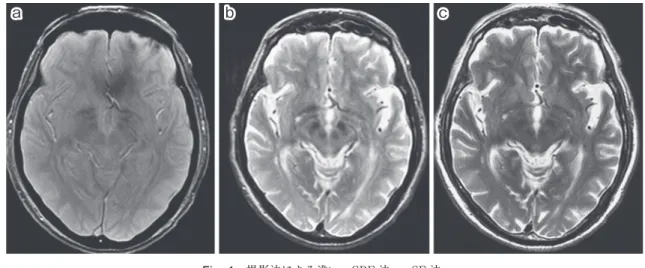
Fig. 1 撮影法による違い―GRE 法 vs. SE 法.
a.GRE 法(T2*強調画像),b.CSE 法(T2強調画像),c.FSE 法(T2強調画像).赤核,黒質の 鉄沈着は,GRE 法より CSE 法の方が明瞭にみえる.
a
b
c
<シンポジウム(1)―5―4>鉄と神経疾患
鉄沈着をきたす疾患の画像診断
百島 祐貴
(臨床神経 2012;52:955-958) Key words:MRI,鉄沈着,T2*強調画像 1.鉄沈着を診断するための MRI 撮像法 脳内に沈着する非ヘム鉄の多くは,フェリチン,ヘモシデリ ンの形で存在し,MRI はこれらの物質の常磁性による T2短 縮を利用して鉄沈着を診断する1). a.GRE 法 vs. SE 法 一般に,鉄沈着の診断には勾配磁場法(Gradient Echo, GRE)法による T2*強調画像が有用である.現在,T2強調画像の多くは高速スピンエコー(Fast Spin Echo,FSE)法で撮影 されるが,この原型である(古典的な)スピンエコー(Conven-tional Spin Echo,CSE)法は鉄沈着の検出能が高く,大脳基 底核,中脳などの鉄沈着は GRE 法より CSE 法の方が明瞭に みとめられるばあいがある(Fig. 1).一方,陳旧性血腫などに よる鉄沈着は,GRE 法がすぐれている.これは,大脳基底核 に沈着するフェリチンは,粒子が小さく,均等に分布するため に磁化率効果が相対的に少なく,一方ヘモシデリン粒子は大 きいために T2*緩和効果が明瞭に現われるためと考えられ る2). b.GRE 法 vs. SWI 法 最近,多くの装置で利用可能となっている磁化率強調画像 法(Susceptibility-weighted Imaging)は,位相画像に後処理 を加えることにより,磁化率のことなる部位をより明瞭に描 出する方法で,脳内鉄沈着については通常の T2*強調画像よ りも感度が高い事が知られており,積極的に利用する価値が ある(Fig. 2)3). c.磁場強度の影響 T2(T2*)緩和効果は,撮影装置の静磁場強度に依存して大 きくなる.現在のところ 1.5T 装置がもっとも多く,3T 装置も かなり普及している.一般に前述のような適切な撮影法を選 択するかぎり,1.5T 以上の装置では診断上の問題はない.し かし,0.2∼1.0T の低磁場装置のばあいは,検出率が低下する ので注意が必要である4). 2.陳旧性微小出血巣の診断 諸疾患における鉄沈着の画像診断については他稿に譲り, 本稿では陳旧性微小出血巣(microbleeds)の MRI 診断につい て述べる. a.陳旧性微小出血巣の画像所見 陳旧性微小出血巣は,様々な病態でみとめられるが,無症候 性出血巣としてみとめられるばあいが多く,長期にわたる高 慶應義塾大学医学部放射線診断科〔〒160―8582 新宿区信濃町 35〕 (受付日:2012 年 5 月 23 日)
臨床神経学 52巻11号(2012:11) 52:956
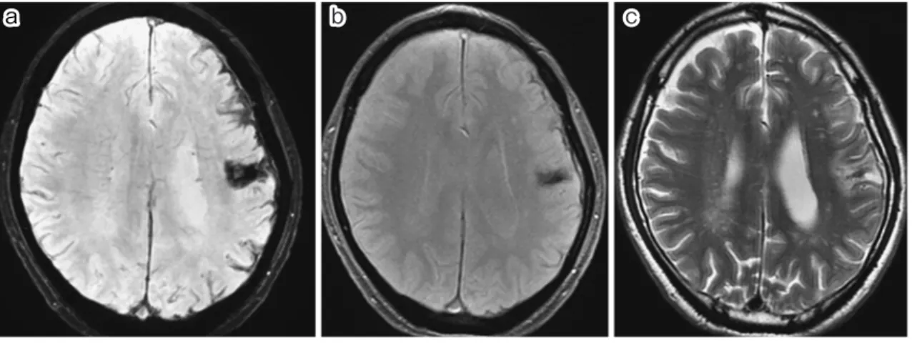
Fig. 2 撮影法による違い―GRE 法 vs. SWI 法.
a.SWI 法(磁化率強調画像),b.GRE 法(T2*強調画像),c.FSE 法(T2強調画像)
外傷性クモ膜下出血後の限局性脳表ヘモシデローシス.脳表の低信号が,SWI 法では GRE 法より明 瞭である.
a
b
c
Fig. 3 陳旧性微小出血巣―慢性高血圧. a, b.T2*強調画像.70 歳男性.無症状.20 年前より高血圧,服薬無し.T2*強調画像でテント上下に 小さな低信号が多発している.a
b
Table 1 陳旧性微小出血巣 をきたす疾患. Hypertensive angiopathy Amyloid angiopathy Hemorrhagic thromboemboli Diffuse axonal injury Cavernous malformation Post-radiation teleangiectasia CADASIL Parry-Romberg syndrome 血圧の症例に多いことが知られている.T2*強調画像,磁化率 強調画像では,数 mm 大の多発低信号としてみとめられる が,その一部が T2強調画像でもみえることがある(Fig. 3). b.鑑別診断 この所見がみとめられる病態としては,Table 1 のような ものが挙げられる4).多くのばあい,病歴,臨床所見から鑑別 が可能であるが,無症候性に偶然発見される例では,慢性高血 圧に続発するものと,多発海綿状血管奇形(血管腫)との鑑別 が問題となることがある.このようなばあい,海綿状血管奇形 では,T2*強調画像,T2強調画像でしばしば低信号に加えて高 信号の混在による不均一な内部構造がみとめられること,T2 強調画像で高信号をともなうことが多いことが鑑別の参考に なる(Fig. 4)4)5). びまん性軸索損傷でも慢性期に多発低信号がみとめられ, 外傷との関連が問題となることがあるが,びまん性軸索損傷 のばあいは,大脳半球の皮髄境界,脳幹,視床など深部構造, 硬膜近傍など,外傷による物理的ストレスがかかりやすい部鉄沈着をきたす疾患の画像診断 52:957 Fig. 4 陳旧性微小出血巣―海綿状血管奇形. a.T2*強調画像,b.T2強調画像,c.T1強調画像.75 歳男性.無症状.30 年前に後頭葉の脳出血 切除術,海綿状血管奇形と診断されている.大脳半球に多発する微小出血巣があるが,T2強調画像 で高信号をともなう内部構造があり,T1強調画像では高信号がみとめられる(矢印).
a
b
c
Fig. 5 陳旧性微小出血巣―びまん性軸索損傷. a, b, c.T2*強調画像.32 歳男性.交通事故後,高次脳機能障害.微小低信号が多発しているが,皮 髄境界,脳幹,硬膜(大脳鎌,小脳テント)近傍に限局しており,外傷との関連が示唆される(矢印).a
b
c
位に限局することが参考となる(Fig. 5)6). ※本論文に関連し,開示すべき COI 状態にある企業,組織,団体 はいずれも有りません. 文 献 1)藤原広和, 百島祐貴. 鉄沈着の画像診断. 神経内科 2010;73: 110-116.2)Haque TL, Miki Y, Kanagaki M, et al. MR contrast of ferriin and hemosiderin in the brain i comparison among gradient-echo, conventional spin-echo and fast spin-echo sequences. Eur J Radiol 2003;48:230-236.
3)Wanga J, Gong X. Superficial siderosis of the central
nervous system : MR findings with susceptibility-weighted imaging. Clin Imag 2011;35:217-221.
4)Blitstein MK, Tung GA. MRI of cerebral microhemor-rhages. Am J Roentgenol 2007;189:720-725.
5)Hegde AN, Mohan S, Lim CC. CNS cavernous haeman-gioma: popcorn in the brain and spinal cord. Clin Radiol 2012;67:380-388.
6)Luccichenti G, Giugni E, Péran P, et al. 3 Tesla is twice as sensitive as 1.5 Tesla magnetic resonance imaging in the assessment of diffuse axonal injury in traumatic brain in-jury patients. Funct Neurol 2010;25:109-114.
臨床神経学 52巻11号(2012:11) 52:958
Abstract
Imaging of diseases with iron deposition
Suketaka Momoshima, M.D.
Diagnostic Radiology, Keio University School of Medicine
Some fundamental technical aspects of magnetic resonance imaging (MRI) in evaluation of iron deposition were discussed. MRI is an imaging modality sensitive to iron deposition of the brain tissue. T2weighted imaging (T2WI), T2*
weighted imaging (T2*
WI), and susceptibility-weighted imaging (SWI) are particularly available for this purpose. They are different in sensitivity and availability, and should be used in the right places respectively. Sus-ceptibility to iron deposition is also dependent on the strength of static magnetic field, which should be taken into account in the interpretation of the images.
(Clin Neurol 2012;52:955-958)
Key words: magnetic resonance imaging, iron deposition, T2 *