博士学位論文
Studies on the mode of action of neurotoxic insecticides
and insecticide resistance
博士学位論文
Studies on the mode of action of neurotoxic insecticides
and insecticide resistance
平成29 年 11 月 6 日
Contents
General Introduction 1 Chapter 1 8
A single crossing-over event in voltage-sensitive Na+ channel genes may cause critical failure of dengue mosquito control by insecticides
Chapter 2 30
Association between the R81T mutation in the nicotinic acetylcholine receptor β1 subunit of Aphis gossypii and the differential resistance to acetamiprid and imidacloprid
Chapter 3 49
The R81T mutation in the nicotinic acetylcholine receptor of Aphis gossypii is associated with neonicotinoid insecticide resistance with differential effects for cyano- and nitro-substituted neonicotinoids
Chapter 4 75
Studies on Aphis gossypii cytochrome P450s CYP6CY22 and CYP6CY13 using an in vitro system
Summary 94 Reference 97 Acknowledgements Publications
General Introduction
Crop protection and insecticides
By 2050, the United Nations estimates that the global population will reach 9.7 billion people (World Population Prospects 2015). It is estimated that only slight increases are to be expected in the amount of arable farmland over the next several decades (World Population Prospects 2015). Insecticides are essential tools for
preventing or minimizing insect damage to, and significantly increasing the quality and quantity of crops, as well as for improving the quality of life of humans. There are currently more than 20 different mechanisms, or modes of action, by which various commercial insecticides control insects by disrupting specific vital biological processes. Many of the commercially available insecticides, such as pyrethroids, neonicotinoids, and some other natural products, target the nervous system of insects and affect the voltage-sensitive sodium channel (Vssc) and nicotinic acetylcholine receptor (nAChR), which are transmembrane proteins that play an important role in signal transduction in the nervous system, and are important targets of insecticides.
Voltage-sensitive sodium channel (Vssc)
The Vssc is composed of α- and β-subunits; the α-subunit is composed of four domains, each containing six transmembrane segments (S1–S6) (Figure 1). The α-subunit is the main functional subunit, while the β-subunit supports it (Feng et al., 1995). The Vssc opens due to an increase or decrease in the membrane potential, which causes an influx of sodium ions into the cell and subsequent depolarization. The Vssc is affected by various compounds, and is a target of pyrethroids, as well as tetrodotoxin,
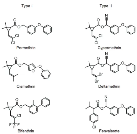
which is also known as fugutoxin. These compounds are classified according to the site of interaction; ten sites of interaction have been identified to date, and the pyrethroids are known to act on site 7 (Zlotkin, 1999). Pyrethroids exhibit insecticidal activity by causing a delay in inactivation and stabilize the open state of the Vssc (Bloomquist, 1996; Narahashi, 1996, 2000). Pyrethroids are grouped into two categories (Type I and Type II) based on their distinct poisoning symptoms, effects on nerve preparations, and chemical structures (Gammon et al., 1981; Lawrence and Casida, 1982; Narahashi, 1986) . Type I pyrethroids lack an α-cyano group, which is present at the
phenoxybenzyl alcohol moiety of Type II pyrethroids (Figure 2). Type I pyrethroids also cause repetitive discharges in response to a single stimulus, whereas Type II pyrethroids cause membrane depolarization accompanied by suppression of cellular excitability (Narahashi, 1986) .
Figure 1. Schematic representation of voltage-sensitive sodium channel (Vssc). The Vssc is composed of α- and β-subunits. The α-subunit is composed of four domains, each containing six transmembrane segments (S1–S6).
Figure 2. Chemical structures of Type I and Type II pyrethroid insecticides.
Nicotinic acetylcholine receptor (nAChR)
nAChR, inhibitory-aminobutyric acid receptor, inhibitory glutamic acid receptor, and glycine receptor belong to the cys-loop ligand-gated ion channel superfamily (Karlin, 2002). nAChR is composed of five subunits having four-transmembrane structures to form homomers or heteromers (Tomizawa and Casida, 2001) (Figure 3); upon binding with its ligand acetylcholine (ACh), this channel opens and allows cations to penetrate. Natural products such as nicotine or neonicotinoids (Figure 4) act on nAChR, bind to it, and exhibit an agonistic effect (Matsuda et al., 1998; Matsuda et al., 2001; Matsuda et al., 2005; Tomizawa and Casida, 2005). Neonicotinoids are a family of insecticides acting on neuronal nAChRs with higher selective toxicity for insects
over vertebrates, and show excellent control for a wide range of insect pests due to their systemic distribution in crop plants (Matsuda et al., 2001).
Figure 3. Schematic representation of the nicotinic acetylcholine receptor (nAChR). The nAChR is composed of five subunits having four-transmembrane structures to form homomers or heteromers.
Figure 4. Chemical structures of nicotine, neonicotinoids and sulfoxaflor
Methods for studying the mode of action of compounds having neurotoxic insecticidal activity
A range of methods are available for studying the mechanism of action of compounds having neurotoxic insecticidal activity, including molecular biology and biochemical approaches, binding assays, optical methods using fluorescent dye, and electrophysiological methods. Among these, electrophysiological methods are the most suitable for detailed testing of the effects of actions or mechanisms of action of
receptors or ion channels because such approaches identify the effects of the compound by detecting changes in current. There are three different types of electrophysiological method. One is the two-electrode voltage-clamp method (TEVC), which uses Xenopus oocytes. This method is suitable for the expression and analysis of receptors of a wide range of biological strains, especially for the analysis of variants (Shimomura et al.,
2006). Another method is the patch clamp method; this enables various measurements, is particularly applicable to the nerve cells of insects, and enables evaluation of the effects or activities of a compound on the expression of receptors in an insect’s body. The third method is the automatic patch clamp method. This was developed to improve the low throughput of electrophysiological methods, which is one of their drawbacks, is labor-saving, and can be used to analyze a large number of compounds, while utilizing the benefits of electrophysiological features.
Insecticide resistance
The development of resistance to insecticides is one of the most important
problems encountered during insect pest management. Since the late 1940s, the number of cases of insecticide resistance, and the number of species and compounds involved has been continually increasing (Sparks and Nauen, 2015). Detoxification by P450 for example, mutation of an amino acid residue at the site of activity, and a decrease in the permeability of an insecticide due to an increased cuticle surface area are known to be involved in such resistance (Bass et al., 2014; Casida and Durkin, 2013; Scott, 1999). In most cases, cytochrome P450-mediated detoxification plays a primary role in
insecticide resistance in a variety of insects (Rauch and Nauen, 2003; Scott, 1999; Zhao et al., 2000) . Cytochrome P450 is found widely in nature and plays roles in many biological processes, including hormone synthesis and the metabolism of exogenous compounds. In insects, cytochrome P450 has been implicated in providing resistance to insecticides via degrading foreign compounds to more soluble and less toxic forms (Scott, 1999) . This is accomplished via either up-regulated cytochrome P450
expression or structural changes that may alter the substrate profile or catalytic activity
of the enzyme (Amichot et al., 2004b; Bergé et al., 1998; Bogwitz et al., 2005; Feyereisen et al., 1989; Nikou et al., 2003; Pridgeon and Liu, 2003).
Aim of this thesis
The author believe that the findings of studies on the mechanisms of action of neurotoxic insecticides and resistance to them will contribute to the research and development of new insecticides, including ones effective against insecticide-resistant insects, and to the establishment of appropriate and effective measures for avoiding resistance. Therefore, in the present study, the author investigated the mechanism of action of the compound that acts on Vssc and nAChR, and the associated mechanism of resistance, mainly using an electrophysiological method. In chapter 1, the author
expressed several types of Aedes aegypti Vssc in Xenopus oocytes and examined the effect of amino acid substitutions in Vssc on pyrethroids susceptibilities. In chapter 2, the author attempted to identify the mechanisms of neonicotinoid resistance of Aphis
gossypii using an insecticidal assay with and without piperonyl butoxide (PBO)
pretreatment, along with sequencing of the nAChR gene and electrophysiology. In chapter 3, two-electrode voltage-clamp electrophysiological analyses and molecular modeling were employed to investigate the differential effects of the R81T mutation of
A. gossypii nAChR on cyano- and nitro-substituted neonicotinoids and sulfoxaflor. In
chapter 4, the author used next-generation sequencing (NGS) to identify P450 family members involved in the metabolism of neonicotinoids insecticides of A. gossypii. Further, molecularly cloned P450 cDNAs and expressed to study P450-mediated insecticide metabolism.
Chapter 1
A single crossing-over event in voltage-sensitive Na+ channel genes may cause critical failure of dengue mosquito control by insecticides
Introduction
Aedes aegypti is the major mosquito vector of dengue fever (DF), yellow fever, and chikungunya fever. DF causes more illness and death in humans than any other arboviral disease (Gubler, 1998). Mosquito control is mainly achieved by pyrethroids because of the high and rapid activity of this class of insecticides in insects, and its low toxicity to mammals. However, intensive and frequent use of these chemicals has resulted in the development of resistance in A. aegypti (Hemingway et al., 2004; Vontas et al., 2012).
Pyrethroid insecticides target the voltage-sensitive Na+ channel (Vssc) (Davies et al., 2007). Some amino acid substitutions alter the affinity of Vssc for pyrethroids and they confer resistance to this class of insecticide in insects (Davies et al., 2007).
Understanding how mutations alter interaction of pyrethroids with Vssc is important for improving our understanding of the pyrethroid mechanism of action and for
constructing appropriate control strategies against pest insects. Previous studies have shown that additional amino acid substitutions concomitantly occurring in Vssc can further reduce an insect's susceptibility to pyrethroids. For instance, houseflies
harboring Vssc with the L1014F substitution cause reduction in the affinity of Vssc for pyrethroids (so-called knockdown resistance or kdr), while the double mutation
L1014F+M918T further reduces the susceptibility of the insects to pyrethroids (so-called super-kdr) (Vais et al., 2000).
In Southeast Asia, one of the largest DF endemic areas, two major Vssc
haplotypes confer pyrethroid resistance to A. aegypti, i.e., S989P+V1016G and F1534C.
A. aegypti with these two haplotypes are widely and sympatrically distributed in this
area (Kasai et al., 2014; Kawada et al., 2009; Stenhouse et al., 2013).
Neurophysiological studies have revealed that V1016G and F1534C single mutations each confer resistance to pyrethroids (Du et al., 2013). The F1534C mutation has also been confirmed from another dengue vector, Aedes albopictus (Kasai et al., 2011). S989P is usually accompanied by V1016G (Srisawat et al., 2010), but its contribution to pyrethroid resistance is unclear. Vssc harboring triple amino acid substitutions (i.e., S989P+V1016G+F1534C) has not been recorded from a field population of A. aegypti although heterozygous form of these two haplotypes was confirmed (Kawada et al., 2009; Stenhouse et al., 2013). Such a haplotype, however, can be generated by a single crossing-over event of the above two major resistance Vssc haplotypes. Moreover, the sensitivity of such a triple-mutated Vssc to pyrethroids has not yet been investigated. In this study, the author analyzed the pyrethroid sensitivity of Vssc harboring triple substitutions, using a two-electrode voltage-clamp method, and we discussed the effect thereof on the control of the dengue mosquito vector by pyrethroids. We also investigated the synergistic effect of S989P on the sensitivity of Vssc harboring the V1016G mutation to pyrethroid insecticides.
Materials and Methods
Mosquito strains
Two strains of A. aegypti were used: SMK, a laboratory reference strain susceptible to pyrethroid insecticides, and SP, originally collected from Singapore in 2009, which has developed a high level of resistance to permethrin (1650-fold in adult females) after permethrin selection for 10 generations (Kasai et al., 2014). Mosquitoes were reared as described previously (Kasai et al., 1998).
In vitro metabolism and synergistic studies revealed that both reduced permethrin susceptibility and enhanced metabolic enzyme activity, via cytochrome P450
monooxygenases, confer the high level of resistance in strain SP (Kasai et al., 2014). Previous study confirmed that this strain possesses both S989P and V1016G mutations in Vssc homozygously (Kasai et al., 2014). The full length of Vssc cDNA in the SP strain, however, has not yet been sequenced.
Chemicals
Permethrin (purity > 99.1%) and deltamethrin (purity > 99.0%) were purchased from LGC Standards (Teddington, UK) and Wako Pure Chemical Industries, Ltd. (Osaka, Japan), respectively. The stock solutions of insecticides (100 mM) were prepared in dimethyl sulfoxide (DMSO). Working solutions were prepared in standard oocyte saline (SOS; 100 mM NaCl, 2 mM KCl, 1.8 mM CaCl2, 1 mM MgCl2, 5 mM HEPES, pH 7.6) immediately prior to experiments. The DMSO concentration in the final solution was < 0.1% and it did not affect the function of Na+ channels in the experiments (data not shown).
Sequencing full-length Vssc cDNA from SMK and SP strains
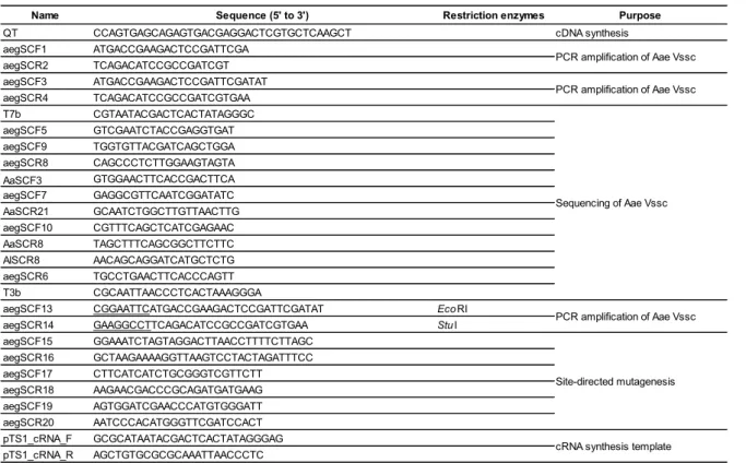
Total RNA was extracted from 10 three-day-old females using ISOGEN (Nippon Gene Co., Ltd., Tokyo, Japan). Genomic DNA was removed by digesting the total RNA samples with DNase I using TURBO DNase (Life Technologies Co., Carlsbad, Ca, USA). cDNA was synthesized using RNA and QT primer (Table 1-1) and reverse transcriptase ReverTra Ace (Toyobo, Osaka, Japan) according to the manufacturer's instructions. The full-length cDNAs of A. aegypti Vssc were amplified by PCR using PrimeStar GXL DNA polymerase (Takara Bio Inc., Shiga, Japan) and the primers aegSCF1 and aegSCR2. Cycling conditions for PCR were as follows: 95 °C for 2 min, followed by 35 cycles of each consisting of 98 °C for 10 s, 60 °C for 15 s, and 68 °C for 6 min. Nested PCR was performed under the same conditions, but using the primers aegSCF3 and aegSCR4. PCR products were purified by agarose gel electrophoresis and inserted into the pTA2 plasmid using the TArget Clone -Plus system (Toyobo, Osaka, Japan). The insert sequence was confirmed using the following primers: T7b, aegSCF5, aegSCF9, aegSCR8, AaSCF3, aegSCF7, AaSCR21, aegSCF10, AaSCR8, AlSCR8, aegSCR6, and T3b. Eight and six clones of full-length Vssc cDNAs from the SMK and the SP strains, respectively, were sequenced. Nucleotide polymorphisms that were confirmed only in a single clone were considered to reflect polymerase errors.
Site-directed mutagenesis and cloning of tipE cDNA
Site-directed mutagenesis was performed by PCR using PrimeStar GXL DNA polymerase and primers that were designed based on the 5' and 3' end sequences of A.
aegypti Vssc (AaNavS2, DDBJ accession number: AB909019), which was isolated
from insecticide susceptible SMK strain as described below. The forward primer
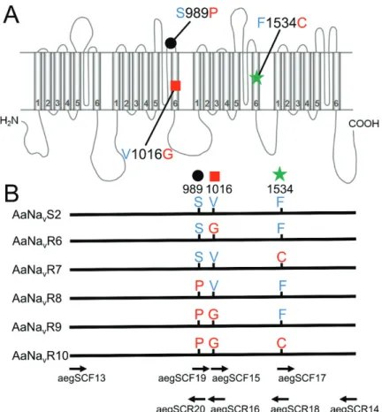
aegSCF13 and reverse primer aegSCR14 included EcoRI and StuI restriction sites (underlined), respectively, to facilitate cloning (Table 1-1). Full-length cDNAs encoding the substitutions V1016G (AaNavR6), F1534C (AaNavR7), S989P (AaNavR8),
S989P+V1016G (AaNavR9), and S989P+V1016G+F1534C (AaNavR10) in Vssc were amplified, as shown in Figure 1-1. In this study, we numbered the amino acid position according to the sequence of the most abundant splice variant of the house fly Vssc (GenBank accession nos. AAB47604 and AAB47605). Starting with AaNavS2 (wild-type), each fragment containing the smallest number of polymorphisms were subjected to oligonucleotide-mediated mutagenesis following a standard protocol (Ho et al., 1989), using the primers aegSCF15, aegSCR16, aegSCF17, aegSCR18, aegSCF19, and aegSCR20. Amplified PCR products were digested with restriction enzymes and cloned into the pTS1 vector (Nippon Gene Co., Ltd, Tokyo, Japan). Clones were fully sequenced and each amino acid substitution was confirmed. Drosophila melanogaster temperature-induced paralytic E (tipE) cDNA was also amplified and cloned into pcDNA3.1(+) (Invitrogen, Carlsbad, CA, USA) using published primer sequences (Feng et al., 1995). The compatibility of Drosophila tipE with Vssc of other insects has been well documented (Burton et al., 2011; Du et al., 2009; Hu et al., 2011; Tan and Tong, 2004).
Table 1-1. Primer sequences used in this study.
Name Sequence (5' to 3') Restriction enzymes Purpose
QT CCAGTGAGCAGAGTGACGAGGACTCGTGCTCAAGCT cDNA synthesis
aegSCF1 ATGACCGAAGACTCCGATTCGA aegSCR2 TCAGACATCCGCCGATCGT aegSCF3 ATGACCGAAGACTCCGATTCGATAT aegSCR4 TCAGACATCCGCCGATCGTGAA T7b CGTAATACGACTCACTATAGGGC aegSCF5 GTCGAATCTACCGAGGTGAT aegSCF9 TGGTGTTACGATCAGCTGGA aegSCR8 CAGCCCTCTTGGAAGTAGTA AaSCF3 GTGGAACTTCACCGACTTCA aegSCF7 GAGGCGTTCAATCGGATATC AaSCR21 GCAATCTGGCTTGTTAACTTG aegSCF10 CGTTTCAGCTCATCGAGAAC AaSCR8 TAGCTTTCAGCGGCTTCTTC AlSCR8 AACAGCAGGATCATGCTCTG aegSCR6 TGCCTGAACTTCACCCAGTT T3b CGCAATTAACCCTCACTAAAGGGA
aegSCF13 CGGAATTCATGACCGAAGACTCCGATTCGATAT EcoRI
aegSCR14 GAAGGCCTTCAGACATCCGCCGATCGTGAA StuI
aegSCF15 GGAAATCTAGTAGGACTTAACCTTTTCTTAGC aegSCR16 GCTAAGAAAAGGTTAAGTCCTACTAGATTTCC aegSCF17 CTTCATCATCTGCGGGTCGTTCTT aegSCR18 AAGAACGACCCGCAGATGATGAAG aegSCF19 AGTGGATCGAACCCATGTGGGATT aegSCR20 AATCCCACATGGGTTCGATCCACT pTS1_cRNA_F GCGCATAATACGACTCACTATAGGGAG pTS1_cRNA_R AGCTGTGCGCGCAAATTAACCCTC Site-directed mutagenesis
cRNA synthesis template PCR amplification of Aae Vssc PCR amplification of Aae Vssc
PCR amplification of Aae Vssc Sequencing of Aae Vssc
Figure 1-1. Mutations of Aedes aegypti Vssc investigated in this study.
(A) Schematic illustration of the locations of the Vssc mutations assessed. Positions are numbered according to the amino acid sequence of the most abundant splice variant of housefly Vssc (GenBank accession nos. AAB47604 and AAB47605). (B) Six Vssc types investigated in this study and the positions of primers used for site-directed mutagenesis are shown. Primer sequences are listed in Methods. AaNavS2 is the wild-type Vssc of which the cDNA was isolated from the insecticide-susceptible SMK strain.
Preparation of cRNA
Templates for in vitro transcription were PCR-amplified from plasmids using KOD plus Neo (Toyobo) and the primers pTS1_cRNA_F and pTS1_cRNA_R (Table 1-1). Capped RNA transcripts were synthesized using the mMessage mMachine T7 Ultra Kit (Life Technologies, Carlsbad, CA, USA). The quantity and quality of cRNAs
were verified by agarose gel electrophoresis and absorption spectroscopy, and cRNAs were stored at −80°C until required for use.
Expression of A. aegypti Vssc in Xenopus oocytes
Female Xenopus laevis were purchased from Hamamatsu Seibutsukyozai (Shizuoka, Japan) and maintained in dechlorinated water at 18 °C. Oocytes were surgically obtained following anesthesia of a Xenopus frog using 0.03% benzocaine (Sigma-Aldrich, Tokyo, Japan), and enzymatically defolliculated by incubating them for 1–1.5 h in a Ca2+-free SOS containing 1 mg/ml collagenase (Wako Pure Chemical Industries, Osaka, Japan). Vssc cRNA and tipE cRNA (0.9 ng each) were co-injected into healthy stage V–VI oocytes, which were incubated at 18 °C in SOS medium
supplemented with 50 µg/ml gentamycin, 100 U/ml penicillin, 100 µg/ml streptomycin, 2.5 mM Na pyruvate, and 4% horse serum, for 2–4 days after injection. The medium was replaced daily and unhealthy oocytes were discarded. It is generally recognized that the knockdown resistance is a recessive trait (Gao et al., 2003; Huang et al., 2004). Since we injected mRNA of a single Vssc haplotype into Xenopus oocytes, theoretically we reproduced the environment of the homozygous Vssc form in the cell.
Electrophysiological recording environment
Na+ currents were recorded using a TEV-200A (Dagan Corporation, Minneapolis, MN, USA) amplifier and digitized at 40 kHz with PowerLab 8/30 (AD Instruments, Nagoya, Japan). Signals were filtered at 2 kHz using a Bessel four-pole filter (Dagan Corporation, Minneapolis, MN, USA). The P/N method was used to subtract capacitive transient currents (Bezanilla and Armstrong, 1977). Recording electrodes were prepared
from borosilicate glass tubes (GC150TF-10; Warner Instruments, Hamden, CT, USA) using a P-1000 puller (Sutter Instruments, Novato, CA, USA). Electrodes were filled with 3 M KCl and had a resistance of 0.5–2.0 MΩ when measured in SOS. Oocytes were continuously perfused with SOS throughout the recording session, at a rate of 5 ml/min using a gravity-fed system. Experiments were performed at 18–19 °C.
Voltage protocols and data analysis
Methods for Na+-current recordings were performed using standard two-electrode voltage clamping, as described by Du et al. (Du et al., 2009). Na+ currents were elicited by a 20-ms test pulse from a holding potential of −120 mV to −20 mV. The peak current ensuing from 20-ms step depolarization, in response to a test potential (V) in the range of −80 mV to +40 mV, in 5-mV increments, from a holding potential, at 10-s intervals was measured. The voltage dependence of activation was then calculated by dividing by (V – Vrev), where Vrev is the reversal potential as determined from the current-voltage curves. Peak conductance values were plotted against V and fitted with the Boltzmann equation to obtain the half-maximal activation voltage. The voltage dependence of Vssc inactivation was determined using a 200-ms pre-pulse in the range of −100 mV to 0 mV, in 5 mV increments, followed by a 20-ms test pulse at −20 mV. Data were plotted as normalized current vs. inactivation pre-pulse voltage, and fitted with the Boltzmann equation to obtain the half-maximal inactivation voltage. Application of permethrin and deltamethrin were conducted using the disposable perfusion system (Tatebayashi and Narahashi, 1994). Pyrethroid-induced tail currents were recorded during a 100-pulse train of 5-ms depolarizations from a holding potential (−120 mV) to −20 mV at 10-ms intervals (Du et al., 2009). The amplitude of tail currents was used to establish the
percentage of modified channels, as previously described (Burton et al., 2011; Tan and Tong, 2004; Tatebayashi and Narahashi, 1994; Vais et al., 2000). The decays of tail currents were fitted with single functions to determine the time constant (Tau values). The sensitivity of Vssc to each insecticide was defined as the concentration of
insecticide at which 25% of channels were modified (EC25).
Statistical analysis
The recorded data were analyzed using GraphPad Prism 5.04 software (GraphPad Software, Inc., La Jolla, CA, USA). Voltage dependences (both activation and
inactivation of V1/2) were compared statistically between wild-type and mutant channels using one-way ANOVA followed by Dunnett's multiple comparison tests. The decays of tail currents were fitted with single functions to determine time constants (Tau values). Statistical comparisons (one-way ANOVA followed by Dunnett's multiple comparison test) were made for time constants of the decays between the wild-type and the mutant channels at P<0.05. Tukey-Kramer HSD (Honestly significant difference) procedure was used to test for the percentage differences of modified channels among Vssc haplotypes after the treatment of insecticides.
Results
Cloning and sequencing the full-length Vssc cDNA from A. aegypti
Eight clones of full-length Vssc cDNA obtained from a pyrethroid-susceptible SMK strain were sequenced. The Vssc cDNA (six clones) of SP was also sequenced to determine if S989P and V1016G were the only substitutions present in Vssc in this strain. The SMK strain harbored two polymorphisms that both generated a substitution at amino acid 427 (R or K). K427 was not confirmed in the Vssc of strain SP. It is known that Vsscs have two mutually exclusive alternative exons (c and d) (Thackeray and Ganetzky, 1995); the expression of both exons was confirmed in both the SMK and SP strains. These two exons are 54 amino acids long and differ at two residues
(V922+T952 and M922+I952 for exons c and d, respectively). In the SMK strain, the ratio of exon c and d were 0.54 and 0.46 (based on 13 clones), and we therefore
concluded that these polymorphisms were not responsible for pyrethroid resistance. For electrophysiology studies, we used SMK-derived AaNavS2 (DDBJ accession number: AB909019, consisted of 2109 amino acid residues), which contains R427 and exon c, as does the wild-type Vssc. We also confirmed that S989P and V1016G are the only amino acid substitutions present in the full-length Vssc cDNA in the SP strain.
Functional analysis of AaNavS2 in Xenopus oocytes
The wild-type A. aegypti Vssc (AaNavS2) and Drosophila tipE, which is required for robust expression of insect Vssc, were co-expressed in Xenopus oocytes. No peak current was detected in oocytes that had not been injected with cRNA (data not shown). A peak current of approximately 0.5–3 µA was detected on days 2–4 in most oocytes
that had been co-injected with wild-type AaNavS2 and tipE cRNA. A representative trace is shown in Figure 1-2A. Currents were completely inhibited by 10 nM
tetrodotoxin (Figure 1-2B), confirming that they were Na+ currents. Oocytes were clamped at a holding potential of −120 mV, then depolarized to test potentials from −80 mV to +40 mV in 5-mV increments (Figure 1-2C). Na+ currents through AaNavS2 were detected at approximately −40 mV, and peaked at −15 mV (Figure 1-2C). The
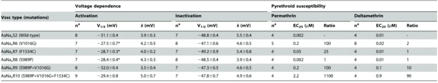
half-maximal activation voltage of wild-type AaNavS2 was −31.1±0.4 mV and the slope factor was 3.9±0.3 mV (Table 1-2). To assess the voltage dependence of
steady-state inactivation, oocytes were held at −120 mV and depolarized with a series of 200-ms inactivating pre-pulses, from −100 mV to 0 mV in 5-mV increments, followed by a 20-ms test pulse at −20 mV. The half-maximal inactivation voltage was −48.8±0.5 mV and the slope factor was 5.5±0.4 mV (Table 1-2, Figure 1-2C).
Figure 1-2. Mutations of Aedes aegypti Vssc investigated in this study.
(A) The cloned AaNavS2 (wild-type) channel was expressed in Xenopus oocytes and a Na+ current trace was recorded. (B) The Na+ current was blocked by the application of 10 nM tetrodotoxin, verifying that this was a Na+ current. (C) Normalized
voltage-conductance and inactivation curves of AaNavS2. The peak current was plotted against the depolarizing voltage (solid circles). The peak current amplitude was plotted as a function of the pre-pulse potential (triangles). Error bars indicate standard errors for 7–9 oocytes (D–I). Na+ current-voltage curves for 6 Vssc types. Error bars indicate standard errors for 7–9 oocytes. Reversal potentials were: (D) wild-type (+35 mV); (E) V1016G single mutation (+35 mV); (F) F1534C single mutation (+35 mV); (G) S989P single mutation (+35 mV); (H) S989P+V1016G double mutation (+25 mV); and (I) S989P+V1016G+F1534C triple mutation (+20 mV).
Table 1-2. Voltage-dependence of activation and inactivation of Aedes aegypti sodium channels and sensitivity of mutant channels to pyrethroids.
Data were fitted with the Boltzmann equation to determine V 1/2, the voltage of
half-maximal conductance or inactivation, and k, the slope factor. Values represent the mean ± standard errors. Voltage dependences were compared statistically between wild-type and mutant channels using one-way ANOVA by Dunnett’s multiple comparison test.
*P<0.05. Ratios were calculated by EC25 (mutant)/EC25 (wild-type).
an, number of biological replications for each insecticide concentration tested.
Effect of Vssc mutations on gating properties of channels
The expression of all five modified Vssc was confirmed in Xenopus oocytes. The effects of three substitutions (S989P, V1016G, and F1534C), alone or in combination, on the voltage dependence of activation, steady-state inactivation, and reversal potential were evaluated. Na+ currents recorded in oocytes expressing mutant channels exhibited voltage-dependent activation and inactivation, similar to those expressing the wild-type AaNavS2 (Figures 1-2D–I, Table 1-2). The reversal potentials of AaNavR9
(S989P+V1016G) and AaNavR10 (S989P+V1016G+F1534C) mutants were slightly shifted, from +35 mV in the wild-type (Figure 1-2D) to +25 mV (Figure 1-2H) and +20 mV (Figure 1-2I) in the respective mutants. Three mutant channels, AaNavR6
(V1016G), AaNavR7 (F1534C), and AaNavR8 (S989P) significantly (P<0.05 by one-way ANOVA followed by Dunnett's multiple comparison test) shifted the
voltage-dependence of activation in the depolarizing direction (Table 1-2). None of the
Vssc mutants changed the voltage dependence of inactivation significantly (Table 1-2).
Effects of amino acid substitutions on the sensitivity of Vssc to permethrin
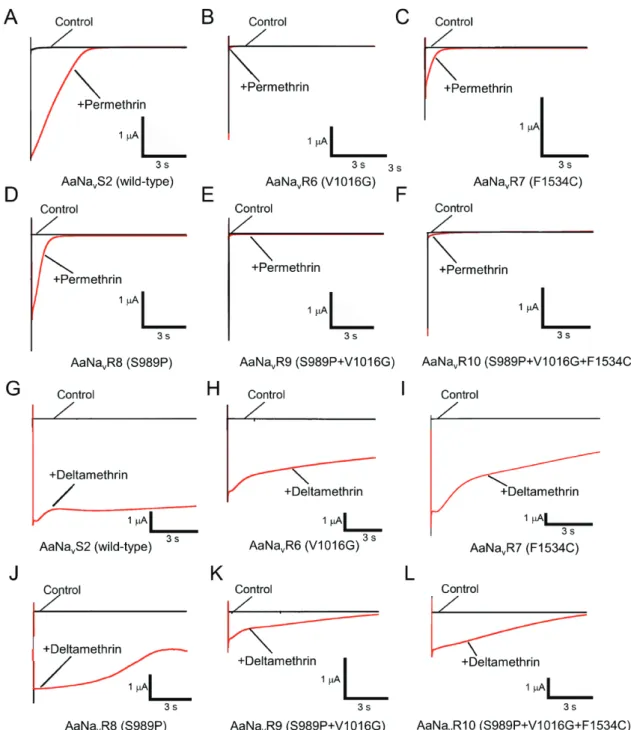
Pyrethroid-induced tail currents were elicited with a train of 100 depolarizing pulses (Burton et al., 2011; Tan et al., 2002; Tatebayashi and Narahashi, 1994; Vais et al., 2000). Currents were recorded 10 min after application of permethrin (Figure 1-3). In the wild-type channel AaNavS2, 100 nM permethrin induced large tail currents that recovered to baseline values within 3 s (Figure 1-3A). In all five mutant channels, the same concentration of permethrin induced only a small tail current that quickly
recovered to baseline (Table 1-3, Figures 1-3B–F). The S989P mutation had no effect on permethrin sensitivity on its own (AaNavR8), or in combination with V1016G
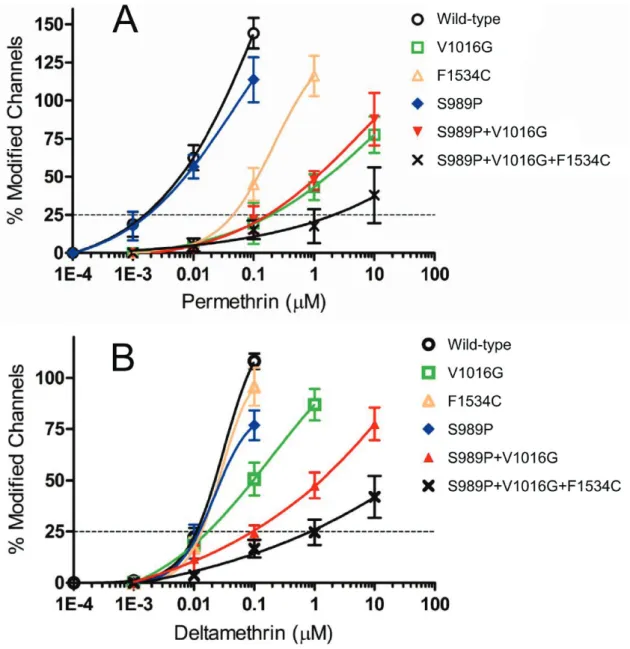
(AaNavR9) (Figure 1-4A). The V1016G and F1534C substitutions independently reduced sensitivity of Vssc to permethrin by 100- and 25-fold, respectively (Table 1-2, Figure 1-4A). The combination of three substitutions, viz., S989P+V1016G+F1534C (AaNavR10), exhibited 1100-fold more resistance, which represents an 11-fold
reduction (1100/100) and a 44-fold (1100/25) reduction in Vssc sensitivity to permethrin as compared to Vssc harboring S989P+V1016G or F1534C, respectively. This
suggested that these substitutions acted synergistically (Table 1-2, Figure 1-4A).
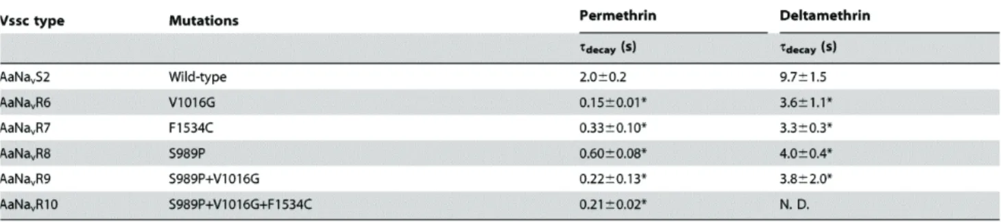
Figure 1-3. Pyrethroid-induced tail currents from oocytes injected with various types of Vssc.
(A, G) AaNavS2 (wild-type); (B, H) AaNavR6 (V1016G); (C, I) AaNavR7 (F1534C); (D, J), AaNavR8 (S989P); (E, K), AaNavR9 (S989P+V1016G); (F, L) AaNavR10 (S989P+V1016G+F1534C) in the absence (control) or presence of 100 nM permethrin (A–F) or 100 nM deltamethrin (G–L).
Table 1-3. Time constants of the decay (τdecay) of pyrethroid-induced tail currents.
The decays of tail currents were fitted with single functions to determine time constants (Tau values). Values represent the mean ± standard errors. Statistical comparisons (one-way ANOVA followed by Dunnett’s multiple comparison test) were made for time constants of the decays between the wild-type and the mutant channels at *P<0.05. N.D., not determined because the tail current tended to keep recovering to base line.
Figure 1-4. Sensitivity of Aedes aegypti Vsscs to permethrin and deltamethrin.
The percentages of modified channels were plotted against different concentrations of permethrin (A) and deltamethrin (B), and fitted to a four-parameter logistic equation. Error bars indicate standard errors for 4–8 oocytes. The percentages of modified channels were significantly different between AaNavR6 (V1016G) and AaNavR9 (S989P+V1016G) at deltamethrin concentration of 1.0 µM (P = 0.0091 by
Tukey-Kramer test) in Figure 1-4B.
Effects of amino acid substitutions on the susceptibility of Vssc to deltamethrin
Deltamethrin-induced tail currents initially increased, and then gradually recovered in the wild-type channel (Figure 1-3G). However, the current in AaNavR10 (S989P+V1016G+F1534C) had a lower amplitude and recovered to baseline more rapidly than did the wild-type (Figure 1-3L). As compared with wild-type Vssc, deltamethrin-induced tail currents of four mutant channels (AaNavR6, -R7, -R8, and -R9) decayed very quickly (Table 1-3, Figures 1-3H–K). We could not estimate the actual time constant for AaNavR10 (S989P+V1016G+F1534C) because the tail current tended to keep recovering to baseline. The deltamethrin-induced tail current of
AaNavR10, however, clearly decayed faster than that of the wild-type. V1016G reduced the sensitivity of Vssc to deltamethrin by 2-fold, while F1534C alone exerted no effect on Vssc sensitivity (Table 1-2, Figure 1-4B). S989P alone (AaNavR8) had no effect on deltamethrin sensitivity, but the combination of S989P and V1016G (AaNavR9) reduced deltamethrin-sensitivity of Vssc by 10-fold (Table 1-2), which was a 5-fold reduction in deltamethrin-sensitivity over that induced by V1016G alone. The percentage of modified channels were significantly different between AaNavR6 and AaNavR9 at deltamethrin concentration of 1.0 µM (P = 0.0091 by Tukey-Kramer test). Furthermore, S989P+V1016G+F1534C (AaNavR10) exhibited a 90-fold greater resistance than wild-type. This represents a 9-fold (90/10) and 90-fold (90/1) greater reduction in sensitivity than that induced by S989P+V1016G and F1534C, respectively, indicating a strong synergistic effect of the triple substitutions (Table 1-2). The
percentage of modified channels were significantly different between AaNavR9 and AaNavR10 at deltamethrin concentration of 10 µM (P = 0.034).
Discussion
To date, the V1016G mutation (usually accompanied by S989P) of Vssc has been confirmed only in A. aegypti distributed in Southeast Asia, including Thailand (Srisawat et al., 2010; Stenhouse et al., 2013), Vietnam (Kawada et al., 2009), Taiwan (Lin et al., 2013), Singapore (Kasai et al., 2014), and the Philippines (Yukiko Higa, personal communications). F1534C mutation of Vssc has also been confirmed from all of above regions except for Taiwan (Kasai et al., 2014; Kawada et al., 2009; Stenhouse et al., 2013). These two haplotypes were confirmed as heterozygous form as well (Kawada et al., 2009; Stenhouse et al., 2013). A previous study revealed that Vssc harboring V1016G exhibited a greater reduction in permethrin susceptibility than did Vssc harboring F1534C, which was in agreement with our findings. Surprisingly, the combination of S989P, V1016G, and F1534C altered the sensitivity of the
S989P+V1016G mutant to permethrin and deltamethrin by 11-fold (1100/100) and 9-fold (90/10), respectively. Theoretically, this haplotype can be generated as the result of a single crossing-over event involving two resistant haplotypes (i.e., S989P+V1016G and F1534C), which are widely and sympatrically distributed throughout Southeast Asia. Pyrethroid insecticides may lose their efficacy in the control of A. aegypti if such a new type of Vssc emerges in the field population of this mosquito species. Therefore, we strongly emphasize the importance of surveying of the triple mutations in Vssc in the field population of this key mosquito vector. Early detection of this unique haplotype may assist in vector control strategies by providing early warning of insecticide insensitivity.
The likelihood of occurrence of such a Vssc with triple mutations may depend on
its fitness cost. In this study, we observed that the reversal potential of AaNavR10 (S989P+V1016G+F1534C) shifted slightly, although there were no statistically significant differences in the voltage dependence of activation or in the voltage
dependence of steady-state inactivation between AaNavR10 and the wild-type AaNavS2. In North and South America, where no V1016G mutation has been confirmed in the Vssc of A. aegypti to date, V1016I is widely distributed (Aponte et al., 2013; Harris et al., 2010; Marcombe et al., 2012). The double mutations V1016I+F1534C have already been detected in some of these regions (Marcombe et al., 2012) and this fact implies the possibility that S989P+V1016G+F1534C triple mutations can also emerge. According to a neurophysiological study, V1016I does not alter the susceptibility of Vssc to pyrethroid (Du et al., 2013) while in another study quantitative trait loci (QTL) that affects permethrin resistance was mapped close to the Vssc gene with this mutation (Saavedra-Rodriguez et al., 2008). Anyway, the synergistic effect of V1016I on the pyrethroid-susceptibility of Vssc with F1534C is intriguing. In our study, we focused on characterization of triple mutations that can be generated by a single crossing-over event between two major resistant Vssc haplotypes, but the pyrethroid-susceptibility of Vssc harboring V1016G+F1534C or S989P+F1534C double mutations may also elucidate the synergistic effects of these mutations.
These results exhibited that S989P has a synergistic effect on the sensitivity of V1016G-harboring Vssc to deltamethrin, but not to permethrin (Figure 1-4). This result contradicts with an earlier observation that S989P does not synergize with V1016G (Du et al., 2013). The discrepancy between these two reports may be due to the use of different evaluation methods. The author measured the Na+ currents of each Vssc at five concentrations of deltamethrin, with at least 4 independent replications, and used EC25
values as a measure of sensitivity. The percentages of modified channels were
significantly different between AaNavR6 (V1016G) and AaNavR9 (S989P+V1016G) at deltamethrin concentration of 1.0 µM (P = 0.0091 by Tukey-Kramer test; Figure 1-4B). In contrast, previous studies used the ratios of modified channels at only one
concentration of the chemical; it is possible that this may have led to an underestimation of the effect of S989P. These findings highlight the need for genotyping S989P mutation as well as V1016G and F1534C for better understanding the pyrethroid susceptibility of the field population of A. aegypti collected especially in Southeast Asia.
Chapter 2
Association between the R81T mutation in the nicotinic acetylcholine receptor β1 subunit of Aphis gossypii and the differential resistance to acetamiprid and imidacloprid
Introduction
The cotton aphid, Aphis gossypii, is an important sucking pest that causes severe crop losses, both in the field and in greenhouses. Neonicotinoid insecticides (Fig. 2-1), which show excellent control of A. gossypii, target insect nicotinic acetylcholine (ACh) receptors (nAChRs) (Matsuda et al., 2001; Matsuda et al., 2005; Thany et al., 2007; Tomizawa and Casida, 2005). nAChRs are members of the cys-loop superfamily of ligand-gated ion channels that conduct fast-moving excitatory cholinergic
neurotransmission (Karlin, 2002). Targeting nAChRs has raised concerns about mammalian toxicity, yet neonicotinoids show highly selective toxicity to insects over vertebrates (Matsuda et al., 2001; Matsuda et al., 2005; Tomizawa and Casida, 2005). Insecticide resistance is a major hindrance to the effective control of insect pests worldwide. Neonicotinoids have been used for more than 20 years to protect
economically important crops (Elbert et al., 2008; Jeschke and Nauen, 2008; Nauen et al., 2008). However, although resistance to neonicotinoids has developed relatively slowly, it is now recognized as an emerging issue (Nauen and Denholm, 2005). In most cases, P450-mediated detoxification plays a primary role in insecticide resistance in a variety of insects, including the whitefly (Bemisia tabaci) and the Colorado potato
beetle (Leptinotarsa decemlineata) (Rauch and Nauen, 2003; Zhao et al., 2000). On the other hand, laboratory selection of Nilaparvata lugens with imidacloprid resulted in an α-subunit Y151S mutation that was responsible for the acquisition of neonicotinoid resistance (Liu et al., 2005; Liu et al., 2006). However, this mutation has not been identified in any pest in the field. Instead, a mutation in the nAChR β1 subunit was found to be a major factor in imidacloprid resistance in a field population of Myzus
persicae (Bass et al., 2011).
Previously reported the emergence of neonicotinoid resistance in the aphid and recently isolated an A. gossypii (Kushima) clone in Miyazaki Prefecture, Japan, that exhibited relatively high neonicotinoid resistance (Matsuura and Nakamura, 2014). Interestingly, the level of the Kushima clone’s resistance to neonicotinoids with nitro substituents (e.g., imidacloprid) was higher than for those with cyano substituents (e.g., acetamiprid; see Figure 1-1 for the structures). However, the mechanisms of
neonicotinoid resistance remain unknown.
In this study, we attempted to identify the mechanisms of neonicotinoid resistance using an insecticidal assay with and without piperonyl butoxide (PBO) pretreatment, along with sequencing of the nAChR gene and voltage-clamp electrophysiology. These results suggest that a mutation in the β1 subunit is the main mechanism of resistance and that differences in resistance levels are probably associated with differential interactions between nitro- and cyano-type neonicotinoids and nAChRs.
Figure 2-1. Chemical structures of neonicotinoid insecticides.
Materials and Methods
Insects
In this study, we used a field-isolated clone of A. gossypii (Kushima) and a susceptible clone that has been maintained since 1993 at the Odawara Research Center, Nippon soda Co., Ltd. (Odawara, Kanagawa, Japan). The Kushima clone was collected from a field in Miyazaki Prefecture, Japan, in 2012. The Kushima clone was reared for four months and used for insecticide bioassays without insecticide selection. These aphid clones were reared on cucumber seedlings at 25 °C and a relative humidity (RH) of 60% under a 16:8-hr light:dark photoperiod.
Chemicals
Acetamiprid and other neonicotinoids were synthesized at the Odawara Research Center. ACh chloride was purchased from Sigma-Aldrich (Tokyo, Japan). The purity of imidacloprid and acetamiprid was >99.9%.
Insecticide bioassays
All compounds were dissolved in a 5% N,N,-dimethylformamide aqueous solution and then diluted with distilled water and 2 × 10⁻⁴% (v/v) of the surfactant RABIDEN 3S (which is a trade name: 1.4% sodium dioctylsulfosuccinate, 8%
polyoxyethylene alkyl ether, and 3% polyoxyethylene fatty acid ester). Wingless female
A. gossypii (n = 5-10) were incubated on cucumber seedlings at the first- to second-leaf
stage of development. One day later, they were removed, and the test solutions were sprayed on the offspring, while the leaves were maintained under the same conditions
(25 °C, 60% RH, and a 16:8-hr light:dark photoperiod). Seventy-two hr after application, the effects of the compounds were assessed using median lethal dose (LC50) values calculated by probit analysis. Each compound was tested in duplicate.
To assess the effects of the synergist PBO, aphids were sprayed with PBO solution (250 ppm, 5% N,N,-dimethylformamide aqueous solution) 5 hr prior to application of the insecticide solution. Each compound was tested in duplicate.
Sequence analysis of the A. gossypii nAChR α1, α2, and β1 subunits
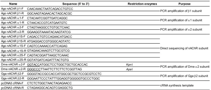
Total RNA was isolated from adult aphids using TRIzol Reagent (Invitrogen Corporation, Carlsbad, CA, USA) in accordance with the manufacturer’s instructions. First strand cDNA was synthesized from total RNA using the Transcriptor First Strand cDNA Synthesis Kit (Roche Diagnostics, K.K., Tokyo, Japan) with oligo-dT primers. PCR amplification was performed using KOD plus Neo polymerase (Toyobo Co., Ltd., Osaka, Japan) and the following gene-specific primers: Ago nAChR β1-F, Ago nAChR β1-R, Ago nAChR α1-F, Ago nAChR α1-R, Ago nAChR a2-F, and Ago nAChR a2-R (Table 2-1). PCR amplification was performed under the following cycling conditions: 2 min at 94 °C, followed by 35 cycles of 10 sec at 98 °C, 30 sec at 56 °C, and 1 min at 68 °C. Amplicons of the expected size were purified using the QIAquick PCR Purification Kit (Qiagen GmbH, Hilden, Germany) and directly sequenced in both directions using the BigDye Terminator V3.1 Cycle Sequencing Kit (Applied Biosystems, Foster City, CA, USA). The direct sequences were confirmed using the following primers: Ago nAChR β1S-F and Ago nAChR β1S-R for subunit β1, Ago nAChR α1S-F and Ago nAChR α1S-R for subunit α1, and Ago nAChR α2S-F and Ago nAChR α2S-R for subunit α2.
Table 2-1. Primer sequences used in this study.
Isolation of cDNA clones encoding nAChRs
cDNA encoding Drosophila melanogaster nAChR α2 subunit (Dα2) was amplified from first strand cDNA synthesized from larval total RNA using the
Transcriptor First Strand cDNA Synthesis Kit with oligo-dT primers. PCR amplification was performed using the following primers: Dme nAChR α2-F and Dme nAChR α2-R (Table 2-1). PCR amplification was performed under the following cycling conditions with KOD plus Neo: 2 min at 94 °C, followed by 30 cycles of 10 sec at 98 °C, 30 sec at 56 °C, and 2 min at 68 °C. Amplicons were purified using the QIAquick Gel Extraction Kit (Qiagen GmbH) and cloned into pTA2 vectors (Toyobo Co., Ltd.). Several clones containing the Dα2 gene were sequenced. One clone identical to a published sequence (GenBank accession number X53583) was excised from the pTA2 vector using the restriction endonucleases KpnI and ApaI and then ligated into the pcDNA3.1(+) vector (Life Technologies, Japan, Tokyo, Japan).
The chicken nAChR β2 subunit was amplified from first strand cDNA
Name Sequence (5' to 3') Restriction enzymes Purpose
Ago nAChR β1-F CAACAAACTAATCAGACCTGTCC Ago nAChR β1-R GGCAAGTAGAACACTAGCACGC Ago nAChR α1-F CTACAATCGGTTGATCAGGC Ago nAChR α1-R CTAACACCGTCATGAATGTC Ago nAChR α2-F CTAGTAAGGCCTGTGCTCAAC Ago nAChR α2-R GGAAGGTAAAATACAAGTATCG Ago nAChR β1S-F CAGACCTGTCCAGAACATGACC Ago nAChR β1S-R ATGAGGACCGTGGGCAGTATC Ago nAChR α1S-F CAGTCCAAAACCATTCAGAG Ago nAChR α1S-R GTAGAACAAAGTCTTGCGTCG Ago nAChR α2S-F CAGTACGGATTAAGCTCAAAC Ago nAChR α2S-R GGTATGATCAGATTTACTGTG
Dme nAChR α2-F GGTACCATGGCTCCTGGCTGCTGCACCAC KpnI
Dme nAChR α2-R GGGCCCTTAATTCTTCTTCTCGGTTAG ApaI
Gga nAChR β2-F GGGGTACCGCCACCATGGCGCTGCTCCGCGTCCTC Gga nAChR β2-R GGGAATTCCCTATTTGGAGGTGGGGGTGCCCTGGC pcDNA3 cRNA-F CTCTCTGGCTAACTAGAGAACC
pcDNA3 cRNA-R CTAGAAGGCACAGTCGAGGCTG
PCR amplification of β1 subunit PCR amplification of α1 subunit PCR amplification of α2 subunit
Direct sequencing of nAChR subunit
PCR amplification of Dme α2 subunit PCR amplification of Gga β2 subunit cRNA synthesis template
synthesized from chicken total RNA obtained from BioChain Institute, Inc. (Newark, CA, USA). PCR was performed using KOD-plus-Neo polymerase with the
gene-specific primers Gga nAChR β2-F and Gga nAChR β2-R. PCR amplification was performed under the following cycling conditions: 2 min at 94 °C, followed by 30 cycles of 10 sec at 98 °C, 30 sec at 56 °C, and 1.5 min at 68 °C. Amplicons were cloned into pTA2 vectors (Toyobo Co., Ltd.) and sequenced, while the β2 gene was cloned into the pcDNA3.1(+) vector.
Preparation of Dα2 and chicken β2 cRNA
Templates for in vitro transcription were obtained by PCR amplification of each plasmid using KOD-plus-Neo polymerase and the gene-specific primers pcDNA3 cRNA-F and pcDNA3 cRNA-R (Table 2-1). The capped RNA transcripts were
synthesized using the T7 polymerase included with the mMessage mMachine T7 Ultra Kit (Life Technologies). The quality and quantity of cRNAs were verified by agarose gel electrophoresis and absorption spectroscopy. The cRNA was stored at −80 °C for further use.
nAChR expression in Xenopus oocytes by cRNA injection and electrophysiology
All experiments were performed at 18–19 °C. Female Xenopus laevis were purchased from Hamamatsu Seibutsukyozai (Shizuoka, Japan) and maintained in tap water. Oocytes were surgically obtained using 0.03% benzocaine (Sigma-Aldrich) and then enzymatically defolliculated by incubation for 1–1.5 hr in Ca2+-free Standard Oocyte Saline (SOS) solution (100 mM NaCl, 2.0 mM KCl, 1.0 mM MgCl2, 5.0 mM HEPES, pH 7.6) (Ihara, 2003) containing 1 mg/mL collagenase (Wako Pure Chemical
Industries, Ltd., Osaka, Japan). Healthy stage V–VI oocytes were injected with equivalent amounts (10 ng each) of cRNA encoding Dα2 and chicken β2 using the Nanoject II Auto-Nanoliter Injector (Drummond Scientific Company, Broomall, PA, USA) and then incubated in SOS solution supplemented with 50 µg/mL gentamycin, 100 U/mL penicillin, 100 µg/mL streptomycin, 2.5 mM sodium pyruvate, and 4% horse serum for 1–4 days. The medium was replaced daily and unhealthy oocytes were discarded.
Two-electrode voltage clamp electrophysiological analysis was performed as described by Ihara et al. (Ihara, 2003) Currents were recorded using the TEV-200A amplifier (Dagan Corporation, Minneapolis, MN, USA) and digitized at 1 kHz using the PowerLab 8/30 data acquisition and analysis system (ADInstruments, Nagoya, Japan). Signals were filtered at 50 Hz using a 4-pole Bessel filter. Recording electrodes were prepared from borosilicate glass tubes (CG150TF-10; Warner Instruments, Hamden, CT, USA) using a P-1000 Puller (Sutter Instruments, Novato, CA, USA). Electrodes were filled with 3 M KCl and had resistance of 0.5–2.0 MΩ when measured in SOS solution. Oocytes were continuously perfused with SOS solution throughout the recording session at a rate of 5 mL/min using a gravity-fed system (Ihara, 2003) and
voltage-clamped at −100 mV, while those with excessive leak currents (>-1000 nA) were not used for further analysis.
Test compounds were first dissolved in dimethyl sulfoxide (DMSO) and later diluted to the appropriate test concentration in SOS solution (100 mM NaCl, 2.0 mM KCl, 1.0 mM MgCl2, 1.8 mM CaCl2, 5.0 mM HEPES, pH 7.6) containing 0.5 µM atropine. The concentration of DMSO was maintained at < 0.1% (v/v) to avoid the effects of the channel or cell. A solution of ACh and SOS was prepared by adding ACh
chloride directly to the atropine-SOS solution immediately before use.
The LabChart 7 (ADInstruments, Nagoya, Japan) and GraphPad Prism 5 (GraphPad Software, Inc., La Jolla, CA, USA) software packages were used for data analysis and the preparation of graphs. Normalized concentration-response data were analyzed by nonlinear regression using GraphPad Prism to obtain Imax (the maximum normalized response), EC50 (the concentration inducing the half-maximal normalized response), and pEC50 (-logEC50). The peak amplitude of the current recorded in response to each challenge was normalized to the maximum amplitude of the response to ACh. Data from the Dα2β2, T77R, and T77R + E79V mutants were normalized to the response to 1 mM ACh.
Results
Neonicotinoid insecticide bioassays
The A. gossypii (Kushima) clone, which was discovered in Miyazaki Prefecture, Japan, exhibited resistance to neonicotinoid insecticides as compared with a susceptible clone (Table 2-2), which was associated with resistance factors (RFs) 23.8–394.
Additionally, the Kushima clone had a high level of resistance (RFs 216–394) to nitro-substituted neonicotinoids (Figure 2-1), such as imidacloprid. In contrast, the Kushima clone exhibited low to moderate levels of resistance (RFs 23.8 and 65.4) to cyano-substituted neonicotinoids (e.g., acetamiprid).
When PBO was used as a pretreatment synergist with the Kushima clone, the LC50 value decreased (Table 2-2). The LC50 values of acetamiprid and imidacloprid to the Kushima clone without PBO pretreatment were 6.54 and 75.7 ppm, but 1.91 and 7.01 ppm with PBO pretreatment, respectively. The synergistic factor (SF) of
acetamiprid was lower than that for imidacloprid (3.4 vs. 11, respectively). These results indicate that the Kushima clone acquired a resistance mechanism other than an
enhanced oxidative metabolism.
Table 2-2. Insecticidal activity of neonicotinoids to susceptible aphids and Kushima clone with and without pretreatment with piperonyl butoxide (PBO)
Experiments were performed in duplicate.
a) Resistance factor (LC50 Kushima clone/LC50 susceptible clone).
b) Synergistic factor (LC50 Kushima clone/LC50 Kushima clone with PBO).
Sequence analysis of A. gossypii nAChR subunit genes
The N-terminal regions of the A. gossypii (Kushima clone) nAChR β1, α1, and α2 subunits, which encompass the conserved domains (loop A-F) that comprise the ACh and neonicotinoid binding sites, were PCR-amplified and then sequenced. Although a limited number of silent single-nucleotide polymorphisms were detected, no amino acid changes were observed in the α1 and α2 subunits. In contrast, a single point mutation, Arg81 (AGA) to Thr (ACA), was detected in the β1 subunit (Figure 2-2). Additionally, the mutation was detected heterozygously (data not shown).
Compound LC50 95% confidense LC50 95% confidense Rfb) (ppm) limit of LC50 (ppm) limit of LC50 Acetamiprid 0.10 0.080-0.12 6.54 4.62-9.73 65.4 Thiacloprid 0.30 0.20-0.61 7.15 4.40-11.8 23.8 Imidacloprid 0.35 0.20-0.85 75.7 44.7-193 216 Clothianidin 0.10 0.061-0.18 39.4 28.0-58.5 394 Thiamethoxam 0.19 0.12-0.31 56.0 40.6-73.7 295 Dinotefuran 0.66 0.40-1.04 167 114-285 253 Nitenpyram 0.30 0.19-0.54 71.0 27.5-103 237 Acetamiprid+PBO 0.028 0.016-0.048 1.91 1.03-3.08 3.4 Imidacloprid+PBO 0.050 0.028-0.079 7.01 3.46-12.0 11
Susceptoble strain Kushima strain
Figure 2-2. Alignment of amino acid sequences in loop D of the agonist binding region of vertebrate and insect nicotinic ACh receptor subunits (nAChRs). Homo sapiens (human), Gallus gallus (chicken), Rattus norvegicus (rat), Drosophila melanogaster (fruit fly), Bemisia tabaci (sweet potato whitefly), Myzus persicae (peach-potato aphid),
Aphis gossypii (cotton aphid).
Agonist activity of ACh
A concentration-response curve of ACh to Drosophila melanogaster Dα2-chicken β2 nAChRs was used as a control in this study. We used the Dα2-vertebrate β2 hybrid receptor for the following reasons: Insect nAChR functional expression is difficult, but the Dα2-chicken β2 hybrid receptor has been well established for assaying
neonicotinoids. The Imax and pEC50 values of ACh for Dα2β2 nAChR were 1.08 ± 0.03 and 4.60 ± 0.04, respectively. The ACh values of the Dα2β2 nAChR were similar to those in previous reports (Ihara, 2003; Shimomura et al., 2005; Shimomura et al., 2006) (Figure 2-3, Table 2-3).
Figure 2-3. Response to acetamiprid of Dα2Gβ2 nicotinic ACh receptors (nAChRs). (A, B, C) Responses of oocytes expressing WT Dα2β2 nAChRs (A), the T77R single mutation (B), and the T77R + E79V double mutation (C) to 100 µM ACh and 100 µM acetamiprid. (D, E, F) Responses of oocytes expressing the WT (D), the T77R single mutation (E), and the T77R + E79V double mutation Dα2β2 nAChRs (F) to 100 µM ACh and 30 µM imidacloprid. Concentration-response curves of ACh, acetamiprid, and imidacloprid of the WT (G), the T77R single mutation (H), and the T77R + E79V double mutation (I) Dα2Gβ2 nAChRs. Each point represents the mean ± S.E.M. of four experiments (n = 4). The peak amplitude of the current recorded in response to each challenge was normalized to the maximum amplitude of the response to ACh. Data from the Dα2β2, T77R, and T77R + E79V mutants were normalized to the response to 1 mM ACh.
Table 2-3. pEC50 and Imax values of ACh, acetamiprid and imidacloprid for wild-type, T77R, T77R+E79V Dα2β2 nAChRs.
Values shown are the results of a fit of the concentration-response data (mean ± S.E.M,
n = 4) illustrated in Figure 2-3. Statistical test (one-way ANOVA, Dunnett’s
multiple-comparison test) is for significant differences from the wild-type data. *p < 0.05. The peak amplitude of the current recorded in the response to each challenge was normalized to the maximum amplitude of the response to ACh. Data from the Dα2β2, T77R, and T77R + E79V mutants were normalized to the response to 1 mM ACh.
Effect of mutation in the nAChR β subunit on neonicotinoid sensitivity
To examine the effects of the R81T mutation in loop D of the β1 subunit on neonicotinoid agonist activity, we constructed an equivalent single mutant (T77R) of the chicken β2 subunit (Figure 2-2). We also evaluated the T77R + E79V double mutant because the cotton aphid nAChR β1 subunit has Val (Val83) at the equivalent amino acid residue of the chicken β 2 subunit (E79). The E79 residue of the chicken β 2 subunit may suppress interactions between the nitro group of neonicotinoids and Arg 77 through the negative electrostatic forces of Glu79 (Shimomura et al., 2006). The Imax and pEC50 values of ACh for Dα2β2 nAChR T77R were 1.03 ± 0.01 and 4.64 ± 0.07, respectively. The Imax and pEC50 values of ACh for Dα2β2 nAChR with the double mutation T77R + E79V were 1.04 ± 0.02 and 4.47 ± 0.03, respectively. The T77R and T77R + E79V mutations did not significantly affect the pEC50 and Imax values for the natural agonist ACh (Table 2-3, Figure 2-3), as compared with the wild type (WT) Dα2β2 nAChR, in accordance with the findings of previous report (Shimomura et al.,
Compounds
pEC50 Imax pEC50 Imax pEC50 Imax
ACh 4.60±0.04 1.08±0.03 4.64±0.07 1.03±0.01 4.47±0.03 1.04±0.02 Acetamiprid 5.26±0.09 0.49±0.03 5.42±0.06 0.99±0.04* 5.38±0.04 0.97±0.03* Imidacloprid 5.61±0.06 0.77±0.03 6.35±0.06* 0.79±0.03 6.18±0.02* 0.96±0.02*
Dα2β2 wild-type Dα2β2 T77R Dα2β2 T77R+E79V
2006). The pEC50 values of acetamiprid and imidacloprid for WT Dα2β2 nAChR were 5.26 ± 0.09 and 5.61 ± 0.06, respectively. On the other hand, the pEC50 values of acetamiprid and imidacloprid for the Dα2β2 nAChR single mutant (T77R) were 5.42 ± 0.06 and 6.35 ± 0.06, and 5.38 ± 0.04 and 6.18 ± 0.02 for the Dα2β2 nAChR double mutant (T77R + E79V), respectively. The pEC50 value of imidacloprid was significantly shifted relative to that of the WT, but that for acetamiprid exhibited no significant change. The Imax values of acetamiprid and imidacloprid for the WT, T77R, and T77R + E79V Dα2β2 nAChRs were, respectively, 0.49 ± 0.03 and 0.77 ± 0.03, 0.99 ± 0.04 and 0.79 ± 0.03, and 0.97 ± 0.03 and 0.96 ± 0.02. Unexpectedly, and importantly, neither the T77R nor the T77R + E79V mutations induced significant changes in the pEC50 values of acetamiprid, whereas significant changes were observed in the Imax values (Figure 2-3, Table 2-3). On the other hand, the T77R + E79V double mutant induced significant changes in both the pEC50 and the Imax values of imidacloprid. In addition, the T77R and T77R + E79V mutations did not significantly affect the maximum amplitudes of the ACh-induced current as compared with the WT (data not shown).
Discussion
In this study, we characterized a field isolate of A. gossypii (Kushima clone) that exhibited levels of neonicotinoid resistance (23.8–394-fold) high enough to impair field performance. Sequencing of the nAChR gene of the Kushima clone showed that it had at least one point mutation (R81T) in the loop D region of the nAChR β1 subunit. The ACh binding site contains loop D (Grutter and Changeux, 2001) and many experimental studies have indicated that the amino acid residue at position 81 within this loop is a key determinant of neonicotinoid binding to nAChRs (Ihara et al., 2008; Shimomura et al., 2006; Talley et al., 2008). In addition, an R81T mutation in Myzus persicae was previously reported to be responsible for reduced sensitivity to neonicotinoids and affinity of nAChR to imidacloprid (Bass et al., 2011).
P450-mediated detoxification has been implicated in resistance mechanisms through the use of PBO. The LC50 values of acetamiprid and imidacloprid to the Kushima strain pretreated with PBO were 1.91 and 7.01 ppm, respectively, which were reduced by 3.4–11-fold. PBO pretreatment also reduced the LC50 values of the
susceptible clone to acetamiprid and imidacloprid (0.028 and 0.050 ppm, respectively). The synergistic factors of acetamiprid and imidacloprid were 3.6 and 6.0, respectively. PBO reduced the LC50 values of acetamiprid and imidacloprid to the Kushima clone as well as to the susceptible clone. These findings suggest that enhanced oxidative
metabolism is not the main mechanism of neonicotinoid resistance (Puinean et al., 2010).
The Kushima clone exhibited high levels of neonicotinoid resistance and different RFs between the nitro- and cyano-substituted neonicotinoids. Specifically, the
cyano-substituted neonicotinoid (Figure 2-1) had smaller RFs than did the
nitro-substituted ones (Table 2-2). We assessed the effect of the R81T mutation (WT Dα2β2, T77R single mutant, and T77R + E79V double mutant nAChRs) on the activity of a neonicotinoid agonist using electrophysiological methods. The Thr 77 residue of the chicken β2 subunit corresponds to the Arg 81 residue of the cotton aphid β1 subunit (Figure 2-2). Interestingly, the T77R and T77R + E79V mutations did not significantly affect the pEC50 value of the natural agonist ACh (Table2-3, Figure 2-3), as previously reported (Shimomura et al., 2006). The R81T mutation, which had no effect on the pEC50 values of ACh, may enable both a high level of resistance to neonicotinoids and a low fitness cost.
The pEC50 value of imidacloprid to the Dα2β2 nAChR T77R single mutant significantly shifted compared with that of the WT Dα2β2 nAChR. Furthermore, the T77R + E79V double mutant induced significant changes in the pEC50 and Imax values of imidacloprid (Figure 2-3,Table 2-3). The pEC50 value of imidacloprid for the Dα2β2 nAChR T77R mutant was 443 nM, which is close to the value determined for native nAChRs on the native neurons of the cockroach (Periplaneta americana) (Ihara et al., 2006) On the other hand, the T77R and T77R + E79V mutants did not significantly change the pEC50 value of acetamiprid, whereas a significant change was observed in the Imax value. Although the effect of the T77R + E79V double mutation on
imidacloprid agonist activity has been previously reported (Shimomura et al., 2006; Toshima et al., 2009) the present study is the first to show its effect on acetamiprid activity. Although it remains unclear whether insecticidal activity is dependent on either the pEC50 or Imax value, differences in the RFs between nitro- and cyano-substituted neonicotinoids were likely due to the effect of the R81T mutation on agonist affinity. The crystal structures of imidacloprid and thiacloprid complexed with AChBP (Ihara et
al., 2014b; Talley et al., 2008) showed that the nitro- and cyano-substituted neonicotinoids had almost identical interactions. Homology modeling of Myzus
persicae nAChR with imidacloprid showed its similarity to the crystal structure of
AChBP with imidacloprid (Matsuda et al., 2009). However, the cyano-substituted neonicotinoid (thiacloprid) had an additional water bridge with the cyano group, which presumably enhanced interactions with loop C (Talley et al., 2008). Recent studies have indicated that the binding characteristics of thiacloprid are distinct from those of
nitro-substituted neonicotinoids (Ihara et al., 2014a), and the binding characteristic of acetamiprid are probably the same as those of thiacloprid. In addition,
electrophysiological studies, photoaffinity labeling experiments, and homology
modeling have indicated that the cyano group of neonicotinoids may interact with loop C in the agonist binding domain of the α subunit (Matsuda et al., 2009; Shimomura et al., 2004). Furthermore, recent electrophysiological experiments have indicated that the cyano group of thiacloprid may have unique interactions as compared with the nitro group of imidacloprid (Toshima et al., 2008). Accordingly, the different effects of acetamiprid and imidacloprid, as indicated by the pEC50 values, are probably due to their slightly different interactions with aphid nAChRs.
In conclusion, we have shown that the Kushima clone harbors an R81T mutation that conveys a high level of resistance to neonicotinoids. In addition, we investigated the effect of the R81T mutation on neonicotinoid affinity. An important finding of this study was the significant changes in the pEC50 values of imidacloprid for the Dα2β2 nAChR T77R single mutant and the T77R + E79V double mutant, whereas those of acetamiprid showed no change. In this study, we used Dα2 and chicken β2 hybrid nAChRs. To elucidate further the effect of mutations on neonicotinoid agonist activities,
further studies with aphid nAChR or complete insect nAChRs are required. Here, we have analyzed the sequences of the aphid nAChR α1, α2, and β1 subunits. However, aphids may carry additional mutations in other nAChR subunit genes or different genes altogether. Therefore, whole-genome analyses of both susceptible aphid strains and the Kushima clone are necessary to investigate other possible resistance mechanisms. Additionally, the mutation of Arg81 (AGA) to Thr (ACA) was detected heterozygously. However, the inheritance pattern remains unclear. Elucidation of this is necessary for understanding the mechanism of resistance. We trust that the findings obtained in this study will contribute to a better understanding of the different modes of action of nitro- and cyano-substituted neonicotinoids to improve resistance management.
Chapter 3
The R81T mutation in the nicotinic acetylcholine receptor of Aphis gossypii is associated with neonicotinoid insecticide resistance with differential effects for cyano- and nitro-substituted neonicotinoids
Introduction
The cotton aphid, Aphis gossypii Glover, is one of the most agriculturally
important insect pests. Neonicotinoid insecticides and sulfoxaflor (Figure 4) excellently control A. gossypii and function via selective interaction with insect nicotinic
acetylcholine receptors (nAChRs) (Jeschke and Nauen, 2008; Wang et al., 2016). The nAChRs are members of the Cys-loop superfamily of ligand-gated ion channels, and neuronal nAChRs play a central role in fast cholinergic neurotransmission in both vertebrates and invertebrates (Changeux, 2012; Hurst et al., 2013). The nAChRs are composed of five-subunit proteins and form a pentagonal assembly. Subunit proteins consist of an extracellular ligand-binding domain (LBD) and four transmembrane regions (TMs), the latter of which line integral ion channels. Agonist binding sites are formed at the subunit interfaces of the LBD, where they are involved in agonist binding; these sites are named loops A–F (Corringer et al., 2000). The binding of ACh to
nAChRs triggers gating of the integral cation channel to mediate cholinergic neurotransmission (Unwin and Fujiyoshi, 2012). The functional properties of neonicotinoid insecticides have been studied using both native and cloned nAChR subunits. For example, commercially available neonicotinoid insecticides have been
shown to be agonists or partial agonists of insect nAChRs (Bai et al., 1991; Kagabu et al., 2002; Matsuda et al., 2001; Nishiwaki et al., 2003; Tan et al., 2007).
Resistance to neonicotinoid insecticides has developed relatively slowly, but it is now a major problem (Nauen and Denholm, 2005). It has been reported in a number of species, such as whiteflies (Bemisia tabaci and Trialeurodes vaporariorum) (Elbert and Nauen, 2000; Gorman et al., 2007; Nauen and Denholm, 2005), the brown planthopper (Nilaparvata lugens) (Zewen et al., 2003), the Colorado potato beetle (Leptinotarsa
decemlineata) (Zhao et al., 2000), and the western flower thrips (Fraklinealla
occidentalis) (Zhao et al., 1995). Resistance to neonicotinoid insecticides has in some
cases been attributed to mutations in nAChR or to increased rates of insecticide
detoxification. With regard to the target site, resistance to neonicotinoid insecticides in
Myzus persicae and Aphis gossypii may be associated with a point mutation (R81T) in
the β1 subunit of nAChR (Bass et al., 2011; Hirata et al., 2015). Many experimental studies have indicated that the amino acid residue affected by this mutation is a key determinant of the binding of neonicotinoid insecticides to nAChR (Ihara et al., 2008; Shimomura et al., 2006; Talley et al., 2008). In most cases, cytochrome P450-mediated detoxification plays a primary role in insecticide resistance in a variety of insects (Rauch and Nauen, 2003; Scott, 1999; Zhao et al., 2000). Cytochrome P450 is found widely in nature and plays roles in many biological processes, including hormone synthesis and the metabolism of exogenous compounds. In insects, cytochrome P450 has been implicated in providing resistance to insecticides via degrading foreign
compounds to more soluble and less toxic forms (Scott, 1999). This is accomplished via either up-regulated cytochrome P450 expression or structural changes that may alter the substrate profile or catalytic activity of the enzyme (Amichot et al., 2004a; Bergé et al.,