Title
[症例報告]Focal fatty change in the liver : Report of two cases
which were difficult to distinguish from a neoplastic tumor in
the liver
Author(s)
Nozato, Eiji; Shiraishi, Masayuki; Miyaguni, Takao; Oushiro,
Takashi; Shimoji, Hideaki; Muto, Yoshihiro
Citation
琉球医学会誌 = Ryukyu Medical Journal, 17(4): 223-227
Issue Date
1997
URL
http://hdl.handle.net/20.500.12001/3353
Focal fatty change in the liver: Report of two cases which were
difficult to distinguish from a neoplastic tumor in the止Ver
Eiji Nozato, Masayuki Shiraishi, Takao Miyaguni, Takashi
Oushiro, Hideaki Shimoji and Yoshihiro Muto
First Department of Surgery, Faculty of Medicine, University of the Ryukyus
(Received on September 2 , 1997, accepted on December 22, 1997)
ABSTRACT
The presence of fatty change in the liver can be diagnosed by routine screening exami-nations, such as ultrasonography or computed tomography. However, when it takes a focal shape (focal fatty liver), an accurate diagnosis is not as easy as for diffuse fatty changes in the liver. In this report, we present two cases of focal fatty change of the liver which were finally diagnosed by biopsies. Ultrasonography showed an irregular hyperechoic area (2.5×2.2cm) located in the left median lobe (segment 4) of the liver in the first case, and an irregular hypoechoic area (2.3cm in diameter) located in segment 7 in the second
case. Computed torn耶hy I(CT) and hepatic arterialangiography in case 1, CT and
mag-netic resonance imaging (MRI) in case 2 could not establish a final diagnosis. A final
diag-nosis of focal fatty change was only possible by means of a percutaneous fine needle biopsy in the first case and an open needle biopsy in the second. Regarding pathology, both regions contained various sized lipid droplets in the cytoplasm of the hepatocytes. In conclusion, these cases indicated that focal fatty change may not demonstrate any particu-lar appearance using conventional diagnostic modalities, and therefore a needle biopsy isthought to be mandatory for making a differential diagnosis. Ryukyu Med. J., 17(4)223
-227, 1997
Key words: focal fatty change, liver tumor, hepatocellular carcinoma, cholangiocellular car-cinoma
INTRODUCTION
Diffuse fatty change of the liver is a well recognized en-tity and is usually detected by either ultrasonography (US) or computed tomography (CT). On the other hand,
focal fatty change of the liver tends to be a poorly rec-ognized entityl . Focal fatty change has been diag-nosed in patients ranging in age from 20 to 80 years-old with no difference in gender3 0. The imaging patterns of this entity in CT or US vary among the cases, depend-ing on the amount of fat deposits and its distribution in
the liver. When focal fatty change is irregularly
distrib-uted, it demonstrates a segmental geographic pattern or occasionally appears as a space occupying region4 61. These regions are often difficult to differentiate from neoplastic tumors using conventional diagnosticproce-dures. We herein report two cases of focal fatty
change of the liver which could not be diagnosed with conventional diagnostic procedures.
CASE REPORTS Casel
A 37-year-old-Caucasian male was referred to our department, on September 19, 1996 for evaluation of a hyperchoic area of the liver which had been inciden-tally pointed out at a US screening. He had no history of liver disease, alcohol or drug abuse. On admission, he did not show any abnormal physical signs or abdomi-nal symptoms. He was 168cm tall and weighed 71kg with a BMI (body mass index) of 25.1. The labora-tory data were all normal, and HBs-antigen, HCV-antibody, tumor-markers (AFP, CEA and CA19-9) were also within the normal range.
Abdominal US showed an irregular hyperechoic area (2.5×2.2cm) in the left medial segment (segment 4) (Fig. 1). Dynamic CT and CT angiography also showed an irregular low density area ( 2 X2.5cm) with an enhanced rim at its margin (Fig. 2, 3). Hepatic angiography showed neither tumor staining nor any vascular abnor-mahty. Since a definite diagnosis could not be made
224 Focal fatty change of the liver
Fig. 1 Ultrasonography showing an irregular hyperechoic area lying below the left medial segment (Case 1).
Fig. 2 Dynamic CT in early phase (above) showing slightly marginal enhancement effect of the low density region, while in the delayed phase (below), enhance-ment effect was unclear (Case 1).
based on these diagnostic measures, an echo-guided percutaneous fine needle biopsy was thus performed. The histologic examination showed the hepatocytes to contain van-ous sized lipid droplets (Fig. 4).
Case2
A hypoechoic region of the liver was pointed out in
Fig. 3 CT angiography findings were identical to the dy-namic CT findings (Case 1).
Fig. 4 A microphotograph of the biopsied specimen show-ing various sized lipid droplets (Case 1). (HE, ×25)
a 69-year-old female by US screening. She was diagnosed as having hepatic malignancy and was thus referred to our department on October 25, 1996. She had no his-tory of liver disease, alcohol or drug abuse. Regarding family history, her elder brother had died of liver cir-rhosis accompanied with hepatoma at 70 years of age.
On admission, she did not demonstrate any particu-lar clinical symptoms or abnormal physical signs. She
was 147cm tall and weighed 48kg with a BMI of 22.2.
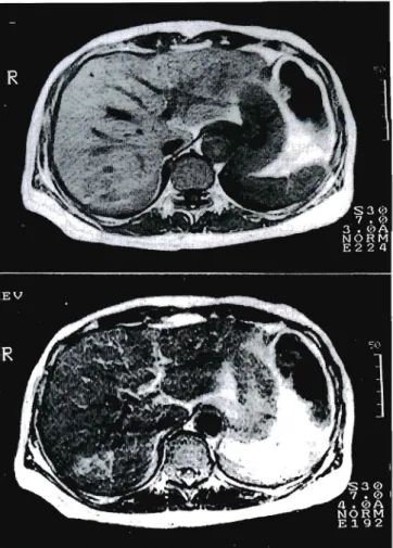
Re-garding the laboratory data, she had hyperlipidemia (total cholesterol 258mg/dl, triglyceride 369mg/dl) and normal tumor marker levels (AFP, CEA and CA19-9). HBs-antigen and HCV-antibody were negative.US showed an irregular hypoechoic region (2.3 cm in diameter) in the posterosuperior segment of the liver without any apparent finding of fatty change around the hypoechoic area (Fig. 5). Since she was hy-persensitive to iodine, enhanced CT could not be per formed. Plain CT showed an irregular low density area in segment 7 of the liver (Fig. 6). This region ap-peared as a low-intensity area on the Tl weighted MR images, and as a high-intensity area on T2 (Fig. 7). Since we could not rule out either cholangiocellular carcinoma
Fig. 5 Ultrasonography showing an irregular hypoechoic area in the posterosuperior segment (Case 2).
Fig. 6 A plainCT showing an irregular low density area Case 2).
or some other hepatic malignancy in the differential diag-nosis, a laparotomy was performed on November 12, 1996. At laparotomy, the liver demonstrated a dark red color, and no tumor mass was palpable. There was a fibrotic scar tissue on the surface of the posterior supe-nor segment. By an intra-operative US, the hypoechoic region was detected just beneath the scar tissue which was the same finding as that observed in the pre-operative US. An echo-guided needle biopsy was thus per-formed in this region, and the histology of the frozen section revealed fatty change in the liver. In a patho-logic study of the biopsied specimen, the hepatocytes con-tamed various sized lipid droplets and hepatocellular ne crosis was also observed around the areas of fatty change (Fig. 8).
DISC USSION
Two cases of focal fatty change of the liver, which could not be differentiated from the neoplastic region, have been presented in this report. In both of these cases, irregular shaped hepatic regions were incidentally
Fig. 7 In MRI, the region shows low-intensity on the Tl weighted image (above) and iso-intensity on the T2 weighted image (below) (Case 2).
Fig. 8 A microphotograph of thebiopsiedspecimen show-ing various sized lipid droplets (Case 2). (HE, ×50)
found by routine abdominal US screening. However, the US images of these two cases were not similar, namely, the regions were hyperechoic in case 1 and hypoechoic in case 2. On abdominal CT, the regions were recog-nized as low density areas.
Kiran et al. classified the US appearances of fatty infiltration of the liver into a geographic pattern, focal involvement by fatty infiltration and focal sparing by
226 Focal fatty change of the liver
fatty infiltration6'. According to this classification, our cases might thus be classified into the focal involve-ment category since no surrounding fatty change was observed. The regional shapes, segmental or lobar wedge distribution without any mass effects, were also char-acteristic in these regions61. In our cases, these regions were also located peripherally close to the liver cap-sule. and thus we could not evaluate the mass effects correctly based on the observed dislocation of the he-patic vascular structures.
On CT imaging, focal fatty change is known to be visualized as an irregular low density area without being enhanced by contrast medium3 6'. In case 1, how-ever, there was an atypical enhanced rim around the re-gion which suggested some hypervascular change to have occurred around the region. Previous reports have described an MR image of focal fatty change to be rec-ognized as a high-intensity area on the Tl weighted image, and as an iso-intensity area on T2 . However, in case 2, the region was detected as a low-intensity area on the Tl weighted image, and as a high-intensity area on T2. These findings of MR thus indicate that the region
might contain more vascular components than usual. These series of imaging features in our cases showed a tumor-like region in the liver, but a differential diagno-sis with a true tumor such as hepatocellular carcinoma (HCC), cholangiocellular carcinoma or some benign tumor was difficult. HCC is the most frequent malig-nancy in the liver, and sometimes HCC demonstrates fatty metamorphosis in cancer cells. From previous re-ports, HCC with fatty metamorphosis has been charac-tenzed by a mosaic pattern in US, CT and MRI, the existence of capsular formation in the US, angiogram, CT and dynamic MRI findings8 10>. In our cases, an
atypical enhanced rim around the region was observed on CT in case 1, but neither a mosaic pattern nor a septum in the tumor could be observed in either case.
Fatty change in the liver usually occurs in a
dif-fuse form throughout the liver3 "'は This homogenous
fatty change of the liver, commonly called "fatty liver , can often be detected in daily practice and is as-sociated with many pathogenetic factors such as alco-hoi, obesity, drugs (e.g. steroids, tetracycline, anticancer agents), malnutrition (e,,g. kwashiorkor, short bowel syn-drome), congestive heart failure, diabetes, hyperlipidemia, pregnancyand so on2 However, our cases did not demonstrate any of these pathogenetic factors except for hyperlipidemia in case 2. As a result, no pathogenetic fac-tors could be specified as a causative factor of the atypical pattern of the focal fatty change in our cases.
Brawer et al. considered the focal tissue hypoxia to be the cause of focal fatty change, because the focal fatty change mainly occurs at the site of the subcapsular region or median segment of the liver which is the site periphery to the portal venous and hepatic arterial circulation4'. In case 1, the region was located in the median segment of
the liver that is watershed of the right and left hepatic artery4'. These anatomic features might be the reason why the regional tissue hypoxia leads to focal fatty change. As a result, the hypervasculanty around the focal fatty change in the CT image might thus be thought to occurr secondary to tissue hypoxia. On the other hand, in case 2, hepatocellular necrosis was recog-nized in a pathologic study and scar tissue formation
was observed macroscopically. We thus hypothesized
that these inflammatory processes might have induced hypervascularity, which resulted in the imbalance of local blood per fusion, and finally contributed to the ap-pearance of focal fatty change in specific areas of the liver.In conclusion, we have presented two cases of focal fatty change in the liver which demonstrated unusual features on various imaging studies. One case revealed in-flammatory changes in macroscopic and pathological studies. Our cases indicated that focal fatty change may sometimes not demonstrate any particular appearance in conventional diagnostic measures, and therefore a needle biopsy is thought to be mandatory for the dif-ferential diagnosis.
ACKNOWLEDGEMENT
We thank Mr. Brian T. Quinn for comments on
the manuscript.REFERENCES
1 ) Mulhern C.B. Jr., Arger P.H. and Coleman B.G.: Nonuniform attenuation in computed tomography study of the cirrhotic liver. Radiology 132: 399-402, 1979.
2 ) Scott W.W. Jr., Sanders R.C. and Siegelman S.S.:
Irregular fatty infiltration of the liver: diagnostic dilemmas. AJR 135: 67-71, 1980.
3) Robert A.H., Melvyn K., Panol C.R. and William M.T.: CT Appearance of Focal Fatty Infiltration of the Liver. AJR 139: 277-281, 1982.
4 ) Michael K.B., Garth E.A. and Klaus J.L.: Focal Fatty Change of the Liver, a Hitherto Poorly Rec-ognized Entity. Gastroenterology 78: 247-252, 1980. 5 ) Kimura K.: irregular fatty change: Fukubu-echo no ABC Takehara Y., Akimoto S. and Kimura K., 204-205, Nihon Ishikai, Tokyo, 1992 (in Japanese). 6) Kiran A.J. and John P.M.: Spectrum of CT and
Sonographic Appearance of Fatty Infiltration of the Liver. Clinical Imaging 17: 162-168, 1993. 7) Banba T. and Fukano M.: Focal fatty change of
the liver. Ryoikibetsu-Shokogun-Shirizu 8: 14-17, 1995 (in Japanese).
8 ) Tanaka S., Kitamura T., Sasaki Y., Taniguchi H. and Ishiguro S. : Hepatocellular Carcinoma: Sonographic and Histologic Correlation. AJR 140: 701-707, 1983.
9) Yoshikawa J., Matsui O., Takashima T., Ida M.,
Takanaka T., Kawamura I., Kakuda K. and
Miyata S.: Fatty Metamorphosis in Hepatocellular
Carcinoma: Radiologic Features in 10 Cases. AJR
151: 717-720, 1988.
10) Ohashi I., Sasaki Y‥ Imaoka S‥ Masutani S.,
Iwamoto S., Furukawa H., Ishikawa OH Kabuto
T., Kameyama M., Koyama H., Iwanaga T., Inoue
EリFujita M. and Ishiguro S.: Three cases of primary
he-patic hpomatous tumors. Acta Hepatologica Japonica 33: 636-642, 1992 (in Japanese).
ll) Tanaka N∴ Fatty Liver. Ryoikibetsu-Shokogun-Shirizu 8: 18-20, 1995 (in Japanese).
12) Clain J.E., Stephens D.H. and Charboneau J.W.:
Ultrasonography and Computed Tomography in
Focal Fatty Liver Report of Two Cases With
Spe-cial Emphasis on Changing Appearances Over Time.
Gastroenterology 87: 948-952, 1984.
13) Matsui O., Takahashi S., Kadoya M., Yoshikawa
J., Gabata T., Takashima T. and Kitagawa K∴
Pseudolesion in Segment of the Liver at CT during Arterial Portography: Correlation with Aberrant Gastric Venous Drainage. Radiology 193: 31-35, 1994.
14) Nishimoto H., Obara S., Morita K., Uechi 0.,
NishiokaM., Yoshida J. and Maeda C∴ A Case of
Focal Fatty Liver Difficult to Distinguish from the Liver Metastasis of Rectal Cancer on CT. Rinsho-Hoshasen 34: 367-70,1989 (in Japanese).