Crystal structure of nesquehonite, MgCO
3
·3H(D)
2
O
by neutron di
ffraction and effect of pH
on structural formulas of nesquehonite
Gen–ichiro YAMAMOTO*, Atsushi KYONO*, Jun ABE**, Asami SANO–FURUKAWA***and Takanori HATTORI***
*Division of Earth Evolution Sciences, Graduate School of Life and Environmental Sciences, University of Tsukuba,
Tsukuba 305–8572, Japan
**Neutron Science and Technology Center, Comprehensive Research Organization for Science and Society,
Ibaraki 319–1106, Japan
***J–PARC Center, Japan Atomic Energy Agency, Ibaraki 319–1195, Japan
Neutron diffraction, Raman spectroscopy, and thermal analysis were performed to investigate the composition, structure, and formation conditions of the magnesium carbonate hydrate nesquehonite. The crystal structure of deuterated nesquehonite was analyzed by Rietveld refinement of the time–of–flight neutron powder diffraction pattern. The crystal structure possessed the monoclinic space group P21/n with lattice parameters of a =
7.72100(12) Å, b = 5.37518(7) Å, c = 12.1430(3) Å,β = 90.165(4)°, and V = 503.956(13) Å3. The refinement with afinal crystal structure model of deuterated nesquehonite converged to wRp = 4.22% and Rp = 3.50%. The result of structure refinement showed that two deuterium atoms are coordinated to the O1, O2, and O6 atoms as a water molecule in the nesquehonite. The fact that the three water molecules were included in the structure suggests the structural formula of the nesquehonite obtained in the study should be written as MgCO3·3H2O not
Mg(HCO3)(OH)·2H2O.
Keywords: Nesquehonite, Magnesium carbonate hydrate, Neutron diffraction, Hydrogen–bonding network
INTRODUCTION
The mineral carbonation of CO2is one of the promising
technology for reducing the amount of carbon dioxide in the atmosphere (e.g., Oelkers et al., 2008; Matter and Ke-lemen, 2009; Sanna et al., 2014). Numerous approaches to
CO2 sequestration have extensively been studied (Park
and Fan, 2004; Rendek et al., 2006; Kelemen and Matter, 2008; Assayag et al., 2009; Montes–Hernandez et al., 2009; Felmy et al., 2012; Farhang et al., 2016; Noiriel and Daval, 2017). Magnesium carbonates, therefore, offer attractive possibilities for the permanent and safe geolog-ical storage of CO2(Hänchen et al., 2008; Ferrini et al.,
2009; Ballirano et al., 2010; Harrison et al., 2019). In the MgO–CO2–H2O system at temperatures ranging from 0 to
60 °C, the thermodynamically stable phases are brucite
Mg(OH)2 and magnesite MgCO3 (Königsberger et al.,
1999). The aqueous phase reaction, nevertheless, yields exclusively hydrated magnesium carbonates. The inhibi-tion of magnesite formainhibi-tion has been ascribed to the strong hydration status of Mg2+ ions (e.g., Hänchen et al., 2008). Instead of magnesite, a variety of magnesium carbonate hydrates are formed depending on the temper-ature, the partial pressure of CO2, and pH (Perchiazzi and
Merlino, 2006; Back and Mandarino, 2008; Frost et al., 2008; Hänchen et al., 2008; Beinlich and Austrheim, 2012; Kristova et al., 2014). A large number of
magnesi-um carbonate hydrates are known in the Mg–CO2–H2O
system (Fig. 1). They can be classified into three types:
the nesquehonite group (MgCO3·nH2O), the
hydromag-nesite group [Mg5(CO3)4(OH)2·nH2O], and the artinite
group [Mg2CO3(OH)2·nH2O]. Nesquehonite is
precipitat-ed in the temperature range from room temperature to 55 °C and at a partial pressure of CO2close to or below
am-bient pressure (Zhang et al., 2006; Morgan et al., 2015). It subsequently transforms to hydromagnesite at moderate doi:10.2465/jmps.200117
G. Yamamoto, g–[email protected] Corresponding author
A. Kyono, [email protected]
temperatures and CO2 pressures depending on the
reac-tion time (Davies and Bubela, 1973; Zhang et al., 2006; Hopkinson et al., 2008). Nesquehonite, therefore, acts as a precursor for hydromagnesite, which is by far the most common naturally occurring magnesium carbonate hy-drate mineral at atmospheric CO2pressure within the
tem-perature interval typical of most surface environments (Langmuir, 1965; Hopkinson et al., 2012).
The structural formula of nesquehonite has been long
recognized as MgCO3·3H2O, which has been approved
by the International Mineralogical Association, Commis-sion on Minerals and Mineral Names (IMA–CNMMN) (Genth and Penfield, 1890; Giester et al., 2000). However, recent studies using IR spectroscopy (Frost and Palmer, 2011; Hopkinson et al., 2012), infrared emission spectros-copy (Frost and Palmer, 2011), and Raman spectrosspectros-copy (Hales et al., 2008; Hopkinson et al., 2012) have strongly
suggested that bicarbonate (HCO3) and hydroxyl (OH)
groups exist in the structure. These results enable the structural formula of nesquehonite to be better defined as Mg(HCO3)(OH)·2H2O (Hales et al., 2008; Frost and
Palmer, 2011; Hopkinson et al., 2012). As a result, the structural formula of nesquehonite has become a contro-versial issue that needs further investigation. In order to interpret accurately the behavior of nesquehonite in an aqueous solution, it is critically important to clarify the
difference between MgCO3·3H2O and Mg(HCO3)(OH)·
2H2O. In previous studies (Stephan and MacGillavry,
1972; Giester et al., 2000), the crystal structure of nesque-honite was examined using an X–ray diffraction tech-nique. As a result, six hydrogen positions were deter-mined by difference electron density syntheses (Giester et al., 2000). The structural factors measured by neutron diffraction are heavily weighted by the contribution from the hydrogen (or deuterium) atoms. We, therefore, inves-tigated the crystal structure of deuterated nesquehonite by
a neutron diffraction technique. It was further character-ized by Raman spectroscopy and thermal analysis. In this paper, we discuss the structural formula of nesquehonite and the possibility that nesquehonite can possess two structural formulas depending on the pH in a solution.
EXPERIMENTAL METHODS
Commercially available MgCl2 (Wako Pure Chemicals
Co., Inc., purity,≥97.0%) and Na2CO3(Wako Pure
Chem-icals Co., Inc., purity,≥99.5%) were used as starting ma-terials for nesquehonite. For the deuterated nesquehonite, solutions containing 20 ml of 0.5 M MgCl2and 20 ml of
0.5 M Na2CO3werefirst prepared with distilled water. A
white slurry immediately appeared when the solutions were mixed in a 1:1 molar proportion. The solution was then stirred at room temperature under vigorous magnetic stirring for 3 h in a closed system while sealed with a paraffin film. The pH value in the solution was ultimately stabilized at 10.9. The precipitate was subsequently fil-tered and washed on the filter with distilled water. The precipitate was collected and dried in the air overnight. The deuterated nesquehonite was also prepared for
neu-tron diffraction measurements by dissolving the MgCl2
and Na2CO3in deuterium oxide (D2O) (Wako Pure
Chem-icals Co., Inc., purity, ≥99.9%). The subsequent steps
were the same as those described above. The precipitate
wasfiltered and washed with deuterium oxide.
In order to identify the precipitates resulting from
mixing MgCl2 and Na2CO3 in water, X–ray diffraction
measurements were performed by powder X–ray diffrac-tion (Bruker AXS GmbH, D8 Advance TSM) equipped with a CuKα rotating–anode generator operated at 40 kV and 40 mA. Powder samples were mounted on a non– reflective silicon plate. Data were collected in step–scan mode in the region of 10°≤ 2θ ≤ 70° with a step size of 0.02° and a counting time of 0.1 s per step.
Raman spectra of undeuterated and deuterated nes-quehonites were recorded by a micro–Raman spectrome-ter (NRS–5100, Japan Spectroscopic Co., Japan) equip-ped with a grating of 1800 lines/mm and a high sensi-tivity cooled CCD detector. Samples were excited with a green laser operating at 532.12 nm. The incident laser power was kept at less than 5.4 mW to prevent dehydra-tion of nesquehonite. A 100× objective lens was used to focus the laser beam onto a 1–µm area. Raman spectra were collected with an integration time of 1 min, and the two accumulations were averaged. The resolution of the spectra was below 4.1 cm−1. The spectra were correct-ed by the emission lines of a neon lamp. The laser beam produced no visible damage on the surface of the samples in the measurements. The least–squares peak–fitting soft-Figure 1. Magnesium carbonate hydrates in the MgO–CO2–H2O
ware Peak–Fit (AISN Software Inc., Chicago) was used to perform the analysis of the Raman spectra data. Data
smoothing and baseline fitting were conducted using a
Savitzky–Golay method and hyperbolic model, respec-tively. Band positions were determined byfitting the Ra-man peaks with a Lorentzian peak shape function.
Thermogravimetric (TG) and differential thermal
analysis (DTA) were performed to characterize the
ther-mal behavior using a therther-mal analyzer (TG/DTA–7300,
Seiko Instruments Inc., Japan). Approximately 10 mg of
undeuterated nesquehonite andα–alumina used as a
ref-erence were placed in Al pans. They were subsequently heated over a 60–550 °C temperature range with a heat-ing rate of 5 °C min−1in argonflowing at 200 mL min−1. Neutron diffraction measurements were conducted using a time–of–flight neutron diffractometer at the BL11 beamline PLANET (Hattori et al., 2015) in the Ma-terials and Life Science Experimental Facility (MLF) of J–PARC, Japan. The deuterated nesquehonite synthesized was loaded into a cylindrical vanadium can with 3.0 mm outer diameter and 0.1 mm wall thickness. The incident beam size was adjusted by slits to approximately 5 mm in the horizontal direction and 10 mm in the vertical direc-tion. To obtain data of sufficient statistical quality for Rietveld structure refinement, the sample was exposed to the neutron beam for 2 h at an accelerator power of approximately 500 kW. The data for the vanadium rod, empty vanadium container, and instrumental background were also separately measured for intensity corrections. The crystal structure of nesquehonite was investigated by the Rietveld structure refinement analysis using the Gen-eral Structure Analysis System (GSAS) package (Larson and Von Dreele, 2004) with the EXPGUI interface (Toby 2001). The lattice parameters, atomic positional parame-ters, isotropic displacement parameparame-ters, profile function, background function, and scale factor were refined using TOF data in the 3.786–40.000 ms range, corresponding to d–spacings of 0.400 < d < 4.227 Å. Regarding site occu-pancy, all sites were assumed to be fully occupied. In the analysis, the peak profile was modeled with an exponen-tial pseudo–Voigt function (Larson and Von Dreele, 2004). The peak cut–off was set to 0.1% of the intensity of the strongest reflection of nesquehonite, and the
back-ground was fitted with an 18–term Chebyshev
polyno-mial (Larson and Von Dreele, 2004). No constraints on bond lengths and angles were applied in the refinements.
RESULTS AND DISCUSSION Characteristics of synthesized nesquehonite
Figure 2 shows the X–ray diffraction patterns of the
pre-cipitates obtained in the study. The peak positions of the precipitates collected from the distilled water were in ap-proximate agreement with that of nesquehonite (PDF 01– 070–6308). Although there is a small peak shift due to the isotope effect, the XRD patterns of the precipitates col-lected from the deuterium oxide were entirely consistent with that of precipitates obtained in the distilled water. Therefore, it appears that undeuterated and deuterated nesquehonite were successively synthesized. The XRD measurements, however, showed a small amount of im-purity that could not be identified.
Figure 3 shows the Raman spectra of the undeuter-ated and deuterundeuter-ated nesquehonite. Compared with the OH stretching vibration modes in the undeuterated nes-quehonite, the OD stretching vibration modes in the deu-terated nesquehonite were significantly shifted to the low-er wavenumblow-er region due to the isotope effect (Fig. 3). In the deuterated nesquehonite, weak OH stretching vi-brations were observed in the range from 3000 to 3700 cm−1 (Fig. 3d), indicating slight atmospheric hydrogen contamination.
Figure 4 shows TG, DTG, and DTA curves for the undeuterated nesquehonite. We could clearly observe three endothermic peaks below 300 °C (i.e., 105, 144, and 195 °C) and two endothermic peaks between 300 and 550 °C (i.e., 435 and 478 °C). The thermogravimetric data for the undeuterated nesquehonite are given in Table 1. The total mass loss of 36.4% experimentally deter-mined between 60 and 320 °C was slightly smaller than the theoretical value of 39.1% that corresponds to the elimination of 3 mol H2O from nesquehonite. On the oth-Figure 2. X–ray diffraction patterns of precipitates collected from (a) deuterium oxide and (b) distilled water. The diffraction peak positions are in agreement with those of nesquehonite (PDF 01– 070–6308). The short vertical lines above the pattern represent unidentifiable peaks. The enlarged view shows a degree of peak shift due to the isotope effect.
G. Yamamoto, A. Kyono, J. Abe, A. Sano–Furukawa and T. Hattori 98
er hand, a total mass loss of 34.1% between 320 and 550 °C was higher than the theoretical value of 31.8% that
corresponds to 1 mol CO2 of nesquehonite. The total
mass loss of 70.5% observed by the thermal analysis was close to the theoretical mass loss of 70.9%.
Neutron diffraction
The crystal structure of the deuterated nesquehonite was determined by Rietveld analysis of the time–of–flight neutron powder diffraction pattern. The results of struc-tural refinements and strucstruc-tural parameters are presented in Table 2. Selected interatomic distances and angles are listed in Table 3. Initial atomic positional parameters were taken from the data of Giester et al. (2000), in which the crystal structure of nesquehonite was defined as MgCO3·3H2O. In order to obtain information on the
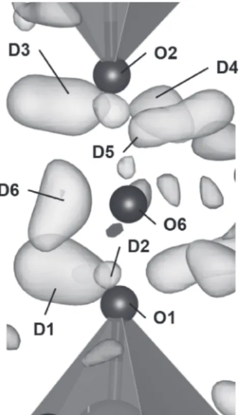
po-sitions of deuterium (D) atoms,first, the crystal structure of deuterated nesquehonite was refined without deuteri-um (D) atoms. As mentioned above in the discussion of the Raman spectra, since the effect of the hydrogen con-tamination was negligible, all residuals were treated as deuterium atom positions. The three–dimensional differ-ence–Fourier map of the residual nuclear scattering length density distribution was visualized by the VESTA pro-gram (Momma and Izumi, 2011) (Fig. 5), which shows residual densities near the O1, O2, and O6 atoms. Two deuterium (D) atoms were then placed near each of the Figure 3. Raman spectra in the 2100–2800 cm−1region of (a)
un-deuterated and (b) un-deuterated nesquehonites, and Raman spectra in the 3000–3700 cm−1region of (c) undeuterated and (d) deu-terated nesquehonites.
Figure 4. TG, DTG, and DTA curves for undeuterated nesqueho-nite.
Table 1. Thermogravimetric data for undeuterated nesquehonite
Table 2. Data collection parameters, refinement details, crystal structural data, and atomic coordinates and displacement param-eters (Å2) for deuterated nesquehonite
oxygen atoms. As a result, all atomic positions and equivalent isotropic displacement parameters were con-verged to the residual factors of wRp = 4.22% and Rp = 3.50% (Table 2). The neutron diffraction data were ade-quately fitted with the crystal structure model (Fig. 6). Figure 7 shows the crystal structure of deuterated nesque-honite refined in the study, implying that deuterium (D) atoms are coordinated to O1, O2, and O6 atoms, forming a water molecule in the nesquehonite. In the crystal struc-ture of nesquehonite, the infinite ribbons running along theb–axis consist of MgO6octahedra, in which each Mg
atom is coordinated to two O atoms in water molecules and four O atoms in CO3groups. The ribbons are
inter-connected by the hydrogen bonding of water molecules between the ribbons (Fig. 7a).
The interatomic distances and angles refined in the study are in satisfactory agreement with those reported by Giester et al. (2000) (Table 3). The D3 and D4 atoms coordinated to O2 form a symmetric configuration with the water molecules, but the D1 and D2 atoms coordinat-ed to O1 exhibit a highly asymmetric configuration, which may be ascribed to the asymmetric charge distribution on the water molecule. The O1–D2 distance has the shortest bond distance of 0.891(9) Å (Table 3). This shortest bond
among all O–D bonds was observed in the previous study as well (Giester et al., 2000). The water molecules con-sisting of O6, D5, and D6 locate between the ribbons and are included in the hydrogen–bonding network (Fig. 7a). The hydrogen bonds range between 1.752(12) Å and 1.953(11) Å, except for the D5£O1 and D5£O2 bonds with distances of 2.346(14) Å and 2.483(13) Å, respec-tively. All hydrogen bonds were shorter than those deter-mined by Giester et al. (2000), which is due to the longer O–D bonds. The water molecules coordinated to Mg atoms and located between ribbons form a three–dimen-sional hydrogen–bonding network (Fig. 7a). It is, there-fore, concluded that three water molecules are included in the structural formula. Consequently, the structural
for-mula of nesquehonite is clearly described as MgCO3·
3H2O. Based on the empirical parameters of Brese and
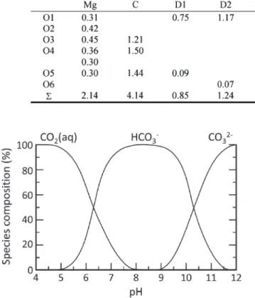
O’Keeffe (1991), the bond valence sums are listed in Table 4. The Mg and C atoms showed reasonable bond
valence sums. Although the Mg–O3 bond distance of
1.988(7) Å is considerably shorter than the average bond distance (Table 3), the bond valence sum of the O3atom
gives a significantly lower bond valence than the ideal value of 2.0 (Table 4). This bond valence deficiency would be a structural feature of nesquehonite, and it sug-Table 3. Selected interatomic distances (Å) and angles (°) in nesquehonite
G. Yamamoto, A. Kyono, J. Abe, A. Sano–Furukawa and T. Hattori 100
gests that the O3 atom can act as a tridentate ligand link-ing three different atoms to form the bicarbonate (HCO3)
species. That is, the O3 atom can potentially be coordi-nated not only to Mg and C atoms but also to the H atom. Consequently, nesquehonite could have the structural
for-mula of Mg(HCO3)(OH)·2H2O due to this bond valence
deficiency. The larger bond valence sums of the O1 and D2 atoms are due to the short O1–D2 bond, whereas the lower bond valence sums of D1 and D6 atoms are
ascrib-ed to the longer O1–D1 and O6–D6 bonds. These
devia-tions from the ideal values are likely caused by the highly asymmetric configuration, which may be ascribed to the asymmetric charge distribution on the water molecule (Soper, 2005), but further study is required to understand the details.
Structural formula of nesquehonite
The structural formula of nesquehonite has been long
rec-ognized as MgCO3·3H2O (Genth and Penfield, 1890;
Giester et al., 2000), but recent studies have strongly sug-gested that its structural formula should be redefined as
Mg(HCO3)(OH)·2H2O (Hales et al., 2008; Frost and
Palmer, 2011; Hopkinson et al., 2012). This controversy may be resolved by accounting for differences in forma-tion condiforma-tions. In this study, we applied an alkali metal carbonate as a starting material, which was the same meth-od as that of Coleyshaw et al. (2003) and Jauffret et al. (2015). However, the pH of alkali metal carbonate solu-tions, such as Na2CO3 and K2CO3, completely differed
from that in Hales et al. (2008) and Hopkinson et al. (2012). The pH value can be considered as a critical factor in the formation of magnesium carbonate hydrates be-cause these minerals exhibit wide chemical variability de-pending on the temperature, the partial pressure of CO2,
and pH (Perchiazzi and Merlino, 2006; Back and Mandar-ino, 2008; Frost et al., 2008; Hänchen et al., 2008; Bein-lich and Austrheim, 2012; Kristova et al., 2014).
It is well known that the relative proportions of car-Figure 6. Neutron powder diffraction pattern and the result of
fit-ting by Rietveld refinement for deuterated nesquehonite.
Figure 7. Hydrogen–bonding interactions (a) between the ribbons and (b) the ribbon structure consisting of MgO6octahedra and
CO3groups. Dotted lines show hydrogen bonds.
Figure 5. Three–dimensional difference–Fourier map of the resid-ual nuclear scattering length density distribution projected along theb–axis. The highest and lowest residual density peaks are 2.3 and−0.18 fm/Å3, respectively. The positive and negative differ-ence peaks are represented as yellow and blue, respectively. Col-or version is available online from https://doi.Col-org/10.2465/jmps. 200117.
bon ion species (CO2, HCO3−, and CO32−) depend on the
alkalinity (Fig. 8). Thus, bicarbonate ion (HCO3−) is the
dominant species between pH 6.35 and 10.33 at 25 °C. In the studies of Hales et al. (2008) and Hopkinson et al. (2012), nesquehonite was synthesized at a pH below 8. It is, therefore, quite reasonable that nesquehonite with
the structural formula of Mg(HCO3)(OH)·2H2O would
be crystallized in a solution with a pH below 8.1. In the present study, on the other hand, nesquehonite was pre-pared from MgCl2 and Na2CO3 solutions, which led to
a high pH value (pH = 10.9). The solution prepared in the study was dominated by the carbonate ion (CO32−).
As a result, nesquehonite with the structural formula of MgCO3·3H2O would be crystallized in the solution. It
has already been reported that nesquehonite of MgCO3·
X H2O phases occurred at pH values between 8.5 and 12.5
(Zhang et al., 2006), whereas the Mg(HCO3)(OH)·2H2O
phase was formed at a pH below 8 (Hopkinson et al., 2012). Consequently, the pH value seems to determine which carbon ion species are incorporated into the nes-quehonite structure. There would, therefore, be two
struc-tural formulas of nesquehonite, MgCO3·3H2O and
Mg(HCO3)(OH)·2H2O, which are formed separately
de-pending on the pH value in an aqueous solution.
ACKNOWLEDGMENTS
The authors must acknowledge and appreciate two anon-ymous reviewers for their constructive comments and in-sightful suggestions to improve the original manuscript. The neutron diffraction experiment was approved by the Neutron Science Proposal Review Committee of J– PARC MLF (proposal no. 2018B0038). This work was partially supported by JSPS KAKENHI Grant Number JP17K05702.
SUPPLEMENTARY MATERIAL
Color version of Figure 5 is available online from https:// doi.org/10.2465/jmps.200117.
REFERENCES
Assayag, N., Matter, J., Ader, M., Goldberg, D. and Agrinier, P. (2009) Water–rock interactions during a CO2 injectionfield–
test: Implications on host rock dissolution and alteration ef-fects. Chemical Geology, 265, 227–235.
Back, M.E. and Mandarino, J.A. (2008) Fleischer’s glossary of mineral species. pp. 346, Mineralogical Record Inc., Tucson. Ballirano, P., De Vito, C., Ferrini, V. and Mignardi, S. (2010) The thermal behaviour and structural stability of nesquehonite, MgCO3·3H2O, evaluated by in situ laboratory parallel–beam
X–ray powder diffraction: New constraints on CO2
sequestra-tion within minerals. Journal of Hazardous Materials, 178, 522– 528.
Beinlich, A. and Austrheim, H. (2012) In situ sequestration of at-mospheric CO2 at low temperature and surface cracking of
serpentinized peridotite in mine shafts. Chemical Geology, 332, 32–44.
Brese, N.E. and O’Keeffe, M. (1991) Bond–valence parameters for solids. Acta Crystallographica, B47: 192–197.
Coleyshaw, E.E., Crump, G. and Griffith, W.P. (2003) Vibrational spectra of the hydrated carbonate minerals ikaite, monohydro-calcite, landsfordite and nesquehonite. Spectrochimica Acta A, 59, 2231–2239.
Davies, P.J. and Bubela, B. (1973) The transformation of nesque-honite into hydromagnesite. Chemical Geology, 12, 289–300. Drever, J.I. (1997) The geochemistry of natural waters: surface and groundwater environments. pp. 436, Prentice Hall, New Jersey. Table 4. Bond valence sums for deuterated nesquehonite
Figure 8. Carbon species in pure water at 25 °C as a function of pH. The pK1and pK2are 6.35 and 10.33, respectively (Drever, 1997).
G. Yamamoto, A. Kyono, J. Abe, A. Sano–Furukawa and T. Hattori 102
Farhang, F., Oliver, T.K., Rayson, M., Bren, G., et al. (2016) Ex-perimental study on the precipitation of magnesite from ther-mally activated serpentine for CO2 sequestration. Chemical
Engineering Journal, 303, 439–449.
Felmy, A.R., Qafoku, O., Arey, B.W., Hu, J.Z., et al. (2012) Re-action of water–saturated supercritical CO2 with forsterite:
Evidence for magnesite formation at low temperatures. Geo-chimica et CosmoGeo-chimica Acta, 91, 271–282.
Ferrini, V., De Vito, C. and Mignardi, S. (2009) Synthesis of nes-quehonite by reaction of gaseous CO2with Mg chloride
solu-tion: its potential role in the sequestration of carbon dioxide. Journal of Hazardous Materials, 168, 832–837.
Frost, R.L., Bahfenne, S., Graham, J. and Reddy, B.J. (2008) The structure of selected magnesium carbonate minerals– A near infrared and mid–infrared spectroscopic study. Polyhedron, 27, 2069–2076.
Frost, R.L. and Palmer, S.J. (2011) Infrared and infrared emission spectroscopy of nesquehonite Mg(OH)(HCO3)·2H2O–
implica-tions for the formula of nesquehonite. Spectrochimica Acta A, 78, 1255–60.
Genth, F.A. and Penfield, S.L. (1890) On lansfordite, nesquehonite, a new mineral, and pseudomorphs of nesquehonite after lans-fordite. American Journal of Science 39, 121–137.
Giester, G., Lengauer, C.L. and Rieck, B. (2000) The crystal struc-ture of nesquehonite, MgCO3·3H2O, from Lavrion, Greece.
Mineralogy and petrology, 70, 153–163.
Hales, M.C., Frost, R.L. and Martens, W.N. (2008) Thermo–Ra-man spectroscopy of synthetic nesquehonite– implication for the geosequestration of greenhouse gases. Journal of Raman Spectroscopy, 39, 1141–1149.
Hänchen, M., Prigiobbe, V., Baciocchi, R. and Mazzotti, M. (2008) Precipitation in the Mg–carbonate system – effects of temper-ature and CO2pressure. Chemical Engineering Science, 63,
1012–1028.
Harrison, A.L., Mavromatis, V., Oelkers, E.H. and Benezeth, P. (2019) Solubility of the hydrated Mg–carbonates nesqueho-nite and dypingite from 5 to 35 °C: Implications for CO2
stor-age and the relative stability of Mg–carbonates. Chemical Ge-ology, 504, 123–135.
Hattori, T., Sano–Furukawa, A., Arima, H., Komatsu, K., et al. (2015) Design and performance of high–pressure PLANET beamline at pulsed neutron source at J–PARC. Nuclear Instru-ments and Methods in Physics Research Section A, 780, 55–67. Hopkinson, L., Rutt, K. and Cressey, G. (2008) The transformation of nesquehonite to hydromagnesite transition in the system MgO–CaO–H2O–CO2: an experimental spectroscopic study.
Journal of Geology, 116, 387–400.
Hopkinson, L., Kristova, P., Rutt, K. and Cressey, G. (2012) Phase transitions in the system MgO–CO2–H2O during CO2
degass-ing of Mg–beardegass-ing solutions. Geochimica et Cosmochimica Acta, 76, 1–13.
Jauffret, G., Morrison, J. and Glasser, F.P. (2015) On the thermal decomposition of nesquehonite. Journal of Thermal Analysis and Calorimetry, 122, 601–609.
Kelemen, P.B. and Matter, J. (2008) In situ carbonation of perido-tite for CO2storage. Proceedings of the National Academy of
Sciences of the United States of America, 105, 17295–17300. Königsberger, E., Königsberger, LC. and Gamsjäger, H. (1999) Low–temperature thermodynamic model for the system Na2CO3–MgCO3–CaCO3–H2O. Geochimica et Cosmochimica
Acta, 63, 3105–3119.
Kristova, P., Hopkinson, L.J., Rutt, K.J., Hunter, H.M.A. and Cres-sey, G. (2014) Carbonate mineral paragenesis and reaction kinetics in the system MgO–CaO–CO2–H2O in presence of
chloride or nitrate ions at near surface ambient temperatures. Applied Geochemistry, 50, 16–24.
Langmuir, D. (1965) Stability of carbonates in the system MgO– CO2–H2O. Journal of Geology, 73, 730–754.
Larson, A.C. and Von Dreele, R.B. (2004) General Structure Anal-ysis System, (GSAS). Los Alamos National Laboratory Re-port LAUR, 86–784.
Matter, J.M. and Kelemen, P.B. (2009) Permanent storage of car-bon dioxide in geological reservoirs by mineral carcar-bonation. Nature Geoscience, 2, 837–841.
Momma, K. and Izumi, F. (2011) VESTA 3 for three–dimensional visualization of crystal, volumetric and morphology data. Journal of Applied Crystallography, 44, 1272–1276. Montes–Hernandez, G., Pérez–López, R., Renard, F., Nieto, J.M.
and Charlet, L. (2009) Mineral sequestration of CO2by
aque-ous carbonation of coal combustionfly–ash. Journal of Haz-ardous Materials, 161, 1347–1354.
Morgan, B., Wilson, S.A., Madsen, I.C., Gozukara, Y.M. and Hab-suda, J. (2015) Increased thermal stability of nesquehonite (MgCO3·3H2O) in the presence of humidity and CO2:
Impli-cations for low–temperature CO2storage. International
Jour-nal of Greenhouse Gas Control, 39, 366–376.
Noiriel, C. and Daval, D. (2017) Pore–scale geochemical reactivity associated with CO2storage: New frontiers at thefluid–solid
interface. Accounts of Chemical Research, 50, 759–768. Oelkers, E.H., Gislason, S.R. and Matter, J. (2008) Mineral
carbo-nation of CO2. Elements, 4, 333–337.
Park, A.H.A. and Fan, L.S. (2004) CO2 mineral sequestration:
physically activated dissolution of serpentine and pH swing process. Chemical Engineering Science, 59, 5241–5247. Perchiazzi, N. and Merlino, S. (2006) The malachite–rosasite
group: crystal structures of glaukosphaerite and pokrovskite. European Journal of Mineralogy, 18, 787–792.
Rendek, E., Ducom, G. and Germain, P. (2006) Carbon dioxide sequestration in municipal solid waste incinerator (MSWI) bottom ash. Journal of Hazardous Materials, 128, 73–79. Sanna, A., Uibu, M., Caramanna, G., Kuusik, R. and Maroto–
Valer, M.M. (2014) A review of mineral carbonation technol-ogies to sequester CO2. Chemical Society Reviews, 43, 8049–
8080.
Soper, A.K. (2005) An asymmetric model for water structure. Jour-nal of Physics: Condensed Matter, 17, S3273–S3282. Stephan, G.W. and MacGillavry, C.H. (1972) The crystal structure
of nesquehonite, MgCO3.3H2O. Acta Crystallographica, B28,
1031–1033.
Toby, BH (2001) EXPGUI, a graphical user interface for GSAS. Journal of Applied Crystallography, 34, 210–213.
Zhang, Z.P., Zheng, Y.J., Ni, Y.W., Liu, Z.M., et al. (2006) Tem-perature– and pH–dependent morphology and FT–IR analysis of magnesium carbonate hydrates. The Journal of Physical Chemistry B, 110, 12969–12973.
Manuscript received January 17, 2020 Manuscript accepted March 9, 2021 Published online April 27, 2021 Manuscript handled by Takaya Nagai