原 著
ブタガラス化保存胚盤胞を用いた非外科的移植方法の改善
三角浩司
1・江川紗智子
2・御澤弘靖
3・平山祐理
2 1日本大学生物資源科学部,神奈川県藤沢市,252-0880 2(独)家畜改良センター,福島県西白河郡西郷村,961-8511 3ミサワ医科工業株式会社,茨城県笠間市,309-1717 (2020 年 4 月 3 日受付,2020 年 5 月 29 日受理) 要 約 本研究は,生産現場でのブタ胚移植を実施することを目的に,ガラス化保存胚の非 外科的移植について検討を行った。供試胚はガラス化保存した胚盤胞∼拡張胚盤胞を用いた。 受胚豚は未経産豚(8∼10ヶ月齢)35 頭を用いた。非外科的移植器具には,市販の深部注入用移 植器(器具 A),深部注入用の試作品(器具 B)およびウシ用胚移植器を改良した子宮体移植用 の試作品(器具 C)を用いた。試験 1では,移植部位の確認のため,各移植器をそれぞれ受胚豚 5 頭に挿入した後,全身麻酔下で開腹手術を行い移植器具の先端部の位置(移植部位)を確認し た。その後,ガラス化保存胚を移植した。1頭あたりの移植胚数は 1腹採取分(14∼16 個)で 実施した。試験 2 では,各移植器をそれぞれ立位無麻酔の受胚豚 5 頭に挿入した後,ガラス化 保存胚を移植した。1頭あたりの移植胚数は 1腹採取分(15∼17 個)で実施した。試験 1 の結 果,器具 A 区では,1頭に子宮穿孔が見られたため,この受胚豚には移植を実施しなかった。 他の受胚豚において,移植器の先端は子宮体および子宮角分岐部付近であった。胚を移植した 受胚豚 4 頭中 2 頭が受胎・分娩し合計 8 頭の子豚を生産した。器具 B 区では,受胚豚 5 頭にお いて,移植器の先端は子宮角分岐部付近であった。受胚豚 5 頭中 3 頭が受胎・分娩し合計 13 頭 の子豚を生産した。器具 C 区では,受胚豚 5 頭において,移植器の先端は子宮頸管出口付近の 子宮体であった。5 頭すべて不受胎であった。試験 2 の結果,器具 A 区で受胚豚 5 頭中 3 頭が 受胎したがうち 2 頭が流産した。残りの 1頭から 5 頭の子豚を生産した。器具 B 区では,5 頭 中 3 頭が受胎・分娩し合計 16 頭の子豚を生産した。器具 C では,5 頭中 3 頭が受胎・分娩し合 計 14 頭の子豚を生産した。今回の結果から,無麻酔下の立位状態においては,ブタガラス化保 存胚を子宮体へ非外科的に移植することで,外科的移植と同等の効率で産子が得られることが 明らかとなった。 ─────────────────────────緒
言
ブタ胚の超低温保存技術は,遺伝資源の保存およ び豚群の疾病制御に応用できる。特に養豚場へ更新 豚等を導入する際,超低温保存したブタ胚を導入し て,農場内の受胚豚に移植する方法が,現時点で最 も衛生的な導入方法である。大曲ら(2015)は,種 豚場で繋養されている種豚から採取した胚をガラス 化保存し,そのガラス化保存胚を導入農場に輸送し た後,外科的に移植することで 60∼75% の分娩率 が得られることを報告した。しかしながら,ブタ胚 移植で確実に子豚を得るためには,開腹手術による 胚移植を実施する必要があり,手術施設が無い一般 の養豚農家では胚移植が困難な状況である。一方で 受胚豚の開腹手術が不要な非外科的胚移植方法がこ れまで様々な手法で報告されてきたが,いずれの手 法も開腹手術に比べると受胎率が低く,再現性に問 題があった(DAY, 1968 ; GALVINら,1994 ; LIら,1996 ;YONEMURAら,1996 ; DUCRO-STEVERINKら,2004)。 開腹手術により豚の胚盤胞を移植する際,子宮角 先端部分に移植した方が子宮角分岐部付近に移植す るより受胎率が高かったことが報告されている (WALLENHORSTら,1999)。この報告から,近年では 深部注入移植器具による非外科的胚移植の成功例が 報告されてきた(SUZUKIら,2004 ; CUELLOら,2005 ;
BATHGATEら,2007 ; NAKAZAWAら,2008 ; YOSHIOKA
ら,2012 ; GOMISら,2012 ; MARTINEZら,2014, 2016)。 しかしながら,深部注入器具の内管を受胚豚の子宮 角に挿入するには,内管挿入ガイドの役割をする外 管を子宮頸管の深部まで挿入する必要があり,頸管 がある程度弛緩している経産豚を受胚豚として用い られるのがほとんどであった。そのため,未経産豚 にも使用できる移植器具の開発および移植器具先端 の到着部位,すなわち胚の移植部位について再検討 が必要であると我々は考えた。 本研究は,市販されている深部注入移植器具の他 に深部注入する内管の材質を市販品より柔らかくし た試作移植器具さらにはウシ用子宮深部移植器を改 良した試作移植器具を用いてガラス化保存胚の非外 科的移植を行ない,それぞれの移植部位の確認およ び移植後の受胎成績について調査した。
材料および方法
本試験に使用したブタは,独立行政法人家畜改良 センター動物実験指針に準じて飼養管理および供試 された(承認番号 24-15, 25-14, 26-16)。また,本試 験で用いた試作移植器具作製に関しては,独立行政 法人家畜改良センターおよびミサワ医科工業株式会 社との間で利益相反が生じないよう適正な管理下で 実施された。 胚の採取方法 胚の採取には,月齢 8ヶ月齢以上のデュロック種 30 頭の性成熟雌豚を用いた。交配用の精液は,12ヶ 月齢以上のデュロック種 3 頭から採取した精液をモ デナ液で希釈後,使用時まで 1 5℃で保存した。供胚 豚は人工授精後,20∼40 日目にプロスタグランジン (PGF2α:クロプロステノールとして 0.184 mg,株式 会社インターベット)を頸部筋肉内投与し,その 24 時間後に同量の PGF2αと妊馬血清性性腺刺激ホル モン(eCG,動物専用セロトロピン,あすか製薬株 式会社)1500 IU を頸部筋肉内投与した。eCG投与 72 時間後に人胎盤性性腺刺激ホルモン(hCG,動物 専用プベローゲン 1,500 単位,三共エール株式会 社)500 IU を頸部筋肉内投与した。ホルモン投与は 午後 4 時に実施した。hCG投与翌日の午前 1 0 時と 午後 4 時および翌々日の午前 1 0 時に人工授精を 行った後,胚の採取は,hCG投与翌日を 0 日として 6 日目の午後 10 時に実施し,おおよそ胚日齢 5.5 日 の時期に胚を採取した。供胚豚を 4% イソフルラン による吸入麻酔下で開腹手術を行い,両子宮角を M2 液(QUINNら,1982)で灌流することにより胚を 採取した。 胚のガラス化および加温希釈方法 胚のガラス化保存方法は,MISUMIら(2013)の方 法に準じた。胚の平衡,ガラス化及び加温希釈液の 基本液は,20 mM HEPES を添加した PZM-5(H-PZM-5)(YOSHIOKAら,2008)を使用した。平衡液 1は,1.8 M エチレングリコール(EG: Wako Pure Chemical)を基本液に添加し,平衡液 2 は,1.8 M EG,0.3 M トレハロース(ナカライテスク株式会社) および 1%(w/v)ポリエチレングリコール(PEG: #6000 分子量 7300∼9000,ナカライテスク株式会社) を添加した。ガラス化液は 6 M EG,0.6 M トレハ ロースおよび 2%(w/v)PEG を添加した。加温希釈 液は 1.8 M EG,0.3 M トレハロースを基本液に添加 した。すべての溶液は使用時に 38℃に保温した。 ガラス化器具は,胚スティック(ミサワ医科工業株 式会社)を用いた。液体窒素が入った発泡スチロー ル内に試験管立てを使用して,キャップをした状態 の冷却用ストローを液体窒素に浸漬し,キャップを 上向きにした状態でストロー内の空気を冷却した。 採取した胚を平衡液 1に移して 5 分間平衡し,続い て,平衡液 2 に移して 5 分間平衡した。平衡終了 後,ガラス化液に胚を移した後,胚をパスツールピ ペットで吸い,ガラス化器具の金属性先端部分に胚 を含むガラス化液(約 1 μl)を薄く付着させた。ガラ ス化器具に胚を付着した後,直ちに冷却用ストロー のキャップを外し,ガラス化器具を金属部分から冷 却用ストローに差し込み,冷却用ストローにしっか りとはめ込んだ。胚を保存したガラス化器具はスト ローケーンに移して液体窒素タンク内で保存した。 胚の加温・希釈は,38℃に加温した加温希釈液 3 ml が入った 35 mm のシャーレにガラス化器具の胚を 付着させた部分を浸漬し,胚をガラス化器具から遊 離させた後,そのまま 3 分間希釈した。希釈後に H-PZM-5 に移して数回洗浄した胚を,0.25 ml スト ローに封入した後,ストロー開口部分をヒートシー ルした状態で 38℃に保温し,移植時まで保持した。
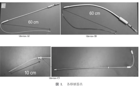
受胚豚の準備 受胚豚は,月齢 8ヶ月∼10ヶ月齢のランドレース 種およびデュロック種合計 35 頭を使用した。人工 授精後 20∼40 日目の受胚豚に PGF2α(クロプロス テノールとして 0.184 mg)を頸部筋肉内投与し,そ の 24 時間後に同量の PGF2αと eCG 1000 IU を頸部 筋肉内投与した。eCG投与 72 時間後に hCG 500 IU を頸部筋肉内投与した後,雄許容を確認した。hCG 投与翌日を 0 日として 5 日目の受胚豚に胚移植を実 施した。 統計処理 各試験区の子豚生産率(分娩子豚総数/総移植胚 数)は,χ2 乗検定を実施し,有意性を検討した。 実験設定 実験 1 市販されているブタ用深部注入用カテーテルセッ ト(器具 A:匠,富士平工業株式会社),NAKAZAWA ら(2008)が報告した移植器具をベースに内管の材 質を柔らかい素材に変更した試作深部注入移植器具 (器具 B)およびウシ用子宮深部胚移植器(モ 4 号, ミサワ医科工業株式会社)の先端に穿孔防止用プラ スチック製部品を装着した移植器具(器具 C)(図 1)を用いて頸管経由で非外科的移植を実施した時 の各移植器具の内管先端部分を開腹手術により確認 した。確認後は,胚移植を行い受胎成績の比較を 行った。まず,受胚豚を移植用の柵に入れた後,飼 料を与えながら無麻酔立位の状態で各移植器具を挿 入した。器具 A および器具 B は外管を外陰部に挿 入し,外管が子宮頸管に達したら外管を左に廻しな がら可能な限り挿入した。その後,外管に内管を挿 入し,内管のストローホルダーが外管の挿入口から 5 cm に達するまで挿入した。器具 C は,先端から 豚の外陰部に挿入し,先端が子宮頸管に達した後, 器具をゆっくり上下に動かしながら子宮頸管内に可 能な限り挿入した。その後,移植器具の内管を最後 まで押し出した。次に受胚豚に移植器具を挿入した 状態で,4% イソフルランの吸入で全身麻酔した後, 手術台に仰向けに乗せて開腹手術を行った。その 際,移植器具と外陰部周辺は,手術時の洗浄水等が かからないようにビニール袋とテーピングテープで 図 1. 各移植器具 Fig. 1. Embryo transfer devices.
Device A : Commercially available device, “Takumi,” for porcine embryo transfer. Inner and outer tubes are separated. The inner tube is made of polyethylene, and the outer tube is made of polyethylene/polypropylene.
Device B : The prototype device for porcine embryo transfer. The inner and outer tubes are sepa-rated. The inner tube is made of polyvinyl chloride, and the outer tube is made of synthetic rubber. Device C : Remolded commercial device, “Mo4,” Mo4 was developed for bovine embryo transfer to the deep parts of the uterus. Inner and outer tubes are integrated. The inner cylinder is made of polyvinyl chloride, and the outer cylinder is made of metal.
カバーした(図 2)。開腹後,子宮角を体外に露出し て各移植器具内管の先端部分を確認した。内管先端 が子宮体および子宮角内に位置している場合は,胚 を封入したストローのヒートシール部分を切り取 り,挿入している移植器具のストローホルダー部分 にヒートシールした方からストローを差し込んだ。 その後,ストローの綿線部分を切り,38℃に加温し た H-PZM-5 3 ml が入った 5 ml のシリンジと胚を 封入したストローの綿線側をストローアダプターを 用いて接続し,シリンジ内の H-PZM-5 を押し出し て胚を子宮内に注入した。また,対照区として,開 腹手術により子宮角先端部分に PP カテーテル(PP カテーテル猫用先穴式,富士平工業株式会社)を用 いてガラス化保存胚を移植した(大曲ら,2015)。各 移植器具および対照区で 5 頭ずつの移植を行い,各 受胚豚の移植器具の位置および受胎成績を比較し た。 実験 2 各移植器具の非外科的胚移植成績を比較するた め,無麻酔立位状態での実験 1と同様の方法で各移 植器具を挿入した後,立位状態で胚移植を実施し た。胚の注入方法は実験 1と同様に行い,対照区 は,実験 1の対照区の成績を用いて,各試験区の受 胎,分娩成績を比較した。
結
果
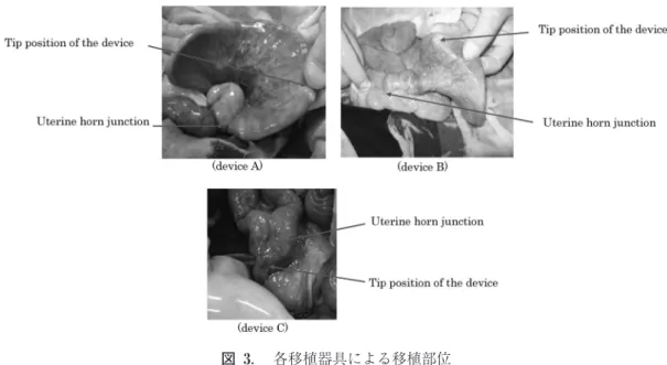
実験 1 各試験区の移植器具内管の先端部分の位置および 受胎・分娩成績は,表 1のとおりである。器具 A 区 の 1頭は,子宮体で移植器具内管が穿孔し,先端が 腹腔内に位置したため,胚移植は行わなかった。穿 孔が見られなかった器具 A 区および B 区の豚での 内管先端部分は,子宮体から子宮角分岐部より 10 cm の場所にあった。いずれも内管が途中で折れ曲 がり U ターンした状態で子宮体あるいは子宮角内 に収まっており,本来期待されていた子宮角中片部 に達していた豚はいなかった(図 3)。器具 C の内 管先端部分は,すべての豚で,子宮体内の子宮頸管 出口付近にあった。受胎成績は,器具 C 区で,すべ ての豚が不受胎であり,他の試験区に比べ有意に低 い成績となった(p<0.01)。器具 A 区,器具 B 区お よび対照区の間には有意な差は認められなかった。 実験 2 器具 A 区は,受胚豚 5 頭に合計 80 個(平均 16.0 個/1腹)の胚を移植したところ 3 頭が受胎した。そ のうち 2 頭が移植後 24,30 日目に流産した。残り の 1頭は,5 頭の子豚を分娩した。器具 B 区は,受 胚豚 5 頭に合計 78 個(平均 15.6 個/1腹)の胚を移 植したところ 3 頭が受胎分娩し,合計 16 頭の子豚 を生産した。器具 C 区は,受胚豚 5 頭に合計 78 個 (平均 15.6 個/1腹)の胚を移植したところ 3 頭が受 胎分娩し,合計 14 頭の子豚を生産した。対照区は, 合計 78 個(平均 15.6 個/1腹)の胚を移植したとこ ろ 4 頭が受胎分娩し,合計 20 頭の子豚を生産した。 器具 A 区の子豚生産率が 6.3% で器具 B 区(20.5 %),器具 C 区(1 7.9%)および対照区(25.6%)に対 して有意に低い成績となった(器具 A vs 対照区:p <0.01,器具 A vs 器具 B, C:p<0.05)。器具 B 区, 器具 C 区および対照区の間に有意な差は認められ なかった(表 2)。考
察
今まで,豚の非外科的胚移植に用いられる受胚豚 は経産豚がほとんどであったことから,今回我々は 未経産豚でも胚移植可能な非外科的移植器具の検討 を行なった。その結果,試作した移植器具を用いて ガラス化保存胚を無麻酔立位の状態で非外科的胚移 植を実施したところ 60% の受胎率が得られ,子宮 図 2. カテーテル挿入後の開腹手術 Fig. 2. Laparotomy after device insertion.The embryo transfer device inserted into the vulva was covered using vinyl and taped se-curely to prevent contamination.
体に胚を移植する非外科的移植器具においても外科 移植した対照区と子豚生産率で差がない成績を得る ことができた。このことから,子宮角分岐部付近お よび子宮体に非外科的胚移植を実施しても効率的に 子豚が得られることが明らかとなった。 最近のブタにおける非外科的胚移植の報告は,経 産豚に深部注入移植器具を用いて子豚生産に成功し た事例が主であった(SUZUKIら,2004 ; CUELLOら, 2005 ; BATHGATEら,2007 ; NAKAZAWAら,2008 ;
YOSHIOKAら,2012 ; GOMISら,2012 ; MARTINNEZら,
2014, 2016)。深部注入移植が検討された背景には, WALLENHORSTら(1999)が,外科的に子宮角先端部 分に胚を移植した際の受胎成績が子宮角分岐部およ び子宮体に移植した際の成績よりも高かったと報告 したことが起因している。一方,ブタの非外科的胚 移植手技は,ウシ胚移植時の様に直腸経由で移植器 具先端を確認することが困難な上,子宮角の長さが 1 m を超えるため,移植器具を確実に子宮角深部に 挿入するには熟練が必要な技術であった。そのた め,実験 1では,無麻酔立位状態の受胚豚に市販器 具および試作器具を挿入し,それら移植器具を挿入 した状態で開腹手術を行ない,実際の移植器具の挿 入状態を調べた。その結果,2 種類の深部注入器具 のいずれも子宮体から子宮角分岐部付近に内管の先 端が位置していた。今回,受胚豚に用いた未経産豚 は,既報で用いられた経産豚に比べ子宮頸管および 子宮角の径が小さいと思われる。このことが,器具 A および B の内管を子宮角深部へ挿入できなかっ た原因であると推察できる。しかしながら,その位 置でガラス化保存胚を移植して双方の移植器具から 子豚を得ることができた。一方,器具 C の内管先端 部分はすべて子宮頸管出口付近の子宮体に位置して おり,そこに胚移植をした結果,受胎例を得られな かった。このことから,開腹手術時の仰向け状態で 胚を移植した場合は,既報の報告とおり子宮体での 受胎率は低くなる結果となった。また,器具 A に 関しては子宮体での穿孔が見られたことから,子宮 深部注入移植器具の材質によっては,子宮角深部に 移植器具を挿入する際に子宮体および子宮角を傷つ ける可能性があることが示された。 図 3. 各移植器具による移植部位 Fig. 3. Each transplantation site facilitated b y the device.
Device A : 0ne recipient’s uterus was accidentally penetrated b y insertion of the device, but in other recipients, the tips of the devices were confirmed at the uterine body or near the uterine horn junction. In addition, it was observed that the inner tube was reversed at the uterine horn or uterine body of all the recipients.
Device B : The tips of the device were positioned near the uterine horn junction in all the re-cipients. In addition, it was observed that the inner tube was reversed at the uterus horn junction in all the recipients.
Device C : The tips of the devices were positioned near the cervical outlet of the uterine body in all the recipients.
実験 2 では,立位無麻酔状態の受胚豚に,各移植 器具を用いて非外科的胚移植を行った。その結果, 実験 1の仰向け状態では,受胎例が得られなかった 器具 C でも受胎,分娩し,子豚が生産された。この ことは,立位状態であれば,子宮体に胚を移植して も受胎・分娩が可能であることを示している。同様 な報告として YONEMURAら(1996)は,人工授精用 スパイラルカテーテルを用いて,非外科的胚移植を 行い,60% の受胎率を得たことを報告している。こ の手法では,スパイラルカテーテルの先端が子宮頸 管を通過した付近で胚を移植して,移植液量と重力 によって,胚を子宮角まで移行させて,子豚を得る ことに成功している。今回,我々が用いた器具 C は,この報告と同様な原理で子豚が得られたと考え られる。一方,子宮深部移植器具である器具 A で 移植した結果,2 頭が流産し,不受胎の 1頭は移植 後に陰部から膿を流出した。また,移植器具 B にお いても不受胎豚の 1頭は移植後に陰部から膿を流出 した。この原因として,試験 1の受胚豚は開腹手術 後に感染防止のため抗生物質を筋肉内に投与した が,試験 2 の受胚豚については抗生物質の投与を行 わなかったことも関連していると考えられる。さら に,器具 A および器具 B は,内管を子宮深部に挿入 するため,挿入ガイドとなる外管を子宮頸管内に挿 表 1. 開腹手術による非外科的移植部位観察後の胚移植成績
Table 1. The result of non-surgical embryo transfer after observing the transplantation site. Device of ET Tip of device
No. of transferred embryos Pregnant (pregnant rate) No. of delivered piglets Piglet production rate device A
Penetrated uterus not ET − − −
Uterine body 15 − 0 0.0%
Uterine horn (5 cm to uterine junction) 16 + 6 37.5% Uterine horn (5 cm to uterine junction) 14 + 2 1 4.3% Uterine horn (10 cm to uterine junction) 14 − 0 0.0%
Total 59 50% 8 1 3.6%a
device B
Uterine horn (5 cm to uterine junction) 14 − 0 0.0% Uterine horn (5 cm to uterine junction) 14 + 4 28.6% Uterine horn (10 cm to uterine junction) 14 − 0 0.0% Uterine horn (10 cm to uterine junction) 16 + 5 31.3% Uterine horn (10 cm to uterine junction) 15 + 4 26.7%
Total 73 60% 1 3 1 7.8%a device C Uterine body 16 − 0 0.0% Uterine body 16 − 0 0.0% Uterine body 16 − 0 0.0% Uterine body 15 − 0 0.0% Uterine body 15 − 0 0.0% Total 78 0% 0 0.0%b Control
Tip of uterine horn 16 + 4 25.0%
Tip of uterine horn 15 + 5 33.3%
Tip of uterine horn 16 + 5 31.3%
Tip of uterine horn 15 − 0 0.0%
Tip of uterine horn 16 + 6 37.5%
Total 78 80% 20 25.6%a
The piglet production rate (number of delivered piglets/number of transferred embryos). Significant difference between different signs (p<0.01).
入する必要があり,外管の径が試作器具 C より太く なっている。今回,我々が移植に用いた受胚豚は未 経産であったため,外管挿入時に子宮頸管等を傷つ けた可能性が考えられる。さらに,実験 1の結果か ら内管が子宮を傷つけた可能性も考えられる。一 方,試作器具 C は移植器具自体の直径が細いため, 未経産豚であっても子宮頸管を傷つけずに挿入でき たと考えられる。 以上の結果から,無麻酔下の立位状態であれば, ブタガラス化保存胚を子宮体へ非外科的に移植する ことで,外科移植と同等の効率で産子が得られるこ とが明らかとなった。さらに,今回試作した器具 C は,未経産豚への非外科的胚移植器具として有効で ある可能性が示唆された。
文
献
BATHGATE, R., K.M. MORTON, B.M. ERIKSSON, D. RATH,
B. SEIG, W.M. MAXWELL and G . EVANS: 2007,
Non-surgical deep intra-uterine transfer of in vitro produced porcine embryos derived from sex-sorted frozen-thawed boar sperm, Anim. Reprod. Sci., 99, 82-92.
表 2. 無麻酔立位での非外科的移植成績
Table 2. The results of non-surgical embryo transfer on the standing state of the recipient without anesthesia administration. Device of ET No. of transferred embryos Pregnant (Pregnant rate) Delivery (Delivery rate) No. of delivered piglets Piglet production rate Remarks device A
15 + − 0 0.0% Abortion (30 day after ET. 3 Fetus)
16 − − 0 0.0%
17 + + 5 29.4%
17 + − 0 0.0% Abortion (24 day after ET. 2 Fetus) 15 − − 0 0.0% Pus outflow from vuiva 8 day after ET
Total 80 60% 20% 5 6.3%a device B 15 + + 7 46.7% 16 + + 4 25.0% 16 + + 5 31.3% 15 − − 0 0.0%
16 − − 0 0.0% Pus outflow from vuiva 6 day after ET
Total 78 60% 60% 1 6 20.5%b device C 16 − − 0 0.0% 16 + + 8 50.0% 16 + + 4 25.0% 15 + + 2 1 3.3% 15 − − 0 0.0% Total 78 60% 60% 1 4 1 7.9%b Control 16 + + 4 25.0% 15 + + 5 33.3% 16 + + 5 31.3% 15 − − 0 0.0% 16 + + 6 37.5% Total 78 80% 80% 20 25.6%bc
The piglet production rate (number of delivered piglets/number of transferred embryos). Significant difference between signs (a vs b, p<0.05 ; a vs c, p<0.01).
GOMIS, J., C. CUELLO, J. SANCHEZ-OSORIO, M.A. GIL, I.
PARRILLA, M.A. ANGEL, C. MASIDE, D. OLMO, J.M.
VAZQUEZ, J. ROCAand E.A. MARTINEZ : 2012,
Non-surgical deep intrauterine transfer of superfine open pulled straw (SOPS) -vitrified porcine em-bryos: Evaluation of critical steps of the proce-dure, Theriogenology, 78, 1339-1349.
CUELLO, C., F. BERTHELOT, F. MARTINAT-BOTTÉ, E.
VENTURI, P. GUILLOUET, J.M. VÁZQUEZ, J. ROCAand E.
A. MARTÍNEZ: 2005, Piglets born after
non-surgi-cal deep intrauterine transfer of Vitrified blasto-cysts in gilts, Anim. Reprod. Sci., 85, 275-286. DAY, B. N : 1968, Pregnancy following non-surgical
egg transfer in pigs, Vet. Rec., 82, 71 2.
DUCRO-STEVERINK, D.W., C.G. PETERS, C.C. MATERS, W.
HAZELEGER and J.W. MERKS: 2004, Reproduction
results and offspring performance after non-surgical embryo transfer in pigs, Theriogenology,
62, 522-531.
GALVIN, JM., DB. KILLIANand A.N.V. STEWART : 1994,
A procedure for successful non-surgical embryo transfer in swine, Theriogenology, 41, 1279-1289. LI, J., A. RIEKE and B.N. DAY: 1996, Porcine
non-surgical embryo transfer, J. Anim. Sci., 74, 2263-2268.
MARTINEZ, E.A., M.A. ANGEL, C. CUELLO, J. SANCHEZ
-OSORIO, J. GOMIS, I. PARRILLA, J. VILA, I. COLINA, M.
DIAZ, J. REIXACH, J.L. VAZQUEZ, J.M. VAZQUEZ, J.
ROCAand M.A. GIL: 2014, Successful non-surgical
deep uterine transfer of porcine morulae after 24 hours culture in a chemically defined medium, PLOS One, 9, e104696.
MARTINEZ, E.A., A. NOHALEZ, C.A. MARTINEZ, I.
PARRILLA, J. VILA, I. COLINA, M. DIAZ, J. REIXACH, J.L.
VAZQUEZ, J. ROCA, C. CUELLO and M.A. GIL : 2016,
The recipients’ parity does not influence their reproductive performance following non-surgical deep uterine porcine embryo transfer, Reprod. Domest. Anim., 51, 1 23-129.
MISUMI, K., Y. HIRAYAMA, S. EGAWA, S. YAMASHITA, H.
HOSHIand K. IMAI: 2013, Successful production of
piglets derived from expanded blastocysts vit-rified using a micro volume air cooling method without direct exposure to liquid nitrogen, J. Reprod. Dev., 59, 520-524.
NAKAZAWA, Y., H. MISAWA, Y. FUJINO, S. TAJIMA, K.
MISUMI, J. UEDA, Y. NAKAMURA, T. SHIBATA, Y.
HIRAYAMAand K. KIKUCHI: 2008, Effect of volume
of non-surgical embryo transfer medium on ability of porcine embryos to survive to term, J. Reprod. Dev., 54, 30-34. 大曲秀明・三角浩司・宮下美保・永渕成樹・御澤弘 靖・山下祥子・星 宏良・平山祐理・吉岡耕治: 2015, 種豚から個体ごとに採取した胚盤胞および 拡張胚盤胞期ガラス化保存胚の胚移植による子豚 生産効率, 日豚会誌,52,1-7.
QUINN, P., C. BARROUS and G . WHITTINGHAM: 1982,
Preservation of hamster oocytes to assay the fertilizing capacity of human spermatozoa, J. Reprod. Fertil., 66, 1 61 -168.
SUZUKI, C., S. IWAMURAand K. YOSHIOKA: 2004, Birth
of piglets through the non-surgical transfer of blastocysts produced in vitro, J. Reprod. Dev., 50, 487-491.
YONEMURA, I., Y. FUJINO, S. IRIE and Y. MIURA: 1996,
Transcervical transfer of porcine embryos under practical conditions, J. Reprod. Dev., 42, 89-94. YOSHIOKA, K., C. SUZUKI and A ONISHI: 2008, Defined
system for in vitro production of porcine embryos using a single basic medium, J. Reprod. Dev., 54, 2008-2013.
YOSHIOKA, K, M. NOGUCHI and C. SUZUKI: 2012,
Pro-duction of piglets from in vitro-produced embryos following non-surgical transfer, Anim. Reprod. Sci.,
131, 23-29.
WALLENHORST, S. and W. HOLTZ: 1999, Transfer of
pig embryos to different uterine sites, J. Anim. Sci.,
Improved Non-Surgical Embryo Transfer Method
using Porcine Vitrified Blastocysts
Koji M
ISUMI1, Sachiko E
GAWA2, Hiroyasu M
ISAWA3and Yuri H
IRAYAMA21Nihon University College of Bioresource Sciences, Fujisawa Kanagawa 252-0880, Japan 2National livestock Breeding Center, Nishigo, Nishishirakawa, Fukushima 961-8511, Japan
3Misawa Medical Industry Co., Kasama, Ibaragi, 309-1717, Japan
In this study, we investigated the non-surgical transfer of porcine vitrified embryos for practical application in commercial farms. Blastocysts and expanded blastocysts were collected from Duroc and were vitrified using the micro volume air cooling (MVAC) method. Here, 35 Landrace and Duroc Gilts (8-10 months-old) were used as recipients. We investigated three types of devices, including commercially available deep implant device (Device A group), deep implant device prototype (Device B group), and modified bovine embryo transfer device (Device C group). In Test 1, the position of each device’s tip was confirmed b y laparotomy under general anaesthesia following insertion into the recipient to confirm the transplantation site, and then the vitrified embryos were transferred. Each test group had 5 recipients. The number of transferred embryos per recipient was 14-16, corresponding as the average number of ovulated embryos per litter. In Test 2, each device was inserted into a non-anesthetized standing re-cipient, and then the vitrified embryos were transferred. Each test group used had 5 recipients. The number of transferred embryos per recipient was 15-17 embryos. In Test 1 of the A group, one recipient’s uterus was accidentally penetrated b y insertion of the device, and as a result, embryo transfer was not performed. In other recipients, the device tip positions were confirmed at the uterine body or near the uterine horn junction. Two out of four recipients were pregnant and successfully delivered, and a total of eight piglets were born in the A group. In the Device B group, the tips of the devices were positioned near the uterine horn junction in all the recipients. Three out of five recipients were pregnant and successfully delivered, producing a total of 13 piglets. In the Device C group, the tips of the devices were positioned near the cervical outlet of the uterine body in all the recipients. None of the five recipients were pregnant. The result of Test 2 in the Device A group indicated that three out of five recipients were pregnant, but two miscarried. The remaining recipient delivered five piglets. In the Device B group, three out of five recipients became pregnant and delivered a total of 16 piglets. In the Device C group, three out of five recipients became pregnant and delivered a total of 14 piglets. These results show that non-surgical transfer of porcine vitrified embryos into the uterine body of a standing recipient without anaesthesia administration had the same efficiency as surgical embryo transfer in terms of piglet production rate.
Jpn. J. Swine Science, 57, 4 : 129-137