INTRODUCTION
MRI devices and associated technology are im-proving and maturing rapidly. In addition, one of the recent hot topics is the appearance of the 3 Tesla (3T) instruments.
Though its licensing by the Japanese Government was slower than by Western countries, it is consid-ered that its impact on clinical diagnosis will be huge when the high S/N ratio and effect of magnetic susceptibility are used effectively.
In this review, the utility of the“Propeller Technique” which is comparatively resistant to body movement
as one of the areas of progress in recent MRI technology is introduced, and the characteristics of 3T MRI are included concerning the develop-ment of applying metabolic changes and functional evaluations in the clinical diagnosis of the central nervous system.
1. Technology born from clinical need; the clinical utility of the Propeller Technique
The shortening of measurement time is a goal of the recent MRI technology, and the efficient use of not only parallel imaging but also a multi-channel coil will shorten the measuring time drastically.
However, even if the measurement time is short-ened using the new measurement technique, any body movement causes a larger artifact in the rapid MR scan due to there being less compensa-tion for body movement than by classical MR im-aging. Such an artifact can be especially
problem-REVIEW
New trend of MRI diagnosis based on the function and
metabolism in the central nervous system
Masafumi Harada
Department of Radiologic Technology, School of Health Sciences, The University of Tokushima, Japan
Abstract : The movement of a subject is a major problem in MRI experiments and diagnosis. At first, this review introduces a new technology named the “Propeller Technique” which can improve the motion artifact by changing the data sampling method in the K trajectory. Our experience of a case who underwent measurement by Propeller technique is reported and the effect of this technique is explained.
One of the recent hot topics is the appearance of the clinical 3T MR instrument, with its characteristic differences from that at 1.5T. The advantage of 3T is that it facilitates the evaluation of functional and metabolic information using MR spectroscopy (MRS) and functional MRI. The application of proton MRS in clinical cases is shown and the standard method to use proton MRS in a clinical setting is demonstrated. Furthermore, the new techniques, which can measure important metabolites in small amount such as neurotransmitters, was de-veloped using a high signal to noise ratio and frequency resolution, which are advantages of 3T. J. Med. Invest. 53 : 199-203, August, 2006
Keywords : MRI, 3 Tesla, metabolism, function, propeller
Received for publication June 30, 2006 ; accepted July 18, 2006. Address correspondence and reprint requests to Masafumi Harada, Department of Radiologic Technology, School of Health Sciences, The University of Tokushima, Kuramoto-cho, Tokushima 770-8509, Japan and Fax : +81-88-633-9022
The Journal of Medical Investigation Vol. 53 2006
atic for the new technique of parallel imaging. This decline in image quality may lead to difficulty in diagnosis and, furthermore, artifacts may lead to misdiagnose. Thus, any reduction of measure-ment time will be meaningless.
So far, for unstable subjects, priority has been given to diagnostic imaging techniques such as CT, sedating patients when MRI was necessary. Nowadays, the“Propeller Technique” is one method which can be effectively applied for unstable patients. The basic theory behind this technique involves the gathering of much raw data on the center in the K space in which the contrast of the image is decided, and the Propeller Technique can minimizes the influence of any position a de-viations due to body movement via the integration of adequate raw data.
When measurements cannot be completed due to the body movement of a subject, we are now applying this new technique in our facilities before resorting to sedation.
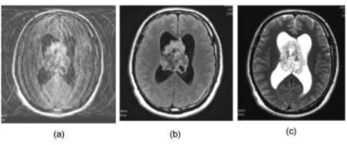
A case in which the Propeller Technique was employed in our facilities is shown in Figure 1, and the effectiveness of this new technique could be observed. This case had a large mass in the lateral ventricle and was complicated by obstructive hy-drocephalus due to the circulatory failure of the cerebrospinal fluid caused by obsession of the Monro foramen. CT demonstrated calcification in the mass.
When a decline in the level of consciousness was recognized, MRI was conducted, but there was a large artifact on conventional T2-WI due to head movement and the details weren’t clear. Then, images of FLAIR and a T2-WI by the Propeller Technique were added and adequate images which could be used for diagnosis were acquired. The state of cystic degeneration, which was observed as a
low intensity signal with FLAIR, could be observed, and still a high intensity signal was thought to be useful for the qualitative diagnosis with T2-WI of the mass. As for the MRI diagnosis of the brain tumor, the examination time including FLAIR imaging and contrast enhanced T1-WI is usually long and it is considered lucky when the examina-tion of an unstable patient can be conducted without sedation. It is also considered that this may greatly benefit radiologists by reducing the number of staff needed in the field of diagnostic radiology. 2. Application of information on cerebral function and metabolism in clinical diagnosis
A method to obtain information on cerebral func-tion and metabolic change was proposed even with an MRI device of 1.5T. In a high magnetic field, it is better to acquire information on function and meta-bolic change due through the increased S/N ratio, improvement in frequency resolution, and so on. Furthermore the examination time can be short-ened by increasing the S/N ratio and an additional measurement for functional of metabolic examina-tion can be afforded. This aspect brought about by high magnetic MRI has significant importance, es-pecially in the clinical setting where time is dis-tinctly limited. The purposes to add information on function and metabolism in clinical diagnosis can be divided roughly into the following :
1) The time when the lesion or abnormality in the brain is known :
The choice of therapy or the therapeutic plan will be improved by understanding the back-ground and mechanism of the disease using functional or metabolic information derived from MRI. In addition, it is considered meaningful that such information will facilitate a prognosis predic-tion including the severity of the disease and the curative effects.
2) The time when the lesion or abnormality in the brain isn’t clear :
Identification of the area or range of disease may be detected by functional or metabolic infor-mation and it will be useful to perform a differen-tial diagnosis.
Although these two purposes cannot be distin-guished strictly in the actual clinical setting, this classification is thought to provide a standard to add functional and metabolic information to ana-tomical information to facilitate easy understand-Fig. 1 : The utility of the “Propeller Technique” in an unstable
subject with central neurocytoma (a) conventional FLAIR image
(b) FLAIR image acquired with the Propeller Technique (c) T2-WI acquired with the Propeller Technique
M. Harada New trend of MRI diagnosis in the CNS
ing. Here, typical examples are introduced to help understand how to use the functional or metabolic information.
3) A case of the evaluation of functional and metabolic changes in MELAS :
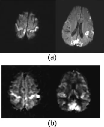
A T2-WI and T1-WI of a 4-year-old child are shown in Figure 2. An abnormal signal can be observed in the occipital lobe containing the visual cortex and the surroundings of the central fissure of the primary motor area. This lesion showed a rather high intensity on diffusion-weighted imaging and hyperperfusion on FAIR imaging, which is one of the spin labeling perfusion MRI techniques (Figure 3).
Proton MRS was measured by setting a region of interest (ROI) in the lesion showing abnormal intensity, revealing the decline of n-acetyl aspar-tate (NAA) and increase in lacaspar-tate, and increase in the signal of macromolecules (MM) in the spec-trum(Figure 4). It was considered that such signal changes in each metabolite suggested neuronal cell damage, an energy metabolism deficiency, and cellular destruction and necrosis.
From these results, it is considered that patho-logical changes in MELAS are based on energy metabolism deficiency in the mitochondria, which leads to a decreased oxygen utilization, and re-gional cerebral blood flow is increased by the va-sogenic reactivity in order to compensate for energy failure. However, because energy failure does not occur via a shortage of blood or oxygen, damage and necrosis of neuronal cells still continues.
Therefore, it can be speculated that the local peripheral circulation and vasogenic reactivity as a function of the glial cells still remains at this stage even though the neuronal cell damage has progressed. 4) A case of multiple sclerosis showing a discrep-ancy between the symptom and anatomical abnor-mality on MRI :
The case was a 27-year-old female who had a past history of multiple sclerosis diagnosis and therapy, and a new symptom of incomplete paraly-sis of the right extremities, as shown in Figure 5. The relapse of multiple sclerosis was suspected and a brain examination by MRI was conducted. How-ever, no remarkable new lesion was found on T2-WI and FLAIR images and a symptomatic cause wasn’t clear with conventional MRI. The multivoxel measurement of proton MR spectroscopy, named Chemical Shift Imaging (CSI), in the brain was
Fig. 3 : Diffusion-weighted images (a) and perfusion-weighted images (b) of a patient with MELAS
The high intensity on DWI indicates a decrease in water diffu-sivity reflecting cytotoxic edema. The high intensity area on perfusion images show high flow rates in the lesion.
Fig. 4 : The ROI setting for acquiring proton MRS (a) and the obtained spectrum from a patient with MELAS
The increase of lactate and decrease of NAA were revealed in this spectrum
Fig. 2 : Anatomical images of a patient with MELAS (a)T2-WI (b)T1-WI
added following conventional measurement by MRI. The result of CSI in this case is shown in Figure 6, and a decrease in NAA and increase in choline were found in the left hemisphere of the cerebrum compared to the maintained NAA peak in the right hemisphere. By constructing a metabolite ratio map by dividing NAA by Cho, a clear decline in the NAA/Cho ratio was noted in the left parietal lobe, as shown in Figure 6.
This result suggested that there was an inflam-matory lesion in the normal appearing white mat-ter which could not be recognized as an abnormal lesion by any conventional imaging modality. It is reported that the lesion is usually found as the ex-istence of inflammatory or degeneration by histo-logical examination, causing symptom or relapse phenomenon(1).
Even in the lesion which is difficult to identify as an anatomical abnormality, proton MRS showed such changes sensitively and with a high de-tectability of the pathological abnormality. As for the metabolite map including NAA, the under-standing of the change is easier when presented visually, and it is considered that the metabolite
map by the CSI is useful for the detection of nor-mal appearing white matter.
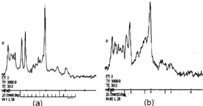
5) For the routine use of the metabolite map by 3 : The spectra of proton MRS of 1.5T and 3T in the same subject, obtained using the same measure-ment parameters, are shown in Figure 7. The spec-trum of 3T showed not only a higher S/N ratio but also a better separation of each signal compared to 1.5T. The characteristics of 3T are a high resolution and high contrast images brought about by the increased S/N ratio, leading to a shortening of the measurement time for routine anatomical exami-nations. It is also expected that this shortening of the measurement time will bring about an environ-ment in which the function or embolic information are easily added in the daily clinical use.
3. New metabolic information measured by the high magnetic field
The advantage of 3T is through its ability to detect a metabolite at a low concentration in tissue which is usually difficult to observe by 1.5T. Here, the ex-ample is introduced though the measurement of γ-aminobutyric acid (GABA), known as an amino acid related to neurotransmitters.
Glutamate is one of the constituent amino acids and it isn’t formed only as a neurotransmitter though there are Glutamate and GABA as an amino acid which relates to the detectable neurotransmission by proton MRS. GABA is not a constituent amino acid and much exists in the neuron. It can be con-sidered that GABA is more related to neurotrans-mitters than Glutamate.
However, Glutamate exists about 10mM in the brain, and only about 1 mM existed in GABA, and the signal of GABA piled up with the signal of the metabolite which concentration is high, and more-over, detailed evaluation was difficult with conven-tional proton MRS. Therefore, some ways of editing the signal of GABA selectively were proposed and we use the J difference method called MEGA-PRESS which takes the GABA signal at 3.0ppm for 3T MRI (2). The GABA signal can be recognized compara-tively clearly in Figure 8 from the region of 27ml for about 10minutes. A change in the spectrum of GABA before and after drug administration in a healthy volunteer is given in Figure 9. On comparison with before medication, the GABAsignal rose after two weeks medication with Li which cures depres-sive psychosis.
As for the intracerebral tubercle with epilepsy, it Fig. 5 : The anatomical images of a patient with multiple sclerosis.
No remarkable abnormality can be found in these images.(a)T 2-WI (b)High resolution and high contrast using the STIR technique
Fig. 6 : Chemical shift imaging of a patient with multiple sclerosis. (a)typical spectra obtained from the right hemisphere of the cerebrum ; (b)typical spectra obtained form the left hemi-sphere of the cerebrum ; (c)NAA/Cho map showing a de-crease in the NAA/Cho ratio in the left hemisphere of the cere-brum.
M. Harada New trend of MRI diagnosis in the CNS
is recognized that GABA is present at high con-centration in patients, with tuberous sclerosis in comparison with the healthy subjects suggesting a relation with the epileptic insult(3).
Because the relation with the neurotransmitter is so intimate, GABA is expected to become a new evaluation index of cerebral function and psychiat-ric disorder.
4. Establishment of a new diagnostic system for brain imaging
The number of diseases recognized as causing functional changes in the brain have recently been increasing, including obsessive-compulsive disor-der and autism, and anatomical observation some-times shows limitations in functional evaluation.
The use of high magnetic field MRI clinically is thought to provide a good opportunity to evaluate metabolic or functional changes for practical use.
In the near future, if MRI facilitate a more de-tailed evaluation of brain function and metabolism, MRI may substitute positron emission tomogra-phy and the magnetoencephalogram or be used to complement these functional measurements.
REFERENCES
1. Chard DT, Griffin CM, Mcleen MA, Kapeller P, Kapoor R, Thompson AJ, Miller DH : Brain metabolite changes in cortical grey and normal appearing white matter in clinically early re-lapsing remitting multiple sclerosis. Brain 125: 2342-2352, 2002
2. Mescher M, Merlde H, Kirsch J, Garwood M, Gruetter R : Simultaneous in vivo spectral editing and water suppression. NMR Biomed 11 : 266 -272, 1998
3. Asaly J : Proton magnetic resonance spectros-copy of brain biopsies from patients with intrac-table epilepsy. Epilepsy Res 35 : 211-217, 1999 Fig. 7 : The difference in spectra between 3T (a) and 1.5T (b). The
signal/noise ratio and resolution of the peaks are markedly dif-ferent.
Fig. 8 : Typical spectrum measured by MEGA-PRESS showing the clear observation of GABA
Fig. 9 : GABA signal changes by drug administration (a)before administration ; (b)after administration An increase in the GABA peak is clearly observed by drug administration.