suppression of AMPK/PGC1 α signaling
著者
富山 恭行
著者(英)
Tomiyama Yasuyuki
学位名
博士(医学)
学位授与機関
川崎医科大学
学位授与年度
平成25年度
学位授与年月日
2014-03-13
学位授与番号
35303甲第603号
URL
http://doi.org/10.15111/00000010
For Review Only
Hepatic oxidative stress in ovariectomized transgenic mice expressing the hepatitis C virus polyprotein is augmented
through inactivation of AMPK/PGC1α signaling
Journal: Hepatology Research Manuscript ID: HEPRES-13-0666 Manuscript Type: Original Article Date Submitted by the Author: 09-Sep-2013
Complete List of Authors: TOMIYAMA, YASUYUKI; Kawasaki Medical School, Hepatology and Pancreatology
Nishina, Sohji; Kawasaki Medical School, Hepatology and Pancreatology Hara, Yuichi; Kawasaki Medical School, Hepatology and Pancreatology Kawase, Tomoya; Kawasaki Medical School, Hepatology and Pancreatology Hino, Keisuke; Kawasaki Medical School, Hepatology and Pancreatology Category: 7. Viral hepatitis
For Review Only
Hepatic oxidative stress in ovariectomized transgenic mice expressing the hepatitis
C virus polyprotein is augmented through inactivation of AMPK/PGC1αααα signaling
Short running title: Oxidative stress in OVX HCV transgenic mice
Yasuyuki Tomiyama, Sohji Nishina, Yuichi Hara, Tomoya Kawase, Keisuke Hino
Department of Hepatology and Pancreatology, Kawasaki Medical School, Kurashiki, Japan
Correspondence to:
Keisuke Hino, M.D., Ph.D.
Department of Hepatology and Pancreatology, Kawasaki Medical School 577 Matsushima, Kurashiki, Okayama
701-0192 Japan Tel: +81-86-462-1111 Fax: +81-86-464-1196
For Review Only
ABSTRACT
Aim: Oxidative stress plays an important role in hepatocarcinogenesis of hepatitis C virus (HCV)-related chronic liver diseases. Despite the evidence of an increased proportion of females among elderly patients with HCV-related hepatocellular
carcinoma (HCC), it remains unknown whether HCV augments hepatic oxidative stress in postmenopausal women. The aim of this study was to determine whether oxidative stress was augmented in ovariectomized transgenic mice expressing the HCV
polyprotein and to investigate its underlying mechanisms.
Methods: Ovariectomized (OVX) and sham-operated female transgenic mice
expressing the HCV polyprotein and nontransgenic littermates were assessed for the production of reactive oxygen species (ROS), expression of inflammatory cytokines, and antioxidant potential in the liver.
Results: Compared with OVX nontransgenic mice, OVX transgenic mice showed
marked hepatic steatosis and ROS production without increased induction of
inflammatory cytokines, but there was no increase in ROS-detoxifying enzymes such as superoxide dismutase 2 and glutathione peroxidase 1. In accordance with these results, OVX transgenic mice showed less activation of peroxisome proliferator-activated receptor γ coactivator 1α (PGC-1α), which is required for the induction of
ROS-detoxifying enzymes, and no activation of adenosine monophosphate–activated protein kinase α (AMPKα), which regulates the activity of PGC-1α.
Conclusions: Our study demonstrated that hepatic oxidative stress was augmented in
For Review Only
potential through inhibition of AMPK/PGC-1α signaling. These results may account in part for the mechanisms by which HCV-infected women are at high risk for HCC development when some period has passed after menopause.
Key words: antioxidant potential, glutathione peroxidase, reactive oxygen species, superoxide dismutase.
For Review Only
INTRODUCTION
Persistent hepatitis C virus (HCV) infection is a major risk factor for the development of HCC in Japan. Approximately 70% of Japanese hepatocellular carcinoma (HCC) patients are currently diagnosed with HCV-associated cirrhosis or chronic hepatitis C1. Nevertheless, the mechanisms underlying HCV-associated hepatocarcinogenesis are incompletely understood. Notably, there is gender disparity in HCC development that is, male gender has been demonstrated to be an independent risk factor associated with HCC development2-4. It is proposed that estrogen-mediated inhibition of IL-6 production by Kupffer cells reduces the HCC risk in females5. In addition, the proportion of females among elderly patients with HCV-related HCC has recently increased in Japan6. These results suggest that menopause may be a risk factor associated with HCC development in female patients with HCV infection.
Numerous studies have shown that oxidative stress is present in chronic hepatitis C to a greater degree than in other inflammatory disease7, 8, and is related to
hepatocarcinogenesis in HCV-associated chronic liver diseases9, 10. We have previously demonstrated that transgenic mice expressing the HCV polyprotein develop liver tumors including HCC, in connection with oxidative stress induced by HCV and iron overload11. Interestingly, such hepatocarcinogenesis was observed only in male transgenic mice, suggesting that females are resistant to oxidative stress in these transgenic mice. On the other hand, it is reported that ovariectomy increases NADPH oxidase activity12 and decreases mitochondrial reduced glutathione levels in rats13. However, it remains unknown how HCV affects ovariectomy-induced oxidative stress.
For Review Only
Investigation of this issue may provide a clue for understanding why the incidence of HCC increases in elderly postmenopausal women with HCV infection. The aim of this study was to determine whether HCV proteins amplify oxidative stress induced by ovariectomy and to investigate the mechanisms underlying this.
For Review Only
MATERIALS AND METHODS Animals
The transgene pAlbSVPA-HCV, containing the full-length polyprotein-coding region under the control of the murine albumin promoter/enhancer, has been described in detail14, 15. Of the 4 transgenic lineages with evidence of RNA transcription of the full-length HCV-N open reading frame (FL-N), the FL-N/35 lineage proved capable of breeding in large numbers. There is no inflammation in the transgenic liver15.
Experimental design
Female FL-N/35 transgenic mice and their normal female C57BL/6 littermates were anesthetized for surgery and underwent either a bilateral ovariectomy or sham operation at the age of 4-6 weeks. We studied ovariectomized (OVX) transgenic mice (n=5), sham-operated transgenic mice (n=5), OVX nontransgenic mice (n=5), and
sham-operated nontransgenic mice (n=5). These mice were fed a normal rodent diet, bred, maintained, and killed by intraperitoneal injection of 10% pentobarbital sodium preceded by 20-hour fasting at the age of 24 weeks. All experimental protocols and animal maintenance procedures used in this study were approved by the Ethics Review Committee for Animal Experimentation of Kawasaki Medical School.
Histological procedures
A portion of liver tissue was immediately snap-frozen in liquid nitrogen for
For Review Only
fixed in 4% paraformaldehyde in PBS and embedded in paraffin for histological analyses. Liver sections were stained with hematoxylin-eosin.
Serum leptin concentration
The serum leptin level was measured using a Rat Leptin Elisa kit (Morinaga Institute of Biological Science Inc. Yokohama, Japan) according to the manufacturer’s instructions.
Hepatic triglyceride content
Lipids were extracted from the homogenized liver tissue by the method of Bligh and Dyer16. The triglyceride level was measured with a TGE-test Wako kit (Wako Pure Chemicals, Tokyo, Japan), according to the manufacturer’s instructions. Protein concentrations in liver were determined by the method of Lowry et al.17, using a DC protein assay kit (Bio-Rad Laboratories, Hercules, California).
In situ detection of reactive oxygen species (ROS)
In situ ROS production in the liver was assessed by staining with dihydroethidium, as
described previously18. In the presence of ROS, dihydroethidium (Invitrogen Corp., Carlsbad, CA)is oxidized to ethidium bromide and stains nuclei bright redby
intercalating with the DNA19. Fluorescence intensity was quantified using NIH image analysis software for 3 randomly selected areas of digital images for each mouse.
For Review Only
potential (BAP)
The levels of dROMs and BAP were measured using a Free Radical Elective Evaluator (Wismerll Co., Ltd., Tokyo, Japan), as described previously20. Measurement of dROMs is based on the ability of the transition metal ions to catalyze the formation of alkoxy and peroxy radicals from hydroperoxides (ROOH) present in serum. The results are expressed in conventional units as U.CARR (Carrtelli units), where 1U.CARR corresponds to 0.8 mg/L H2O2. Measurement of BAP is based on the ability of
antioxidants to reduce ferric (F3+) ions to ferrous (Fe2+) ions.
RNA isolation and real-time reverse transcription polymerase chain reaction
(RT-PCR)
Total RNA was isolated using an RNeasy mini kit (QIAGEN, Hilden, Germany) and reverse-transcribed into cDNA by using a Superscript III reverse transcription kit (Invitrogen Corp.). The PCR reactions were run in the ABI Prism 7700 sequence detection system (Applied Biosystems, Foster, CA, USA). The levels of mRNA were determined using cataloged primers (Applied Biosystems) for mice (TNF-α;
Mm00443258_m1, IL-1β; Mm00434228_m1, IL-6; Mm00446190_m1, superoxide dismutase 2 [SOD2]; Mm01313000 _m1, glutathione peroxidase 1 [GPx1];
Mm00656767_g1, and sirtuin 3 [SIRT3]; Mm00452131_m1). Expression of these genes was normalized to expression of GAPDH mRNA (GAPDH; Mm99999915_g1).
For Review Only
Mitochondrial extraction from liver tissue was performed using a Qproteome Mitochondrial Isolation kit (QIAGEN) according to the manufacturer’s instructions. The nuclear fraction from liver tissue was prepared using a Nuclear Extraction kit (Panomics, Fremont, CA, USA) according to the manufacturer’s instructions.
Immunoblotting
Liver lysates and the mitochondrial and nuclear fractions from liver were separated by sodium dodecyl sulfate-polyacrylamide gel electrophoresis. The proteins were
transferred to polyvinylidene difluoride membranes (Millipore, Bredford, MA, USA), blocked overnight at 4˚C with 5% skim milk and 0.1% Tween 20 in Tris-buffered saline, and subsequently incubated for 1 hour at room temperature with an anti-mouse SOD2 antibody (Santa Cruz Biotechnology Inc., Santa Cruz, CA, USA), anti-rabbit GPx1 antibody (Abcam, Cambridge, MA, USA), anti-rabbit SIRT3 antibody (Abcam), anti-rabbit peroxisome proliferator-activated receptor γ coactivator 1α
(PGC-1α) antibody (Abcam), anti-rabbit adenosine monophosphate–activated protein kinase α (AMPKα) antibody (Cell Signaling Technology Inc., Boston, MA, USA), anti-rabbit phospho-AMPKα (Thr172) antibody (Cell Signaling Technology Inc.), anti-mouse mitochondrial heat shock protein 70 (HSP70) antibody (Thermo Scientific, , Rockford, IL, USA), anti-rabbit β-actin antibody (Cell Signaling Technology, Inc.), or anti-rabbit lamin B1 antibody (Abcam).
For Review Only
Quantitative values are expressed as mean ± standard deviation. Two groups among multiple groups were compared by the rank-based Kruskal-Wallis analysis of variance test followed by Scheffe’s test. The statistical significance of correlation was determined by the use of simple regression analysis. A P value of < 0.05 was considered to be significant.
For Review Only
RESULTS
Ovariectomy enhanced hepatic steatosis in FL-N/35 transgenic mice
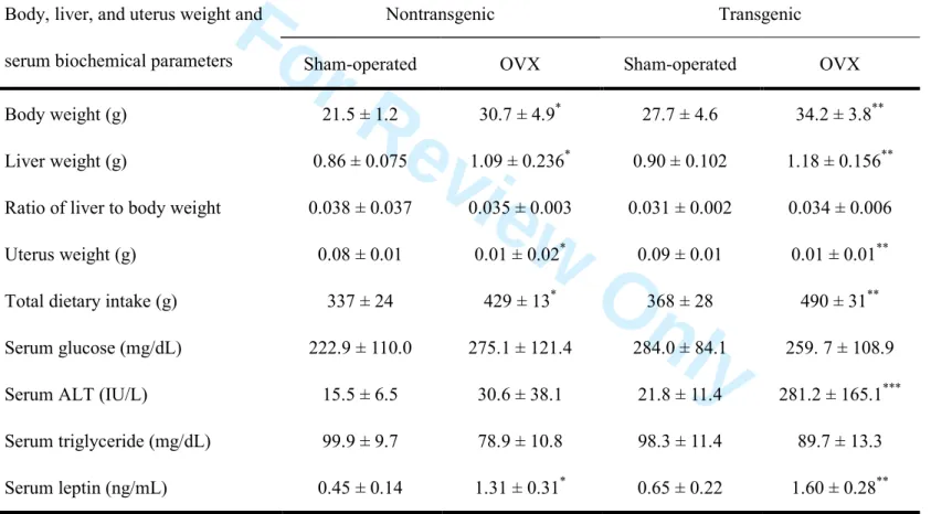
As confirmation of successful ovariectomy-induced suppression of endogenous estrogen production, the uterine weight of OVX mice was significantly decreased compared with that of sham-operated mice (Table 1). Dietary intake, body weight, liver weight and serum leptin levels were significantly greater in OVX mice than in
sham-operated mice regardless of whether they were transgenic or nontransgenic (Table 1). Interestingly, the serum alanine aminotransferase (ALT) level was significantly higher in OVX transgenic mice than in mice in the other three groups, but the levels were comparable in OVX nontransgenic and sham-operated nontransgenic mice (Table 1). To determine why OVX transgenic mice have a higher ALT level, we investigated the liver histology of the mice in the four groups (OVX transgenic, sham-operated transgenic, OVX nontransgenic and sham-operated nontransgenic mice). In contrast to the mild-to-moderate degree of hepatic steatosis noted in OVX nontransgenic mice and sham-operated transgenic mice, OVX transgenic mice developed severe hepatic steatosis (Fig. 1A) without infiltration of inflammatory mononuclear cells. Hepatic triglyceride content was measured to quantify the degree of steatosis. The triglyceride content was significantly greater in OVX transgenic mice than in mice in the other three groups (Fig. 1B), which was consistent with the results for hepatic steatosis. Thus, the increase in the serum ALT level in the OVX transgenic mice was thought to reflect the hepatic steatosis.
For Review Only
Ovariectomy increased ROS and IL-6 production in the liver
Only OVX transgenic mice showed marked hepatic steatosis, regardless of the
comparable diet intake and the ratio of liver to body weight of OVX nontransgenic mice (Table 1). We have previously demonstrated that iron-overloaded male FL-N/35
transgenic mice expressing the HCV polyprotein develop severe hepatic steatosis through increased ROS production11. Therefore, we examined whether ROS production was relevant to the marked hepatic steatosis observed in the OVX transgenic mice. Ovariectomy significantly increased ROS (superoxide) production in both transgenic mice and nontransgenic mice, but the level of ROS production was greater in the OVX transgenic mice than in the OVX nontransgenic mice (Fig. 2A and 2B). We next
measured inflammatory cytokine levels in the liver. Ovariectomy significantly increased hepatic expression of IL-6 mRNA to the same degree in both transgenic mice and nontransgenic mice (Fig. 3). This ovariectomy-induced increase in hepatic IL-6 mRNA was consistent with the results of a previous report that OVX mice produced more hepatic IL-6 than non-OVX mice after chemically induced liver injury5. There also was a trend for increased in TFN-α and IL1β mRNA expression after ovariectomy in both the transgenic mice and nontransgenic mice, but their increases did not reach statistical significance, probably because of the large deviation (Fig. 3). These results suggested that inflammatory cytokines were unlikely to be associated with greater ROS production in OVX transgenic mice than in OVX nontransgenic mice.
For Review Only
FL-N/35 transgenic mice
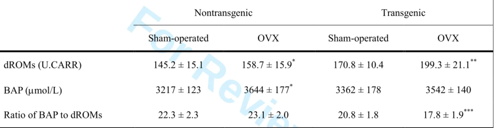
The increases in inflammatory cytokine production, especially that of IL-6, after ovariectomy were comparable in transgenic and nontransgenic mice. Nevertheless, the serum ALT level, hepatic steatosis and ROS production were greater in OVX transgenic mice than in OVX nontransgenic mice. Therefore we measured dROMs and BAP in serum to compare antioxidant potentials in OVX transgenic and OVX nontransgenic mice. We confirmed the significant negative correlation between the ratio of BAP to dROMs and hepatic content of superoxide (Fig. 4). As expected, the values for dROMs were higher in OVX mice than in sham-operated mice, regardless of whether they were transgenic or nontransgenic. However, a significant increase in the BAP value was found in OVX nontransgenic mice but not in OVX transgenic mice, which resulted in a lower ratio of BAP to dROMs in the OVX transgenic mice than in the OVX
nontransgenic mice (Table 2).
The first line of defense against ROS is the detoxifying enzymes that scavenge ROS. These include SODs and GPx1. Therefore we next investigated the expression levels of SOD2 and GPx1. The hepatic expression levels of SOD2 mRNA and GPx1 mRNA were significantly greater in OVX nontransgenic mice than in sham-operated
nontransgenic mice, but were comparable in OVX transgenic mice and sham-operated transgenic mice (Fig. 5A). Western blot analysis of the hepatic mitochondria fractions also showed significant increases of SOD2 and GPx1 expression in OVX nontransgenic mice but not in OVX transgenic mice (Fig. 5B). These results suggested that antioxidant defense mechanisms might be induced against ovariectomy-related ROS production in
For Review Only
nontransgenic mice but not in transgenic mice.
SIRT3 and PGC-1ααααexpression in OVX FL-N/35 transgenic mice
PGC-1α is a master regulator of mitochondrial biogenesis and respiration21 and required for the induction of many ROS-detoxifying enzymes, including SOD2 and GPx1 upon oxidative stress22. SIRT3 is a member of a class III histone deacetylase and is reported to mediate PGC-1α-dependent induction of ROS-detoxifying enzymes23. In accordance with the changes in SOD2 and GRPx1 levels after ovariectomy, the hepatic expression of SIRT3 mRNA was significantly greater in OVX nontransgenic mice than in
sham-operated nontransgenic mice, but comparable in OVX transgenic mice and sham-operated transgenic mice (Fig. 6A). Western blot analysis of hepatic mitochondria showed a significant increase of SIRT3 expression in OVX nontransgenic mice but not in OVX transgenic mice (Fig. 6A).
PGC-1α interacts with various nuclear receptors in addition to peroxisome
proliferator-activated receptor γ (PPARγ) and is docked to the promoter of its target genes by all these nuclear receptors. Therefore, we investigated PGC-1α expression levels not only in liver homogenates but also in the nuclear fraction of mouse liver. The expression levels of PGC-1α in liver homogenates were comparable in sham-operated and OVX nontransgenic mice and in sham-operated and OVX transgenic mice. However, the expression levels of PGC-1α in the nuclear fraction of the liver significantly increased after ovariectomy in both nontransgenic and transgenic mice, and OVX transgenic mice had a lower PGC-1α expression level than OVX
For Review Only
nontransgenic mice (Fig. 6B). These results suggested that the antioxidant potential against ovariectomy-induced ROS production might be reduced in OVX transgenic mice through lesser activation of PGC-1α than in OVX nontransgenic mice.
Suppressed AMPK activation in OVX FL-N/35 transgenic mice
PGC-1α activity is modulated through both transcriptional regulation and regulation of its activity by posttranslational modifications24. AMPK is one of the signaling pathways regulating PGC-1α and acts both through modulation of PGC-1α transcription and by phosphorylation of the PGC-1α protein24. HCV has been shown to reduce the kinase activity of AMPK through Ser485/491 phosphorylation of AMPK25. Therefore, we examined the expression levels of AMPK to investigate the mechanisms underlying the lower PGC-1α expression in the nuclear fraction of the OVX transgenic liver. The expression levels of AMPKα, which is one of the 3 subunits (α, β and γ) of AMPK, were comparable in sham-operated and OVX mice and in nontransgenic and transgenic mice. However, the expression level of phosphorylated AMPKα was significantly greater in OVX nontransgenic mice than in mice in the three other groups, though it was similar in sham-operated transgenic mice and OVX transgenic mice (Fig. 6C). In addition, its levels were significantly greater in nontransgenic mice than in transgenic mice (Fig. 6C). These results suggested that AMPK was activated in OVX
nontransgenic mice, but not in OVX transgenic mice, because AMPK is active only after phosphorylation of the α subunit at a threonine residue within the kinase domain (T172) by upstream kinases26. Taken together, the results in the present study suggested
For Review Only
that OVX FL-N/35 transgenic mice developed marked hepatic steatosis concomitant with increased ROS production via attenuation of antioxidant potential through inactivation of the AMPK/ PGC1α signaling pathway.
For Review Only
DISCUSSION
The OVX mice in the present study were assumed to be a standard model for evaluating the biological effect of ovariectomy because the effects of ovariectomy on dietary intake, body weight, uterine weight, liver weight and serum leptin levels were similar to the results from previous studies27-30. Ovariectomy increased ROS (superoxide) production in both transgenic liver and in nontransgenic liver, which was consistent with the ovariectomy-induced increase in NADPH oxidase activity12 and the protective effect of estrogen against mitochondrial oxidative damage13 found in previous studies. Of note was the much greater degree of ROS production after ovariectomy in transgenic mice than in nontransgenic mice. Interestingly, the effect of ovariectomy on female FL-N/35 transgenic mice in the present study resembled the effect of iron overload on male FL-N/35 transgenic mice noted in our previous study11 in that it generated greater ROS production in the liver compared with OVX or iron-overloaded nontransgenic mice. Considering that ovariectomy and iron overload are both oxidative stressors and that FL-N/35 transgenic mice express the HCV polyprotein in the liver, HCV protein expression has the potential to increase the sensitivity to oxidative stress in the liver. At least two possibilities may account for the increased sensitivity to oxidative stress in FL-N/35 transgenic mice. One possibility is an additive effect of HCV-induced ROS production on ovariectomy-induced oxidative stress. The HCV core protein has been shown to inhibit mitochondrial electron transport31 and to induce ROS production32. In fact, basal ROS production tended to be higher in transgenic mice than in nontransgenic mice, but was not significantly different. These results suggested that additive
For Review Only
HCV-induced ROS production was unlikely to be the cause of the significantly increased ROS production after ovariectomy in the transgenic mice. The other possibility is HCV-associated attenuation of antioxidant potential against
ovariectomy-induced oxidative stress. In this respect OVX transgenic mice had a lower ratio of BAP to dROM than OVX nontransgenic mice and the expression of SOD2 and GPx1 in the liver was not increased. These results suggested that HCV protein
attenuated antioxidant potential against ovariectomy-induced oxidative stress. PGC-1α is required for the induction of many ROS-detoxifying enzymes upon oxidative stress22. SIRT3 has been shown to function as a downstream target gene of PGC-1α and mediate the PGC-1α-dependent induction of ROS-detoxifying enzymes23. Additionally, AMPK, which is a crucial cellular energy sensor, regulates PGC-1α activity through both modulation of PGC-1α transcription and phosphorylation of the PGC-1α protein24, 33. Thus, AMPK/PGC-1α signaling is one of the important pathways that protects cells from oxidative stress through the induction of several key
ROS-detoxifying enzymes. Recent evidence indicating that HCV replication inhibits AMPK activity25 prompted us to investigate whether the antioxidant potential against ovariectomy-induced oxidative stress in FL-N/35 transgenic mice was attenuated through inhibition of this signaling pathway. As expected, upon ovariectomy AMPK was activated in nontransgenic mice, but not in transgenic mice. This, in turn, led to the lower expression of PGC-1α in the nuclear fraction of the liver in OVX transgenic mice than in OVX nontransgenic mice, resulting in the absence of significant induction of SIRT3 in the mitochondrial fraction of the liver in the OVX transgenic mice. Thus, ROS
For Review Only
production in the liver in OVX transgenic mice was increased by attenuation of the antioxidant potential through inhibition of AMPK/PGC-1α signaling. However, it remains unknown why the expression of PGC-1α in the nuclear fraction was
significantly increased in OVX transgenic mice regardless of the lack of activation of AMPK. Various kinases other than AMPK and posttranslational modifications other than phosphorylation have been shown to regulate PGC-1α expression24. Therefore further investigations are required to clarify this issue.
Of particular concern is the relevance of the present results to HCC development in patients with HCV-associated chronic liver diseases. Male gender has been shown to be an independent risk factor for the development of HCC in patients with HCV-related cirrhosis2-3. It is proposed that estrogen-mediated inhibition of IL-6 production by Kupffer cells reduces the HCC risk in females5, and therefore menopause is assumed to be a risk factor for HCC development. In fact, a recent study from Japan demonstrated a higher proportion of females, especially among elderly patients with HCV-related HCC, suggesting that the gender disparity in HCC development becomes less distinct as the patient’s age at HCC diagnosis increases6. On the other hand, in addition to the directly induced generation of ROS by the HCV proteins, chronic inflammation with production of proinflammatory cytokines and/or aging has the potential to deliver an additional burden of ROS. These multiple sources of ROS production create a procarcinogenic environment under which chromosomal damage is likely to occur. In these
circumstances, the present findings that OVX transgenic mice have increased ROS production in the liver via attenuation of antioxidant potential through inhibition of
For Review Only
AMPK/PGC-1α signaling may indicate one of the mechanisms by which women with HCV infection are at high risk for HCC development when some period has passed after menopause.
For Review Only
ACKNOWLEDGMENTS
This research was supported by a Grant-in-Aid for Scientific Research (B) (23390201) from the Japan Society for the Promotion of Science, a Health and Labor Sciences Research Grant for Research on Hepatitis from the Ministry of Health, Labor and Welfare of Japan, and Research Project Grant P2 from Kawasaki Medical School.
For Review Only
REFERENCES
1. Chen DS, Locarnini S, Wait S, et al. Report from a Viral Hepatitis Policy Forum on implementing the WHO framework for global action on viral hepatitis in North Asia. J Hepatol 2013; 10.1016/j.jhep.2013.06.029.
2. Bruno S, Silini E, Crosignani A, et al. Hepatitis C virus genotypes and risk of hepatocellular carcinoma in cirrhosis: a prospective study. Hepatology 1997; 25: 754-8.
3. Degos F, Christidis C, Ganne-Carrie N, et al. Hepatitis C virus related cirrhosis: time to occurrence of hepatocellular carcinoma and death. Gut 2000; 47: 131-6. 4. Bosch FX, Ribes J, Diaz M, Cleries R. Primary liver cancer: worldwide incidence
and trends. Gastroenterology 2004; 127: S5-S16.
5. Naugler WE, Sakurai T, Kim S, et al. Gender disparity in liver cancer due to sex differences in MyD88-dependent IL-6 production. Science 2007; 317: 121-4. 6. Kumada T, Toyoda H, Kiriyama S, et al. Characteristics of elderly hepatitis C
virus-associated hepatocellular carcinoma patients. J Gastroenterol Hepatol 2013; 28: 357-64.
7. Farinati F, Cardin R, De Maria N, et al. Iron storage, lipid peroxidation and glutathione turnover in chronic anti-HCV positive hepatitis. J Hepatol 1995; 22: 449-56.
8. Barbaro G, Di Lorenzo G, Asti A, et al. Hepatocellular mitochondrial alterations in patients with chronic hepatitis C: ultrastructural and biochemical findings. Am J Gastroenterol 1999; 94: 2198-205.
For Review Only
9. Tanaka H, Fujita N, Sugimoto R, et al. Hepatic oxidative DNA damage is associated with increased risk for hepatocellular carcinoma in chronic hepatitis C. Br J Cancer 2008; 98: 580-6.
10. Moriya K, Nakagawa K, Santa T, et al. Oxidative stress in the absence of inflammation in a mouse model for hepatitis C virus-associated
hepatocarcinogenesis. Cancer Res 2001; 61: 4365-70.
11. Furutani T, Hino K, Okuda M, et al. Hepatic iron overload induces hepatocellular carcinoma in transgenic mice expressing the hepatitis C virus polyprotein. Gastroenterology 2006; 130: 2087-98.
12. Ji H, Zheng W, Menini S, et al. Female protection in progressive renal disease is associated with estradiol attenuation of superoxide production. Gend Med 2007; 4: 56-71.
13. Borras C, Sastre J, Garcia-Sala D, Lloret A, Pallardo FV, Vina J. Mitochondria from females exhibit higher antioxidant gene expression and lower oxidative damage than males. Free Radic Biol Med 2003; 34: 546-52.
14. Beard MR, Abell G, Honda M, et al. An infectious molecular clone of a Japanese genotype 1b hepatitis C virus. Hepatology 1999; 30: 316-24.
15. Lerat H, Honda M, Beard MR, et al. Steatosis and liver cancer in transgenic mice expressing the structural and nonstructural proteins of hepatitis C virus.
Gastroenterology 2002; 122: 352-65.
16. Bligh EG, Dyer WJ. A rapid method of total lipid extraction and purification. Can J Biochem Physiol 1959; 37: 911-917.
For Review Only
17. Lowry OH, Rosebrough NJ, Farr AL, Randall RJ. Protein measurement with the Folin phenol reagent. J Biol Chem 1951; 193:2 65-75.
18. Nishina S, Hino K, Korenaga M, et al. Hepatitis C virus-induced reactive oxygen species raise hepatic iron level in mice by reducing hepcidin transcription. Gastroenterology 2008; 134: 226-38.
19. Harrison-Findik DD, Schafer D, Klein E, et al. Alcohol metabolism-mediated oxidative stress down-regulates hepcidin transcription and leads to increased duodenal iron transporter expression. J Biol Chem 2006; 281: 22974-82. 20. Cesarone MR, Belcaro G, Carratelli M, et al. A simple test to monitor oxidative
stress. Int Angiol 1999; 18: 127-30.
21. Kelly DP, Scarpulla RC. Transcriptional regulatory circuits controlling mitochondrial biogenesis and function. Genes Dev 2004; 18: 357-68.
22. St-Pierre J, Drori S, Uldry M, et al. Suppression of reactive oxygen species and neurodegeneration by the PGC-1 transcriptional coactivators. Cell 2006; 127: 397-408.
23. Kong X, Wang R, Xue Y, et al. Sirtuin 3, a new target of PGC-1alpha, plays an important role in the suppression of ROS and mitochondrial biogenesis. PLoS One 2010; 5: e11707.
24. Fernandez-Marcos PJ, Auwerx J. Regulation of PGC-1alpha, a nodal regulator of mitochondrial biogenesis. Am J Clin Nutr 2011; 93: 884S-90.
For Review Only
25. Mankouri J, Tedbury PR, Gretton S, et al. Enhanced hepatitis C virus genome replication and lipid accumulation mediated by inhibition of AMP-activated protein kinase. Proc Natl Acad Sci U S A 2010; 107: 11549-54.
26. Hawley SA, Boudeau J, Reid JL, et al. Complexes between the LKB1 tumor suppressor, STRAD alpha/beta and MO25 alpha/beta are upstream kinases in the AMP-activated protein kinase cascade. J Biol 2003; 2: 28.
27. Rogers NH, Perfield JW 2nd, Strissel KJ, Obin MS, Greenberg AS. Reduced energy expenditure and increased inflammation are early events in the development of ovariectomy-induced obesity. Endocrinology 2009; 150: 2161-8.
28. Pighon A, Gutkowska J, Jankowski M, Rabasa-Lhoret R, Lavoie JM. Exercise training in ovariectomized rats stimulates estrogenic-like effects on expression of genes involved in lipid accumulation and subclinical inflammation in liver. Metabolism 2011; 60: 629-39.
29. Hong J, Stubbins RE, Smith RR, Harvey AE, Nunez NP. Differential susceptibility to obesity between male, female and ovariectomized female mice. Nutr J 2009; 8: 11.
30. Kamada Y, Kiso S, Yoshida Y, et al. Pitavastatin ameliorated the progression of steatohepatitis in ovariectomized mice fed a high fat and high cholesterol diet. Hepatol Res 2013; 43: 401-12.
31. Korenaga M, Wang T, Li Y, et al. Hepatitis C virus core protein inhibits mitochondrial electron transport and increases reactive oxygen species (ROS) production. J Biol Chem 2005; 280: 37481-8.
For Review Only
32. Okuda M, Li K, Beard MR, et al. Mitochondrial injury, oxidative stress, and antioxidant gene expression are induced by hepatitis C virus core protein. Gastroenterology 2002; 122: 366-75.
33. Hardie DG, Ross FA, Hawley SA. AMPK: a nutrient and energy sensor that maintains energy homeostasis. Nat Rev Mol Cell Biol 2012; 13: 251-62.
For Review Only
FIGURE LEGENDS
Figure 1. Hepatic steatosis and triglyceride content in sham-operated and OVX
FL-N/35 transgenic and nontransgenic mice. (A) Hepatic steatosis in mice in each group (H&E, original magnification × 100). (B) Hepatic triglyceride content in mice in each group (n=5). The results are shown as box plot profiles. The bottom and top edges of the boxes are the 25th and 75th percentiles, respectively. Median values are shown by the line within each box. *: P<0.05 versus mice in the other three groups.
Figure 2. Reactive oxygen species (ROS) production in sham-operated and OVX
FL-N/35 transgenic and nontransgenic mice. (A) Frozen liver sections from mice in each group were stained with dihydroethidium (DHE). (B) Fluorescence intensity was quantified by NIH image analysis software for three randomly selected areas of digital images for five mice in each group. The results are shown as box plot profiles. The bottom and top edges of the boxes are the 25th and 75th percentiles, respectively. Median values are shown by the line within each box. *: P<0.05 versus sham-operated nontransgenic mice. **: P<0.05 versus OVX nontransgenic mice and sham-operated transgenic mice.
Figure 3. Expression levels of inflammatory cytokines in sham-operated and OVX
FL-N/35 transgenic and nontransgenic mice. The mRNA levels of IL-6, IL-1β, and TNF-α were measured by real-time RT-PCR for five mice in each group. The relative quantities of target mRNA in the liver were normalized with β-actin mRNA. *: P<0.05
For Review Only
versus sham-operated nontransgenic mice. **: P<0.05 versus sham-operated transgenic mice.
Figure 4. Negative correlation between the ratio of BAP to dROMs (BAP/dROMs) and
hepatic content of superoxide. R=−0,453, P<0.05. Hepatic content of superoxide was determined based on the area of dihydroethidium (DHE) fluorescence.
Figure 5. Expression levels of superoxide dismutase 2 (SOD2) and glutathione
peroxidase 1 (GPx1) in sham-operated and OVX FL-N/35 transgenic and nontransgenic mice. (A) The mRNA levels of SOD2 and GPx1 were measured by real-time RT-PCR for five mice in each group. The relative quantities of target mRNA in the liver were normalized with β-actin mRNA. (B) Immunoblots for SOD2 and GPx1 were performed using mitochondrial fractions of liver lysates from five mice in each group. *: P<0.05 versus sham-operated nontransgenic mice.
Figure 6. Expression levels of sirtuin 3 (SIRT3), peroxisome proliferator-activated
receptor γ coactivator 1α (PGC-1α), adenosine monophosphate–activated protein kinase α (AMPKα), and phosphorylated AMPKα (P-AMPKα) in sham-operated and OVX FL-N/35 transgenic and nontransgenic mice. (A) The mRNA levels of SIRT3 were measured by real-time RT-PCR for five mice in each group. The relative quantities of target mRNA in the liver were normalized with β-actin mRNA. Immunoblots for SIRT3 were performed using the mitochondrial fractions of liver
For Review Only
lysates from five mice in each group. (B) Immunoblots for PGC-1α were performed using liver lysates and their nuclear fractions from five mice in each group. *: P<0.05 versus sham-operated nontransgenic mice. **: P<0.05 versus sham-operated transgenic mice. (C) Immunoblots for AMPKα and P-AMPKα were performed using liver lysates from five mice in each group. *: P<0.05 versus mice in the other three groups. **:
For Review Only
Table 1 Body, liver, and uterus weight and serum biochemical parameters
Body, liver, and uterus weight and serum biochemical parameters
Nontransgenic Transgenic
Sham-operated OVX Sham-operated OVX
Body weight (g) 21.5 ± 1.2 30.7 ± 4.9* 27.7 ± 4.6 34.2 ± 3.8**
Liver weight (g) 0.86 ± 0.075 1.09 ± 0.236* 0.90 ± 0.102 1.18 ± 0.156**
Ratio of liver to body weight 0.038 ± 0.037 0.035 ± 0.003 0.031 ± 0.002 0.034 ± 0.006
Uterus weight (g) 0.08 ± 0.01 0.01 ± 0.02* 0.09 ± 0.01 0.01 ± 0.01**
Total dietary intake (g) 337 ± 24 429 ± 13* 368 ± 28 490 ± 31**
Serum glucose (mg/dL) 222.9 ± 110.0 275.1 ± 121.4 284.0 ± 84.1 259. 7 ± 108.9
Serum ALT (IU/L) 15.5 ± 6.5 30.6 ± 38.1 21.8 ± 11.4 281.2 ± 165.1***
Serum triglyceride (mg/dL) 99.9 ± 9.7 78.9 ± 10.8 98.3 ± 11.4 89.7 ± 13.3
For Review Only
Data are mean ± standard deviation. *P<0.05 compared with sham-operated nontransgenic mice. **P<0.05 compared with sham-operated transgenic mice. ***P<0.01 compared with mice in the other three groups.
For Review Only
Table 2 Derivatives of reactive oxygen metabolites (dROMs), biological antioxidant potential (BAP) and ratio of BAP to dROMs
Nontransgenic Transgenic
Sham-operated OVX Sham-operated OVX
dROMs (U.CARR) 145.2 ± 15.1 158.7 ± 15.9* 170.8 ± 10.4 199.3 ± 21.1**
BAP (µmol/L) 3217 ± 123 3644 ± 177* 3362 ± 178 3542 ± 140
Ratio of BAP to dROMs 22.3 ± 2.3 23.1 ± 2.0 20.8 ± 1.8 17.8 ± 1.9***
Data are mean ± standard deviation. *P<0.05 compared with sham-operated nontransgenic mice. **P<0.05 compared with sham-operated transgenic mice. ***P<0.05 compared with OVX nontransgenic mice. OVX; ovariectomized.