学位論文 博士(医科学)甲
Circadian regulation of allergic reaction
by the mast cell clock in mice
(マウスにおけるアレルギー反応の日内変動は
マスト細胞の内在時計によって制御される
)
中村 勇規
clock in mice
Yuki Nakamura,aNobuhiro Nakano, PhD,bKayoko Ishimaru,aMutsuko Hara, PhD,bTakako Ikegami, PhD,c
Yu Tahara, PhD,dRyohei Katoh, MD, PhD,eHideoki Ogawa, MD, PhD,bKo Okumura, MD, PhD,bShigenobu Shibata, PhD,d
Chiharu Nishiyama, PhD,band Atsuhito Nakao, MD, PhDa,b Yamanashi and Tokyo, Japan
Background: It remains elusive how allergic symptoms exhibit prominent 24-hour variations. In mammals the circadian clocks present in nearly all cells, including mast cells, drive the daily rhythms of physiology. Recently, we have shown that the circadian clocks drive the daily rhythms in IgE/mast cell– mediated allergic reactions. However, the precise mechanisms, particularly the specific roles of the mast cell–intrinsic clockwork in temporal regulation, remain unclear.
Objective: We determined whether the mast cell clockwork contributes to the temporal regulation of IgE/mast
cell–mediated allergic reaction.
Methods: The kinetics of a time of day–dependent variation in passive cutaneous anaphylactic reactions were compared between mast cell–deficient mice reconstituted with bone marrow–derived cultured mast cells generated from mice with a wild-type allele and a dominant negative type mutation of the key clock geneClock. We also examined the temporal responses of wild-type andClock-mutated bone marrow–derived cultured mast cells to IgE stimulationin vitro. Furthermore, factors influencing the mast cell clockwork were determined by using in vivo imaging.
Results: TheClock mutation in mast cells resulted in the absence of temporal variations in IgE-mediated degranulation in mast cells bothin vivo and in vitro associated with the loss of temporal regulation of FcεRI expression and signaling. Additionally, adrenalectomy abolished the mast cell clockworkin vivo. Conclusion: The mast cell–intrinsic clockwork, entrained by humoral factors from the adrenal gland, primarily contributes to the temporal regulation of IgE/mast cell–mediated allergic reactions. Our results reveal a novel regulatory mechanism for IgE-mediated mast cell responses that might underlie the circadian pathophysiology in patients with allergic diseases. (J Allergy Clin Immunol 2013;nnn:nnn-nnn.)
Key words: The circadian clock, mast cells, IgE, allergy, mouse
Allergic diseases are characterized by symptoms that exhibit prominent 24-hour variations.1,2For instance, in patients with al-lergic rhinitis, the symptoms are worse overnight or early in the morning (‘‘morning attack’’) and often compromise nighttime sleep, resulting in a poor daytime quality of life.3Although these phenomena have been recognized for decades,4,5 the precise mechanisms remain unclear.
The circadian clocks drive daily rhythms in physiology that enable organisms to keep track of the time of day. In mammals the light-entrained central oscillator located in the suprachiasmatic nucleus of the hypothalamus synchronizes the peripheral oscil-lators present in nearly all cell types, including mast cells, through neural and endocrine pathways.6-11The mechanisms of rhythm generation are based on transcriptional-translational feedback loops, wherein 2 transcription factors, CLOCK and BMAL1, ac-tivate the transcription of the Period (Per) and Cryptochrome (Cry) genes. The PER and CRY proteins in turn inhibit their own expression by repressing CLOCK/BMAL1 activity.6-9
In a classical mouse model of IgE/mast cell–mediated allergic reaction (passive cutaneous anaphylactic [PCA] reaction), we have recently shown that there are time of day–dependent variations that rely on the normal activity of a key clock gene, Period2 (Per2),10suggesting that the circadian clocks drive the daily rhythms in IgE/mast cell–mediated allergic reactions. How-ever, the precise mechanisms, particularly the specific roles of the mast cell–intrinsic clockwork in the temporal regulation, have been unclear. This study aimed to determine whether the mast cell clockwork contributes to the temporal regulation of IgE/ mast cell–mediated allergic reactions.
METHODS
For more information, see theMethodssection in this article’s Online Repository atwww.jacionline.org.
Mice
Male 5- to 6-week-old C57BL/6 mice, mast cell–deficient WBB6F1-W/ Wv mice (Japan SLC, Tokyo, Japan), PER2::Luciferase (PER2::LUC) knock-in mice (C57BL/6 background),12and C57BL/6 ClockD19/D19mice13were bred under specific pathogen-free conditions. ClockD19/D19 mice have an
From the Departments ofaImmunology andePathology, Faculty of Medicine, University of Yamanashi;bthe Atopy Research Center, Juntendo University School of Medicine, Tokyo;cthe Laboratory of Molecular and Biochemical Research, Research Support Center, Juntendo University Graduate School of Medicine, Tokyo; anddthe Depart-ment of Physiology and Pharmacology, School of Advanced Science and Engineering, Waseda University, Tokyo.
Supported in part by grants from the Ministry of Education, Culture, Sports, Science, and Technology, Japan.
Disclosure of potential conflict of interest: The authors declare that they have no relevant conflicts of interest.
Received for publication February 6, 2013; revised July 25, 2013; accepted for publica-tion July 30, 2013.
Corresponding author: Atsuhito Nakao, MD, PhD, Department of Immunology, Faculty of Medicine, University of Yamanashi, 1110 Shimokato, Chuo, Yamanashi 409-3898, Japan. E-mail:[email protected].
0091-6749/$36.00
Ó 2013 American Academy of Allergy, Asthma & Immunology
http://dx.doi.org/10.1016/j.jaci.2013.07.040
Abbreviations used
BMCMC: Bone marrow–derived cultured mast cell ChIP: Chromatin immunoprecipitation mMCP: Mouse mast cell protease
PCA: Passive cutaneous anaphylaxis PER2::LUC: Period2::Luciferase
siRNA: Small interfering RNA ZT: Zeitgeber time
A to T point mutation in the 59 splice site of intron 19 and, as a consequence, an in-frame deletion of the entire exon 19 (ClockD19/D19), which results in loss of normal transcriptional activity.13This autosomal dominant mutation eventu-ally provokes arrhythmicity in mice. All mice were housed under 12-hour light/12-hour dark conditions (the light was turned on at 6AM, which is Zeit-geber time [ZT] 0, and the light was turned off at 6PM, which is ZT12) with ad libitum access to food and water for at least 2 weeks.
Preparation of bone marrow–derived cultured mast cells
Bone marrow–derived cultured mast cells (BMCMCs) were generated from the femoral bone marrow cells of male mice.14
PCA reaction
Mast cell–deficient WBB6F1-W/Wv mice were reconstituted with subcu-taneous injections (dorsal skin) of BMCMCs (1.53 106per mouse) derived from wild-type C57BL/6 mice or C57BL/6 ClockD19/D19mice. Six weeks after reconstitution, the mice were sensitized subcutaneously in the BMCMC-injected dorsal skin region with mouse anti-TNP IgE (0.5 mg/ 20 mL; BD Bio-sciences, San Jose, Calif) to induce a PCA reaction. Saline alone was used as a negative control. The mice were then challenged intravenously 24 hours later with 50 mg of DNP-BSA (Cosmo Bio, Tokyo, Japan) with 0.5% Evans blue dye. Quantitative analysis of the PCA reaction was performed, as previously described.10
Statistical analysis
The statistical analyses were performed by using the unpaired Student t test for 2-group comparisons and ANOVA for comparison of more than 2 groups. Statcel3 software (OMS Publishing, Saitama, Japan) was used for the analysis. A P value of less than .05 was considered significant, unless otherwise indicated.
RESULTS
The mast cell clock times PCA reactions
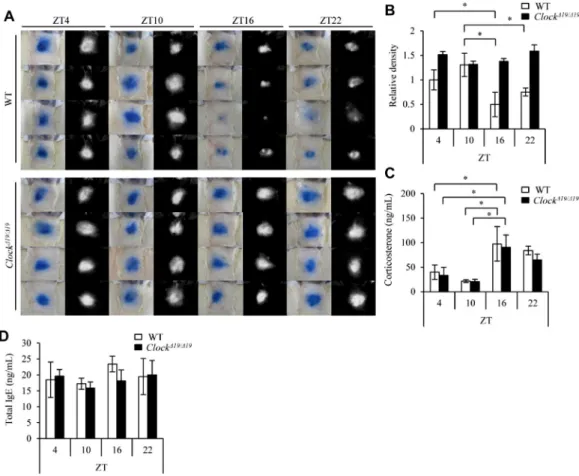
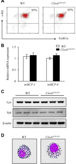
To determine whether the mast cell–intrinsic clockwork con-tributes to the temporal regulation of IgE/mast cell–mediated allergic reactions, we compared the kinetics of a time of day– dependent variation in the PCA reaction between mast cell– deficient W/Wv mice subcutaneously reconstituted with BMCMCs generated from wild-type mice and mice with a dominant negative– type mutation of Clock (ClockD19/D19mice).13There were compa-rable levels of cell-surface FcεRIa and c-kit expression (without synchronization, please see below), mouse mast cell protease (mMCP) 5 and mMCP-6 mRNA expression, and Syk and Lyn pro-tein expression and similar morphology between wild-type and Clock-mutated BMCMCs (see Fig E1 in this article’s Online Repository at www.jacionline.org), suggesting that the Clock mutation did not affect the differentiation of mast cells. We also confirmed that the numbers of mast cells in the skin were compara-ble between mice reconstituted with wild-type BMCMCs and those reconstituted with Clock-mutated BMCMCs (seeFig E2in this article’s Online Repository atwww.jacionline.org).
The extent of PCA reactions showed a time of day–dependent variation in control mice, with a clear nadir around the onset of night (10PM, which was ZT16;Fig 1, A and B), as described
pre-viously in conventional wild-type mice.10This variation was ab-sent in mast cell–specific Clock-mutated mice (Fig 1, A and B). In contrast, the daily profiles and levels of serum corticosterone and IgE were comparable between the mice (Fig 1, C and D). These results suggest that the mast cell–intrinsic clockwork is critical to the daily rhythm generation in the PCA reaction.
IgE-mediated mast cell responses show temporal variationsin vitro
To support the in vivo findings, we examined the temporal re-sponses of mast cells to IgE stimulation by using BMCMCs gener-ated from knock-in mice expressing a PER2::LUC fusion protein (PER2::LUC BMCMCs),12 wild-type mice, and ClockD19/D19 mice13in vitro. We noted that the time window when the mast cell clockwork was functional appeared to be very limited (approx-imately 0-36 hours after medium change) in in vitro culture condi-tions based on the monitoring of bioluminescent emission of PER2::LUC BMCMCs (seeFig E3in this article’s Online Repos-itory atwww.jacionline.org).10This might be due to a lack of oscil-lator coupling in the dissociated cell cultures, leading to damping of the ensemble rhythm at the population level.15Therefore we com-pared the extent of IgE-mediated degranulation in mast cells be-tween 12-hour cultured PER2::LUC BMCMCs (after a medium change) with the nadir of the PER2::LUC protein level and 24-hour cultured PER2::LUC BMCMCs with the peak PER2::LUC protein level. A simple medium change is a trigger to synchronize the circadian clocks in peripheral cells in vitro.6-9We avoided using potent reagents to synchronize peripheral clocks (eg, cyclic AMP activators and dexamethasone) for the in vitro experiments because such reagents affect IgE-mediated signaling in mast cells indepen-dent of ‘‘clock’’ function.16,17
The extent of IgE-mediated b-hexosaminidase release was significantly higher in the 12-hour cultured PER2::LUC BMCMCs than in the 24-hour cultured PER2::LUC BMCMCs (Fig 2, A). More detailed kinetic studies using the 6-, 12-, 18-, and 24-hour cultured BMCMCs after the medium change showed similar find-ings in wild-type BMCMCs but not in Clock-mutated BMCMCs (Fig 2, B). There were little differences in spontaneous b-hexosa-minidase release (ie, without IgE stimulation) between wild-type and Clock-mutated BMCMCs at the representative time points (see Fig E4 in this article’s Online Repository at www. jacionline.org). In contrast, the extent of IgE-mediated b-hexosa-minidase release was comparable between the 48- and 60-hour cul-tured wild-type BMCMCs (Fig 2, C), both of which appeared to no longer have functional clockwork (seeFig E3). The extent of IgE-mediated IL-13 production also showed similar temporal varia-tions in 12- and 24-hour cultured wild-type BMCMCs (Fig 2, D). Consistently, the extent of IgE-dependent intracellular Ca21 mobilization and the total tyrosine phosphorylation levels of cell lysates were higher in the 12-hour cultured wild-type BMCMCs than those in the 24-hour cultured wild-type BMCMCs, which were absent in Clock-mutated BMCMCs (Fig 2, E and F). Interest-ingly, FcεRI-independent stimulation of the 12- and 24-hour cul-tured wild-type BMCMCs (and also Clock-mutated BMCMCs) with calcium ionophore A23187 showed comparable b-hexosa-minidase release (Fig. 2, G). Collectively, the IgE-mediated de-granulation and signaling in mast cells showed temporal variations in vitro, and disruption of the mast cell clockwork by ei-ther a Clock mutation or long-term culture without a medium change (synchronization) abolished the variations.
FcεRIb transcription is under circadian control by the mast cell clock
The high-affinity IgE receptor (FcεRI) on mast cells consists of 3 subunits (FcεRI a, b, and g chains), and IgE binds to FcεRIa with a high affinity, whereas the b and g chains transduce the extracellular signals into the intracellular signaling pathways,
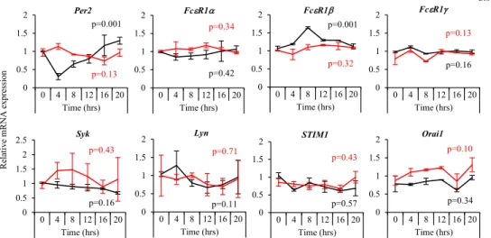
including Ca21 signaling.18-20 Given the findings that IgE-mediated intracellular Ca21mobilization showed temporal varia-tions and that direct stimulation of Ca21signaling with A23187 did not show temporal variations in degranulation in mast cells (Fig 2, E and G), we thought that the mast cell–intrinsic clock-work likely targets FcεRI signaling–associated molecules up-stream of Ca21signaling in mast cells, thereby temporally fine tuning FcεRI signaling.
A quantitative PCR analysis revealed that FcεRIb, but not other FcεRI signaling–associated molecules upstream of Ca21 signal-ing, such as FcεRIa and FcεRIg, Syk, and STIM1,18-20showed circadian mRNA expression with a peak at approximately 8 hours and a nadir at approximately 20 hours after a medium change in wild-type BMCMCs (Fig 3). The circadian oscillation of FcεRIb
mRNA was not observed in Clock-mutated BMCMCs. We con-firmed that Per2 mRNA showed circadian oscillations in wild-type BMCMCs but not in Clock-mutated BMCMCs. These data suggest that FcεRIb transcription is under circadian control by the mast cell–intrinsic clock.
CLOCK binds to the promoter of FcεRIb and modulates its transcription
Several E-box–like elements to which the CLOCK/BMAL1 complex theoretically can bind21 are present in the promoter
regions of the mouse FcεRIb (seeFig E5in this article’s Online Repository at www.jacionline.org).22 To directly show that FcεRIb is a target of the mast cell clockwork, we performed chro-matin immunoprecipitation (ChIP) assays using anti-CLOCK antibody.
CLOCK bound to the promoter of FcεRIb in the 6- and 24-hour, but not 12-hour, cultured wild-type BMCMCs (Fig 4, A), which was largely consistent with the mRNA kinetics of FcεRIb (Fig 3). The integrity of the ChIP samples was confirmed by means of constitutive binding of CLOCK to the promoter of the Per2 gene containing the noncanonical E-box enhancer 2 (E2) sequence, but not the E5 sequence, in wild-type BMCMCs, as previously reported (Fig 4, B).23CLOCK levels were comparable in the 12- and 24-hour cultured wild-type BMCMCs (Fig 4, C).
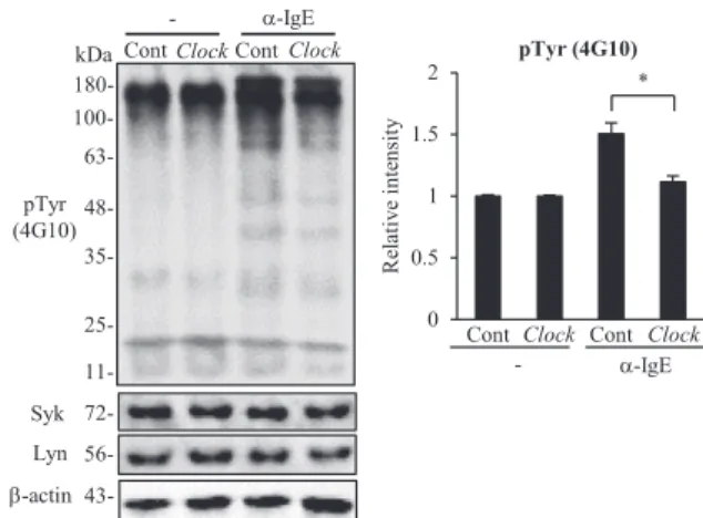
We then determined whether CLOCK affected the transcription of FcεRIb in mast cells. Clock small interfering RNA (siRNA), but not control siRNA, suppressed Clock mRNA and protein levels in wild-type BMCMCs at 48 hours after siRNA transfection (Fig 4, D and E). At this time point, FcεRIb, but not Syk and Lyn, mRNA levels decreased in association with the reduction of IgE-mediated b-hexosaminidase release in Clock, but not control, siRNA-treated wild-type BMCMCs (Fig 4, F and G). Consis-tently, the extent of the IgE-dependent increase in total tyrosine phosphorylation levels of cell lysates was reduced in Clock
FIG 1. The mast cell–intrinsic clockwork is critical for time of day–dependent variations in PCA reactions. A, Representative pictures of skin color reactions in W/Wv mice reconstituted with wild-type BMCMCs (WT) or Clock-mutated BMCMCs (ClockD19/D19, left panels) and digitalized images of density value evaluations (right panels) at the indicated time points. Data shown are results from the same group of wild-type or ClockD19/D19 mice challenged at the indicated time points. B, Quantitative analysis of the data in Fig 1, A. C and D, Serum corticosterone (Fig 1, C) and total IgE (Fig 1, D) levels at the indicated time points. Values represent means6 SDs (n5 4 per group). A representative result of 2 independent experiments is shown. *P < .05.
siRNA–treated wild-type BMCMCs compared with that seen in control siRNA–treated wild-type BMCMCs (seeFig E6in this ar-ticle’s Online Repository at www.jacionline.org). Furthermore, CLOCK overexpression in wild-type BMCMCs enhanced the promoter activity of FcεRIb in a reporter assay (seeFig E7in this article’s Online Repository atwww.jacionline.org).22These results suggest that CLOCK temporally binds to the promoter of FcεRIb and modulates its transcription.
Unfortunately, we were unable to evaluate the FcεRIb protein levels directly because of the lack of a good antibody. Therefore to confirm that temporal regulation of FcεRIb transcription by the mast cell clock was indeed translated into protein levels, we examined cell-surface FcεRIa expression levels in the 12- and 24-hour cultured wild-type BMCMCs because the
cell-surface FcεRIa levels are regulated by FcεRIb protein levels in mast cells.24-26As expected, the cell-surface FcεRIa expres-sion levels increased in the 12-hour cultured wild-type BMCMCs compared with those in the 24-hour cultured wild-type BMCMCs, which was absent in Clock-mutated BMCMCs (Fig 4, H). We also found that the cell-surface FcεRIa expression
levels decreased in Clock, but not control, siRNA–treated wild-type BMCMCs (seeFig E8 in this article’s Online Repository atwww.jacionline.org). Notably, the expression levels of FcεRI at the cell surfaces of peritoneal mast cells from peritoneal lavage fluid performed at different times of day (ZT4 and ZT16) showed a time of day–dependent variation in wild-type mice, whereas this variation was absent in Clock-mutated mice (Fig 4, I). Collectively, it is likely that temporal modulation of FcεRIb
A B C D
E F
G
FIG 2. The mast cell–intrinsic clockwork temporally regulates IgE-dependent degranulation and signaling in mast cells in vitro. A, Release ofb-hexosaminidase from the 12- or 24-hour cultured (after the medium change) BMCMCs derived from PER2::LUC knock-in mice (PER2::LUC). B, Release ofb-hexosaminidase from the 6-, 12-, 18-, or 24-hour cultured (after the medium change) BMCMCs derived from wild-type (WT) and ClockD19/D19mice. C, Release ofb-hexosaminidase from the 48- or 60-hour cultured (after the medium change) wild-type BMCMCs. D, IgE-dependent IL-13 production from the 12- or 24-hour cultured (after the medium change) wild-type BMCMCs. E, IgE-dependent intracellular Ca21mobilization in the 12- or 24-hour cultured (after the medium change) BMCMCs derived from WT and ClockD19/D19mice. F, Left panel, IgE-dependent total tyrosine phosphorylation levels in the 12- or 24-hour cultured (after the medium change) BMCMCs derived from WT and ClockD19/D19mice. Right panel, Quantitative analysis. G, Release ofb-hexosaminidase from the 12- or 24-hour cultured (after the medium change) BMCMCs stimulated with A23187 derived from WT and ClockD19/D19 mice. Values represent means 6 SDs (n5 3 per group). Similar results were obtained in at least 3 independent experiments. The statistical analysis in Fig 2, E, was done with the unpaired Student t test. *P < .05.
transcription by CLOCK is indeed translated into protein levels in mast cells in a circadian manner, thereby timing FcεRI expres-sion and signaling.
The adrenal gland is important to synchronize the mast cell clockwork
The data (Figs 1-4) suggest that if the individual mast cell clockwork does not synchronize with internal zeitgebers (‘‘time givers’’ in German), this would lead to damping of the ensemble rhythm at population levels of mast cells and might result in un-coupled IgE-dependent responses. However, there is no informa-tion available about how mast cells tell time in the body.
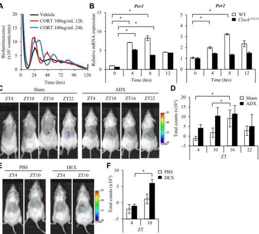
We screened several bioactive substances for this effect by monitoring the bioluminescence emission in consistent cultures of PER2::LUC BMCMCs (see Fig E9in this article’s Online Repository atwww.jacionline.org). The addition of corticoster-one to PER2::LUC BMCMCs before the medium change or at 12 or 24 hours after the medium change either enhanced the am-plitude of or phase shifted the mast cell clockwork, respectively (seeFig E9, A, andFig 5, A). Consistently, corticosterone induced Per1 and Per2 mRNA expression in wild-type BMCMCs (Fig 5, B). Interestingly, corticosterone induced Per1, but not Per2, mRNA in Clock-mutated BMCMCs (Fig 5, B). The biolumines-cence emission from mast cell–deficient W/Wv mice subcutane-ously reconstituted with PER2::LUC BMCMCs showed a time of day–dependent variation in the PER2::LUC protein in sham-operated mice, with a peak at 10 PM (ZT16), as previously described,10whereas adrenalectomy resulted in the absence of variation (Fig 5, C and D). Conversely, dexamethasone treatment of W/Wv mice subcutaneously reconstituted with PER2::LUC BMCMCs at ZT4 enhanced the PER2::LUC protein levels at ZT10 compared with those seen in PBS-treated mice (Fig 5, E and F). These results suggest that humoral factors derived from the adrenal gland (one candidate is corticosterone) might play a critical role in synchronizing (or resetting) the mast cell clock-work in vivo.
DISCUSSION
This study demonstrated that the mast cell–intrinsic clock, entrained by internal zeitgebers originated from the adrenal gland, temporally gated IgE-mediated mast cell responses, at least in part, by timing FcεRI expression and signaling. The results identify a novel regulatory mechanism for IgE-mediated mast cell responses and provide a new insight into the allergic diseases that are regulated by many environmental and internal causes and triggers.27
The extent of the PCA reactions showed a time of day– dependent variation in W/Wv mice reconstituted with wild-type BMCMCs similar to that seen in conventional wild-type mice (Fig 1, A and B),10suggesting that this model of mast cell–specific gene inactivation reliably reflected at least the temporal pheno-type in normal mice. In contrast, this variation was absent in W/Wv mice reconstituted with Clock-mutated BMCMCs (Fig 1, A and B). Additionally, corticosterone did not induce Per2 mRNA in Clock-mutated, but not wild-type, BMCMCs (Fig 5, B), suggesting that the circadian clock in Clock-mutated mast cells cannot reset its clockwork according to a systemic tim-ing cue. Because the daily profiles and levels of serum corticoster-one were comparable between the mice (Fig 1, C), these findings strongly suggest that the mast cell–intrinsic clockwork is critical to the circadian regulation of PCA reactions.
Wang et al11 reported that IgE-mediated degranulation in BMCMCs did not show temporal variations, although they did not show the actual data. We also reported that Per2 mutation did not affect IgE-mediated degranulation in BMCMCs when we used wild-type and Per2-mutated BMCMCs cultured for more than 48 hours after a medium change (synchronization) and evaluated IgE-mediated degranulation at one time point alone.10This study suggests that it is important to use BMCMCs with functional clockwork to evaluate their circadian outputs and that timing of the analysis is also critical. Thus different experi-mental conditions of BMCMCs are likely to explain some differ-ent (contradictory) results between the previous10,11and current studies.
FIG 3. The mRNA expression kinetics of FcεRI-related signaling molecules in wild-type (WT) and Clock-mu-tated BMMCs. Wild-type or Clock-muClock-mu-tated (ClockD19/D19) BMCMCs were consistently cultured in vitro after the medium change, and then, at the indicated time points, mRNA was extracted, and a quantitative PCR analysis was performed for Per2, FcεRIa, FcεRIb, FcεRIg, Syk, Lyn, STIM1, and Orai1 mRNA. The values rep-resent means6 SDs (n 5 3 per group). P values are shown (1-way ANOVA).
We suggest that FcεRIb is a key molecule that links the mast cell clockwork and IgE-mediated mast cell responses. Because FcεRIb is a critical modulator of FcεRI expression and signal-ing,24-26CLOCK targeting FcεRIb might be a reasonable strategy for temporally fine tuning FcεRI signaling in mast cells. However, it remains to be determined precisely how the mast cell clockwork modulates the transcription of FcεRIb in mast cells. Because rhythmic histone modifications (acetylation and methylation) are involved in circadian regulation of transcription and the CLOCK/BMAL1 complex is associated with histone acetyltrans-ferase and methyltransacetyltrans-ferases,28we speculate that the CLOCK/ BMAL1 complex might induce histone modifications that en-compassed the FcεRIb promoter in a circadian manner, thereby temporally controlling FcεRIb transcription.
Indeed, we found that the acetylation status of histone H4 and the methylation status of histone H3K4 (typical markers for active chromatin) showed temporal alterations in the mouse FcεRIb promoter (seeFig E10in this article’s Online Repository atwww. jacionline.org), which was largely associated with temporal CLOCK binding to the promoter (Fig 4, A). How these epigenetic
changes occur in a temporal manner in the promoter region of FcεRIb remains to be determined.
Wang et al11reported that FcεRIa mRNA displayed a circadian oscillation in BMCMCs, which was not observed herein (Fig 3). To synchronize BMCMCs, they used serum shock (50% horse se-rum) that might contain factors strongly influencing the mast cell clockwork (eg, glucocorticoid).16,17 Therefore the discrepancy might be due to different synchronization procedures. In addition, the genetic backgrounds of mice might affect the circadian out-puts29because BMCMCs from ICR mice did not show circadian expression of FcεRIb mRNA10in contrast to this study. Consis-tently, a time of day–dependent variation in the expression levels of FcεRI at the cell surfaces of peritoneal mast cells observed in wild-type C57BL/6 mice (Fig 4, I) was not observed in wild-type ICR mice (data not shown).
Opposite results appeared to be obtained in IgE-mediated degranulation between Clock-mutated (ClockD19/D19) and Clock-deleted (Clock siRNA–treated) BMCMCs: unlike Clock-Clock-deleted BMCMCs (Fig 4), ClockD19/D19BMCMCs did not show reduction of FcεRIb transcription and IgE-mediated degranulation at any
A B C
D E F
G H I
FIG 4. CLOCK binds to the promoter of FcεRIb and modulates its transcription in mast cells. A and B, Detec-tion of CLOCK binding to the promoter region of mouse FcεRIb (Fig 4, A) or E2 or E5 enhancer of mPer2 (Fig 4, B) in the 6-, 12-, and 24-hour cultured (after the medium change) wild-type BMCMCs. C, CLOCK andb-actin protein expression in the 12- and 24-hour cultured wild-type (WT) BMCMCs (from the medium change). D, Clock mRNA expression in wild-type BMCMCs at 24 and 48 hours after Clock siRNA transfection. E, CLOCK protein expression in wild-type BMCMCs at 24 and 48 hours after Clock siRNA transfection and quantitative analysis. F, FcεRIb, Syk, and Lyn mRNA expression in wild-type BMCMCs at 24 and 48 hours after Clock siRNA transfection. G, Release ofb-hexosaminidase from wild-type BMCMCs at 24 and 48 hours after Clock siRNA transfection. H, A quantitative analysis of FcεRIa levels on the 12- and 24-hour cultured wild-type and Clock-mutated BMCMCs (after the medium change) by using fluorescence-activated cell sort-ing stainsort-ing. MFI, Mean fluorescence intensity. I, Quantitative analysis of FcεRIa levels on peritoneal mast cells from peritoneal lavage fluid of wild-type and ClockD19/D19mice performed at different times of the day (ZT4 and ZT16) by using fluorescence-activated cell sorting staining. MFI, Mean fluorescence intensity. Values represent means6 SDs (n 5 3 per group). Similar results were obtained in at least 3 independent experiments. *P < .05.
time of day either in vitro or in vivo (Figs 1-3). ClockD19/D19mice have more severe phenotypes in circadian behavior and physiol-ogy relative to Clock-deficient mice,30which is probably due to a dominant negative–type capability of the mutant ClockD19/D19 protein that can inhibit both CLOCK and CLOCK homolog NPAS2 functions.29 Therefore ClockD19/D19 BMCMCs likely have severe attenuation of the circadian clock machinery com-pared with Clock-deleted BMCMCs, which might result in aber-rant temporal regulation of FcεRIb expression through undefined mechanisms.
This study suggests that corticosterone might function as a timing signal to synchronize (or reset) the mast cell clockwork in vivo, as with other peripheral clocks, such as those in the liver.31 However, we cannot exclude that the net endocrine and metabolic changes after the adrenalectomy (eg, induction of low body tem-perature) might lead to desynchronization of the mast cell clock-work in vivo because peripheral clocks are, in many cases, entrained by several different pathways in vivo.6-9
On the basis of the current findings that the disruption of mast cell– intrinsic clockwork alone results in the absence of daily rhythms in PCA reactions regardless of the intact daily profiles and levels of serum corticosterone (Fig 1), we think that aberrant corticosterone (or glucocorticoid) profiles and levels might indirectly affect the daily rhythms in PCA reactions by influencing the mast cell clock-work. The precise roles of corticosterone (or steroids treatment) in the circadian regulation of PCA reactions remain to be determined. In summary, we suggest that the peripheral, but not systemic, circadian clock in mast cells is a primary driver for the daily rhythm generation in IgE/mast cell–mediated allergic reaction. It remains to be determined whether the current findings in mice are relevant to the mechanisms underlying the prominent 24-hour variations in symptoms characteristic of patients with allergic diseases.
We thank Suguru Fukumoto, Ms Kazuko Nakamura, Tomoko Tohno, and Yuko Ohnuma for their assistance and Professor Hitoshi Okamura (Kyoto University, Japan) for providing us with mouse Clock expression plasmid.
A B
C D
E F
FIG 5. Factors derived from the adrenal glands synchronize or reset the mast cell–intrinsic clockwork. A, Monitoring of PER2::LUC bioluminescence of BMCMCs from PER2::LUC knock-in mice from the medium change for 120 hours. Corticosterone (CORT; 100 ng/mL) was added to the culture at 12 or 24 hours after the start of monitoring. B, Wild-type (WT) or Clock-mutated (ClockD19/D19) BMCMCs were stimulated with or without 100 ng/mL corticosterone for the indicated times, and then quantitative PCR was performed for Per1 and Per2 mRNA detection. C, Representative pictures of in vivo imaging of W/Wv mice reconstituted with subcutaneous injections of BMCMCs derived from PER2::LUC knock-in mice at the indicated time points. Before the in vivo imaging, the mice were subjected to a sham operation (Sham) or adrenalectomy (ADX). D, Quantitative analysis of the data in Fig 5, C. E, Representative pictures of in vivo imaging of W/Wv mice reconstituted with subcutaneous injections of BMCMCs derived from PER2::LUC knock-in mice. The mice were treated with dexamethasone (DEX; 1 mg/kg) or PBS at ZT4. The bioluminescence emission from the mice was recorded at ZT4 (before dexamethasone or PBS administration) and ZT10. F, Quantitative analysis of the data in Fig 5, E (n5 5 per group). Values represent means 6 SDs (n 5 3 per group unless otherwise indicated). Similar results were obtained in at least 2 independent experiments. *P < .05.
Clinical implications: The mast cell–intrinsic clock, entrained by internal zeitgebers originated from the adrenal gland, tem-porally gates IgE/mast cell–mediated allergic reactions, which might underlie the prominent approximately 24-hour varia-tions in allergic symptoms.
REFERENCES
1.Smolensky MH, Lemmer B, Reinberg AE. Chronobiology and chronotherapy of allergic rhinitis and bronchial asthma. Adv Drug Deliv Rev 2007;59:852-82. 2.Maurer M, Ortonne JP, Zuberbier T. Chronic urticaria: an internet survey of health
behaviours, symptom patterns and treatment needs in European adult patients. Br J Dermatol 2009;160:633-41.
3.Gelfand EW. Inflammatory mediators in allergic rhinitis. J Allergy Clin Immunol 2004;114(suppl):S135-8.
4.Reinberg A, Ghata J, Side E. Nocturnal asthma attacks: their relationship to the circadian adrenal cycle. J Allergy 1963;34:323-30.
5.Reinberg A, Zagula-Mally Z, Ghata J, Halberg F. Circadian reactivity rhythm of human skin to house dust, penicillin, and histamine. J Allergy 1969;44:292-306. 6.Reppert SM, Weaver DR. Molecular analysis of mammalian circadian rhythms.
Annu Rev Physiol 2001;63:647-76.
7.Takahashi JS, Hong HK, Ko CH, McDearmon EL. The genetics of mammalian cir-cadian order and disorder: implications for physiology and disease. Nat Rev Genet 2008;9:764-75.
8.Dibner C, Schibler U, Albrecht U. The mammalian circadian timing system: orga-nization and coordination of central and peripheral clocks. Annu Rev Physiol 2010;72:517-49.
9.Kowalska E, Brown SA. Peripheral clocks: keeping up with the master clock. Cold Spring Harb Symp Quant Biol 2007;72:301-5.
10.Nakamura Y, Harama D, Shimokawa N, Hara M, Suzuki R, Tahara Y, et al. Circa-dian clock gene Period2 regulates a time-of-day-dependent variation in cutaneous anaphylactic reaction. J Allergy Clin Immun 2011;127:1038-45.
11.Wang X, Reece SP, Van Scott MR, Brown JM. A circadian clock in murine bone marrow-derived mast cells modulates IgE-dependent activation in vitro. Brain Be-hav Immun 2010;25:127-34.
12.Yoo SH, Yamazaki S, Lowrey PL, Shimomura K, Ko CH, Buhr ED, et al. PERIOD2; LUCIFERASE real-time reporting of circadian dynamics reveals persistent circadian oscillations in mouse peripheral tissues. Proc Natl Acad Sci U S A 2004;101:5339-46.
13.Vitaterna MH, King DP, Chang AM, Kornhauser JM, Lowrey PL, McDonald JD, et al. Mutagenesis and mapping of a mouse gene, Clock, essential for circadian be-havior. Science 1994;264:719-25.
14.Tokura T, Nakano N, Ito T, Matsuda H, Nagasako-Akazome Y, Kanda T, et al. In-hibitory effect of polyphenol-enriched apple extracts on mast cell degranulation in vitro targeting the binding between IgE and FcεRI. Biosci Biotechnol Biochem 2005;69:1974-7.
15.Welsh DK, Yoo SH, Liu AC, Takahashi JS, Kay SA. Bioluminescence imaging of individual fibroblasts reveals persistent independently phased circadian rhythms of clock gene expression. Curr Biol 2004;14:2289-95.
16.Kimura Y, Inoue Y, Honda H. Further studies on rat mast cell degranulation by IgE-anti-IgE and the inhibitory effect of drugs related to cAMP. Immunology 1974;26: 983-8.
17.Yamaguchi M, Hirai K, Komiya A, Miyamasu M, Furumoto Y, Teshima R, et al. Regulation of mouse mast cell surface FcεRI expression by dexamethasone. Int Im-munol 2001;13:843-51.
18.Metcalfe DD, Peavey RD, Gilfillan AM. Mechanisms of mast cell signaling in an-aphylaxis. J Allergy Clin Immunol 2009;124:639-46.
19.Galli SJ, Tsai M. IgE and mast cells in allergic diseases. Nat Med 2012;18: 693-704.
20.Bischoff SC. Role of mast cells in allergic and non-allergic immune responses: comparison of human and murine data. Nat Rev Immunol 2007;7:93-104. 21.Hardin PE. Transcription regulation within the circadian clock: the E-box and
be-yond. J Biol Rhythms 2004;19:348-60.
22.Maeda K, Nishiyama C, Tokura T, Akizawa Y, Nishiyama M, Ogawa H, et al. Reg-ulation of cell type-specific mouse FcεRIb-chain gene expression by GATA-1 via four GATA motifs in the promoter. J Immunol 2003;170:334-40.
23.Yoo SH, Ko CH, Lowrey PL, Buhr ED, Song EJ, Chang S, et al. A noncanonical E-box enhancer drives mouse Period2 circadian oscillations in vivo. Proc Natl Acad Sci U S A 2005;102:2608-13.
24.Kraft S, Rana S, Jouvin MH, Kinet JP. The role of the FcεRIb-chain in allergic dis-eases. Int Arch Allergy Immunol 2004;135:62-72.
25.Ra C, Nunomura S, Okayama Y. Fine-tuning of mast cell activation by FcεRIb chain. Front Immunol 2012;3:112.
26.Rivera J, Fierro NA, Olivera A, Suzuki R. New insights on mast cell activation via the high affinity receptor for IgE. Adv Immunol 2008;98:85-120.
27.Holgate ST. Asthma: a simple concept but in reality a complex disease. Eur J Clin Invest 2011;41:1339-52.
28.Ripperger JA, Merrow M. Perfect timing: epigenetic regulation of the circadian clock. FEBS Lett 2011;585:1406-11.
29.Ripperger JA, Jud C, Albrecht U. The daily rhythm of mice. FEBS Lett 2011;585: 1384-92.
30.Debruyne JP, Noton E, Lambert CM, Maywood ES, Weaver DR, Reppert SM. A clock shock: mouse CLOCK is not required for circadian oscillator function. Neuron 2006;50:465-77.
METHODS Mice
All animal experiments were approved by the Institutional Review Board of the University of Yamanashi and the Committee for Animal Experimen-tation of the School of Science and Engineering at Waseda University.
Preparation of BMCMCs
BMCMCs were generated from the femoral bone marrow cells of male mice and cultured as described previously.E1 Diff-Quik stain (a modified May-Giemsa stain; Sysmex Ltd, Hyogo, Japan) was used to identify mast cells morphologically.
Fluorescence-activated cell sorting staining of BMCMCs
BMCMCs or peritoneal exudates collected from mice were incubated for 15 minutes with rat anti-mouse antibodies to CD16/32 (2.4G, BD Biosciences) to block nonspecific binding and then were stained with fluorescein isothi-ocyanate–conjugated anti-mouse FcεRIa (MAR-1; eBioscience, San Diego, Calif) and a phycoerythrin-conjugated anti-mouse c-Kit antibody (2B8; BD PharMingen, San Diego, Calif) in PBS for 30 minutes on ice. The stained cells (live gated on the basis of forward and side scatter profiles) were analyzed on a FACSCalibur instrument (BD Biosciences), and data were processed with the CellQuest software program (BD Biosciences).
Staining and quantification of mast cells
Mouse tissue samples of back skin were fixed and embedded in paraffin, ensuring a cross-sectional orientation, and 2-mm sections were cut. Slides of paraffinized sections were dewaxed, rehydrated, and stained metachromati-cally with 0.05% toluidine blue (pH 4.1). Mast cells were expressed in numbers per millimeter of horizontal field length.
Evaluation of PCA reactions
Vascular permeability was visualized 30 minutes later by means of blue staining of the injection sites on the reverse side of the skin. These staining sites were digitized with a high-resolution color camera (digital camera LUMIX DE-991; Panasonic, Tokyo, Japan), and the images were saved in Windows photo viewer as 8-bit color-scale JPEG files. The open source ImageJ 1.43 software package (National Institutes of Health, Bethesda, Md) was used for the image analysis, as described previously.E2 Briefly, color-scale images exported from Windows photo viewer were converted to ‘‘hue/saturation/brightness’’ stack-type images by using the Image tool. Thereafter, the hue/saturation/brightness stack images were split into hue, saturation, and brightness images, respectively. Only blue color–stained areas were selected from the hue image by using the thresh-old tool. These images were then combined with the saturation image and the density values for the blue color–stained areas and measured with the analytic tool.
Corticosterone and IgE ELISA
The serum corticosterone and IgE levels at the indicated time points were determined by using the AssayMax Corticosterone ELISA kit (AssayPro, Charles, Mo) or mouse total IgE ELISA kit (Morinaga Institute of Biological Science, Kanagawa, Japan).
Measurement of bioluminescence in BMCMCs
BMCMCs generated from PER2::LUC knock-in mice were placed in a 35-mm Petri dish after centrifugation at 1500 rpm for 5 minutes and incubated at 378C. The bioluminescence was monitored at 10-minute intervals for 120 hours with a dish-type luminometer (Kronos DioAB-2550; ATTO, Tokyo, Japan).
The reagents used were cell membrane–permeable dibutyl cyclic AMP, corticosterone, adrenaline, noradrenaline, and melatonin (Sigma-Aldrich, St Louis, Mo) and human TGF-b1, mouse thymic stromal lymphopoietin, and mouse TNF-a (R&D Systems, Minneapolis, Minn).
Quantitative real-time PCR
Quantitative real-time RT-PCR with specific primers and probes for mouse FcεRIa, FcεRIb, FcεRIg, Syk, Lyn, STIM1, Orai1, Per2, mMCP-5, mMCP-6, and glyceraldehyde-3-phosphate dehydrogenase (GAPDH; Applied Biosys-tems, Foster City, Calif) was performed with the AB7300 real-time PCR system (Applied Biosystems).
b-Hexosaminidase release assay
The b-hexosaminidase release assay was performed and analyzed, as described previously.E1Briefly, BMCMCs (53 106cells/mL) were incubated for 1 hour at 48C with 1 mg/mL anti-DNP mouse IgE mAb (BD Biosciences) and then stimulated for 40 minutes at 378C with 1 mg/mL anti-mouse IgE an-tibody (BD Biosciences). In some experiments BMCMCs were stimulated with calcium ionophore A23187 (3 mmol/L, Sigma-Aldrich).
IL-13 ELISA
The 12- or 24-hour cultured BMCMCs were stimulated with anti-mouse IgE antibody, as described above, in a b-hexosaminidase release assay and then cultured for 24 hours, and the culture supernatants were collected. The concentrations of IL-13 in the culture supernatants were measured by using the mouse IL-13 ELISA kit (R&D Systems).
Measurement of [Ca21]imobilization
Measurement of [Ca21]imobilization was performed, as previously
descri-bed.E3Briefly, the IgE-sensitized BMCMCs (13 106cells/mL) were loaded
with 4 mmol/L Fluo-3 AM (Dojindo, Kumamoto, Japan) for 30 minutes at 378C. The cells were resuspended in 13 Tyrode buffer, and then changes in dye fluorescence on addition of 1 mg/mL anti-mouse IgE were monitored by using flow cytometry. [Ca21]imobilization was expressed as the relative
fluorescence intensity.
Western blot
Western blotting was performed, as previously described.E4Briefly, IgE-sensitized BMCMCs (5 3 106 cell/mL) were stimulated with anti-IgE
(1 mg/mL) for 5 minutes at 378C. After stimulation and centrifugation, the cells were lysed by means of direct addition of sampling buffer (Bio-Rad Laborato-ries, Hercules, Calif). The cell lysates were electrophoretically removed in 10% SDS polyacrylamide gel and transferred onto Bio Trace PVDF membrane (Pall Corporation, Port Washington, NY). The membrane was incubated with a primary antibody and an appropriated secondary horseradish peroxidase– conjugated antibody. The signals were detected by using ECL (GE Healthcare Bioscience, Bucks, United Kingdom). The immunoreactive bands were visual-ized with the ChemiDoc XRS-J imaging system (Bio-Rad Laboratories) and an-alyzed with Quantity One (Bio-Rad Laboratories) to determine their relative intensity. Primary antibodies used are anti–phospho-tyrosine antibody 4G10 (Millipore, Billerica, Mass), anti-mouse Syk antibody and anti-mouse Lyn an-tibody (Cell Signaling Technology, Beverly, Mass), ChIP grade anti-KAT13D/ CLOCK antibody (rabbit polyclonal IgG; Abcam, Cambridge, Mass), and anti– b-actin antibody (Santa Cruz Biotechnology, Santa Cruz, Calif).
ChIP assay
The ChIP assay was performed, as previously described.E5Antibodies used for the immunoprecipitation are ChIP grade anti-mouse/human KAT13D/ CLOCK antibody (rabbit polyclonal IgG, Abcam), control purified rabbit IgG (Invitrogen, Carlsbad, Calif), control purified mouse IgG (Life Technol-ogies, Carlsbad, Calif), anti-dimethyl histone H3 (Lys4) antibody (clone, MABI0303; Cell Signaling Technology, Beverly, Mass), anti–trimethyl-his-tone H3 (Lys9) antibody (clone, MABI0308; Cell Signaling Technology), anti-acetyl histone H3 antibody (Millipore, Billerica, Mass), and anti-acetyl histone H4 antibody (Millipore). Equivalent masses of immunoprecipitated and input DNA were analyzed by using real-time PCR with primers and a TaqMan probe for the promoter region of the FcεRIb chain.E6
mPer2-E2 and mPer2-E5E7primers were as follows: FcεRIb sense, 59-ACAGCAAGAGAAAGGAGTCACTGAT-39;
FcεRIb antisense, 59-CATGCGGAACCTACTTGTCAGA-39; and FcεRIb TaqMan probe, 59-FAM-CAATCAGCCTGGAGACT-MGB-39.
mPer2-E2 primers were as follows: sense, 59-CCACCAATTGATGAGCG TAGC-39; antisense, 59-CGTCGCCCTCCGCTG-39; and TaqMan probe, 59-FAM-TCACGTTTTCCACTATGTG-MGB-39.
mPer2-E5 primers were as follows: sense, 59-TCCTGCCACATTGAG ATTTGG-39; antisense, 59-GTGATTGCCCCACACTCACA-39; and TaqMan probe, 59-AAGAGATGGCACGTTAGT-MGB-39.
Data are presented as the ratio of the cycling threshold value of immunoprecipitated DNA to that of input DNA.
Transfection by means of electroporation
Wild-type BMCMCs were plated at 13 106cells/mL in 10-cm Petri dish
24 hours before transfection. Transfection of BMCMCs with siRNAs or plasmids was performed with the Mouse Macrophage Nucleofector kit (VPA-1009; Amaxa Biosystems, now Lonza, Basel, Switzerland), according to the manufacturer’s protocol.
siRNA reagents
Specific siRNAs against Clock (Stealth RNAi, ClockMS203030 3_RNAI) and the negative control (Stealth RNAi; Negative control Low GC Duplex #2) were purchased from Invitrogen.
Luciferase assay
The reporter plasmid (100 ng)22.4k/pGL3-Basic, containing 2.4 kb 59-upstream from the translation initiation codon of the mouse b-chain geneE6 or pGL3-Basic was transfected into wild-type BMCMCs
(23 106) with the internal control plasmid pRL-CMV (25 ng; Promega,
Madison, Wis) in the presence or absence of mouse Clock expression plasmid (provided by Professor Hitoshi Okamura, Kyoto University) or pcDNA3.1 (Invitrogen; 100 ng). The relative luciferase activity of 22.4k/pGL3-Basic is represented as the ratio to the activity driven by pGL3-Basic after adjustment of the transfection efficiency. After 20 to 24 hours of cultivation, the cells were harvested and treated with a Dual-luciferase assay kit (Promega) for the measurement of luciferase activity. The luminescence was measured with Gene Light 55 (Microtec, Chiba, Japan).
Analysis of peritoneal mast cells
Peritoneal exudates were collected from wild-type or Clock-mutated mice at the indicated time points (ZT4 and ZT16), and the surface FcεRIa levels on mast cells gated by FcεRIa and c-kit were assessed by means of fluorescence-activated cell sorting analysis (histogram).
In vivo imaging
Mast cell–deficient WBB6F1-W/Wv mice were reconstituted with subcutaneous injections of BMCMCs (1.53 106per mouse) derived from PER2::LUC mice or with PBS alone. Six weeks after reconstitution, 20 mL of luciferin (50 mg/mL) was subcutaneously administered at the indicated time points, and then the bioluminescence emission from the mice was recorded, as described previously.E2In our preliminary study we did not find any bioluminescence emission from the W/Wv mice reconstituted with BMCMCs (1.53 106per mouse) derived from wild-type C57BL/6 mice after
the luciferin injection (data not shown). For some experiments, the mice were treated intraperitoneally with water-soluble dexamethasone (1 mg/kg, Sigma-Aldrich) or PBS at ZT4, and the mice were then analyzed by means of in vivo imaging at ZT10.
Adrenalectomy
Bilateral adrenalectomy was carried out, and the adrenalectomized mice were maintained as reported previously.E8
REFERENCES
E1. Tokura T, Nakano N, Ito T, Matsuda H, Nagasako-Akazome Y, Kanda T, et al. Inhibitory effect of polyphenol-enriched apple extracts on mast cell degranulation in vitro targeting the binding between IgE and FcεRI. Biosci Biotechnol Biochem 2005;69:1974-7.
E2. Nakamura Y, Harama D, Shimokawa N, Hara M, Suzuki R, Tahara Y, et al. Cir-cadian clock gene Period2 regulates a time-of-day-dependent variation in cutane-ous anaphylactic reaction. J Allergy Clin Immunol 2011;127:1038-45. E3. Kitahata Y, Nunomura S, Terui T, Ra C. Prolonged culture of mast cells with
high-glucose medium enhances the FcεRI-mediated degranulation response and leukotriene C4 production. Int Arch Allergy Immunol 2010;152:22-31. E4. Nakamura Y, Miyata M, Shimokawa N, Ohnuma Y, Katoh R, Ogawa H, et al.
House dust mite allergen Der f 1 can induce the activation of latent TGF-b via its protease activity. FEBS Lett 2009;583:2088-92.
E5. Maeda K, Nishiyama C, Tokura T, Nakano H, Kanada S, Nishiyama M, et al. FOG-1 represses GATA-1-dependent FcεRIb-chain transcription: transcriptional mechanism of mast-cell-specific gene expression in mice. Blood 2006;108:262-9. E6. Maeda K, Nishiyama C, Tokura T, Akizawa Y, Nishiyama M, Ogawa H, et al. Regulation of cell type-specific mouse FcεRIb-chain gene expression by GATA-1 via four GATA motifs in the promoter. J Immunol 2003;170:334-40. E7. Yoo SH, Ko CH, Lowrey PL, Buhr ED, Song EJ, Chang S, et al. A noncanonical
E-box enhancer drives mouse Period2 circadian oscillations in vivo. Proc Natl Acad Sci U S A 2005;102:2608-13.
E8. Sei H, Oishi K, Chikahisa S, Kitaoka K, Takeda E, Ishida N. Diurnal amplitudes of arterial pressure and heart rate are dampened in Clock mutant mice and adre-nalectomized mice. Endocrinology 2008;149:3576-80.
FIG E1. Clock mutation does not affect the differentiation of mast cells. BMCMCs were generated from wild-type (WT) or Clock-mutated (ClockD19/D19) mice. Five weeks after the start of bone marrow cell culture, flow cytometric analysis with anti-FcεRIa and an anti–c-Kit antibody (A), real-time PCR analysis for mMCP-5 and mMCP-6 mRNA expression (B), Western blot analysis for Lyn and Syk protein expression (C), and Giemsa staining (D) were performed. Representative data from 3 indepen-dent experiments with similar results are shown.
A B
FIG E2. Toluidine blue staining. A, Toluidine blue staining of mast cells (indicated by arrows) on the back skin of mast cell–deficient W/Wv mice reconstituted with subcutaneous injections (back skin) of BMCMCs (1.53 106
per mouse) derived from wild-type (WT) or ClockD19/D19mice. Scale bar5 100 mm. B, Quantitative analysis of Fig E2, A (n5 4).
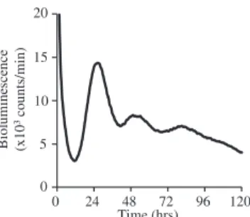
0 24 48 72 96 120 Time (hrs) Bio lumine sce n ce (x1 0 3co unts/min) 0 5 10 15 20
FIG E3. Monitoring of PER2:LUC protein expression in mast cells. Moni-toring of PER2::LUC bioluminescence of BMCMCs derived from PER2::LUC knock-in mice after a medium change (synchronization) for 120 hours is shown.E2
FIG E4. Spontaneousb-hexosaminidase release from wild-type (WT) and Clock-mutated BMCMCs. Release of spontaneous b-hexosaminidase (without IgE stimulation) from the 6-, 12-, 18-, or 24-hour cultured (after the medium change) BMCMCs derived from wild-type and Clock-mutated mice (ClockD19/D19, n5 3).
tcccaggaaa ataactgagg tgatttaggg tactatggat agtatcttgc agttgtgaaa aaccaatcat agaactgaac tgaaatataa gcaaggcaga cctgttctct cttgtgcatt gtgttctggg gtagttactt gtgataaagt cgagtgaaca ctgtgtctgg gcagacatgt
tgctcagtttccactattca gacttgctca caccatttct atgacattca catccctcca
tattttcctt gtcttgttct taccattaca aaaagtgcag cagttgccac acacatgttc agataaagta tatttcataa ttcaaatagt atattgaata attccagtgg catggtttac agcactaaga actctgatgg tgcttttgtc aagatcatct gctgaggata gtaaatacta tgcagaagac aggaagagaa aatgagaaag gggtgaggag agatgggtga gagggagagg gatagcaaga aggaggggag ggggaagaga gatgacataa agttagagat acacatagta tagttttggt cccatacatg aagccaaatgaagagctcat gacataggta tagtagatga
agctgccaaa gagcaccctt gctttttctt ctgctctctt ttatcttctc atttcctaga
tgcttcagta ttagggtcca taaatcccac atctctggca gaattatgct ctaaactgtt cacatttctg tcttcatctt gctctctatt cagtagatgt ctcagttttg cactactttt cattacagca agagaaagga gtcactgata tcaatcagcc tggagacttatctgacaagt aggttccgca tgaagataat cattggtatt cagagccaac ccatcttaac tgcccatcca gagcacaccg catttctgtg taacagtatc tttcattcct ggatagccca attaatgaaa aaATGgacac agaaaatagg agcagagcag atcttgctct cccaaatcca caagaatcct
Antisense primer
TaqMan probe Sense primer
FIG E5. E-box–like elements in the promoter region of the mouse FcεRIb. There are several noncanonical E-box–like elements (‘‘CANNTG’’ or ‘‘CANNTT’’) present in the promoter region of the mouse FcεRIb.E6
CLOCK/ BMAL1-mediated circadian control of a gene transcription can be driven by noncanonical E-box sequences in the promoter region of the gene.E7
‘‘CANNTG’’ or ‘‘CANNTT’’ or their reverse sequences are indicated in green. The translation start site (ATGgacaac..) is indicated in blue. Primers and probe sequence sites used for the ChIP assay (FcεRIb) are also shown in red.
FIG E6. Knockdown of Clock reduces IgE-dependent increases in the total tyrosine phosphorylation in mast cells. IgE-dependent increases in the total tyrosine phosphorylation levels of the cell lysates were evaluated by means of Western blot analysis in Clock or control siRNA–treated wild-type BMCMCs at 48 hours after siRNA transfection. Left panel, Representative pictures of Western blot analysis with anti–phospho-tyrosine antibody 4G10. Right panel, Quantitative analysis (n5 3). *P < .05.
A
B
FIG E7. CLOCK overexpression enhances promoter activity of FcεRIb in mast cells. A, The reporter plasmid22.4k/pGL3-Basic containing 2.4 kb 59-upstream from the translation initiation codon of the mouseb-chain gene or pGL3-Basic was transfected into wild-type BMCMCs with the internal control plasmid pRL-CMV. The relative luciferase activity of 22.4k/pGL3-Basic is represented as the ratio to the activity driven by pGL3-22.4k/pGL3-Basic (n5 3). B, Western blot analysis of the cell lysates described in Fig E7, A, with anti-CLOCK or anti–b-actin antibody. *P < .05.
A B
FIG E8. Knockdown of Clock reduces surface FcεRIa levels on mast cells. Cell-surface FcεRIa expression levels were evaluated by means of fluorescence-activated cell sorting (FACS) analysis in Clock or control siRNA–treated wild-type BMCMCs at 48 hours after siRNA transfection. A, Representative histogram. B, Quantitative analysis (n5 3). *P < .05. MFI, Mean fluorescence intensity.
0 10 20 30 40 0 24 48 72 96 120 0 10 20 30 40 0 24 48 72 96 120 Vehicle CORT 100ng/mL Vehicle cAMP 1mM Bio lumine sce nce Bio lumine sce nce (x1 0 3co unts/min) (x1 0 3co unts/min) Time (hrs) B 0 10 20 30 0 24 48 72 96 120 0 10 20 30 0 24 48 72 96 120 0 10 20 30 0 24 48 72 96 120 Time (hrs) 0 10 20 30 0 24 48 72 96 120 Vehicle CORT 100ng/mL Bio lumine sce n ce (x1 0 3co unts/min) Vehicle Adrenaline 10 M Bio lum ine sc ence (x1 0 3co u n ts/min ) Bio lu m in esce n ce (x1 0 3co unts/min) Bi o lu m ine sce nce (x1 0 3co unts/min) Vehicle Nor-Adrenaline 10 M Vehicle Melatonin 10 M 0 5 10 15 20 0 24 48 72 96 120 Vehicle TSLP 10ng/mL 0 5 10 15 20 0 24 48 72 96 120 Vehicle cAMP 1mM 0 5 10 15 20 0 24 48 72 96 120 Vehicle TNF- 10ng/mL 0 5 10 15 20 0 24 48 72 96 120 Vehicle TGF- 10ng/mL Bio lumine sce nce (x1 0 3co unts/min) Bio lum ine sc ence (x1 0 3co unts/min) Bio lumine sce nce (x1 0 3co unts/min) Bi o lu m ine sce nce (x1 0 3co unts/min) Time (hrs) C μ μ μ β α A
FIG E9. Screening several bioactive molecules for mast cell clockwork modulatory activity. A and B, Indi-cated concentrations of bioactive molecules or vehicles were added to consistent cultures of BMCMCs de-rived from PER2::LUC knock-in mice (PER2::LUC BMCMCs) for 2 hours, and then the PER2::LUC bioluminescence was monitored for 120 hours with a dish-type luminometer. A and B, The experiments were performed independently. Dibutyl cyclic AMP was used as a positive control, and it enhanced the amplitude of the mast cell clockwork (PER2::LUC expression). CORT, Corticosterone. C, Indicated concentra-tions of bioactive molecules or vehicles were added to consistent cultures of BMCMCs derived from PER2::LUC knock-in mice (PER2::LUC BMCMCs) at 24 hours after the medium change. PER2::LUC biolumi-nescence was monitored for 96 hours after stimulation (total of 120 hours), as described above. Note that there were little changes in the kinetics of bioluminescence after the additions of adrenaline, noradrenaline, and melatonin but not corticosterone (CORT).
A B
FIG E10. Epigenetic changes in the promoter region of FcεRIb. A, The acetylation status of histone H3 and H4 in the promoter region of mouse FcεRIb in the 6-, 12-, and 24-hour cultured (after the medium change) wild-type BMCMCs was evaluated by means of ChIP analysis. B, The methylation status of histone H3K4 and H4K9 in the promoter region of mouse FcεRIb in the 6-, 12-, and 24-hour cultured (after the medium change) wild-type BMCMCs was evaluated by means of ChIP analysis (n5 3). *P < .05.