0
抗生物質既治療進行性肺炎患者において、
現在起炎病原体となっている病原体と定着病原体とを
判別する分子技術とその応用
(論文の区分 博士課程)
著者名 FERRY DWI KURNIAWAN
担当指導教員氏名 萩原弘一 教授
自治医科大学大学院医学研究科 地域医療学系
内科系総合医学・総合医学
1 TABLE OF CONTENTS
No Subheading Page
1 Introduction 2 2 Methods 3 3 Results 11 4 Discussion 15 5 Conclusion 18 6 References 19
2 1. Introduction
Pneumonia is one of leading causes in morbidity and mortality globally.[1, 2] Empiric broad-spectrum antibiotics have been used to reduce mortality to anticipate all possible pathogens.[3] This strategy frequently leads to antibiotics misuse and drug-resistant risk.[4] Therefore, adequate antibiotic management based on the prompt identification of the pathogens will guide to rationale antibiotic management. However, identification of causative pathogen may not be performed well due to negative culture result and culture time lag. Thus, rapid and sensitive bacterial diagnostics and antibiotic susceptibility tests are required.[5]
Culture independent diagnostic methods may have advantages over culture methods such as nucleic acid amplification testing. It has significantly reduced turnaround times and enabled high throughput testing for multiple pathogens.[6] Ironically, the highly sensitive nature of PCR may obscure the clear discrimination between pathogens that actually cause pneumonia and those simply colonizing the site. With quantification of both pathogen and human gene into a ratio (HIRA-TAN method) can solve this problem and it has successfully detected pathogens in untreated community-acquired pneumonia (CAP).[7, 8]
One of important clinical questions regarding the HIRA-TAN is whether it shows clinical benefits in pneumonia cases when the first-line antibiotic treatment has failed. Disease progression in the presence of empirical antibiotics is a serious situation. Therefore, rapid and accurate identification of pathogens is required as salvage microbiology.[9] In this circumstance, the HIRA-TAN may have clinical utility. The aim of this study was to preliminarily investigate the utility of the HIRA-TAN in patients who have progressive pneumonia even under treatment with empirical antibiotics.[10] The result will give evidence that the HIRA-TAN provides information on pathogens causing progressive pneumonia, even in the patients for whom culture provides negative results.
3 2. Methods
2.1 Study subjects
This prospective study was carried out between December 2016 and January 2017 in Dr. Zainoel Abidin Hospital, Banda Aceh, Indonesia. The inclusion criteria for patients were to have cough, chest discomfort, productive sputum, fever, and dyspnea on physical examination; leukocytosis on laboratory examination; and progressive pulmonary infiltrates on chest radiology. The patients met criteria of nosocomial pneumonia according to the Indonesian Society of Respirology (ISR) guideline and had antibiotic treatment up to 72 hours prior to specimen collection. Written informed consent was obtained from all patients.
2.2 Sample preparation
The clinical specimens were sputum or pleural fluid that was collected by thoracentesis. All the clinical specimens were collected at Dr. Zainoel Abidin Hospital with sterile screw-top containers, then transported to the laboratory. The specimen was divided into two portions: one was used for conventional culture and the other was used for the HIRA-TAN.
The sputum were expectorated in the morning that enables to collect secretion accumulated during sleep. Patients were instructed to rinse the mouth thoroughly with water to reduce oral contamination. Sputum were collected in a sterile plastic container screw top lid and inspected the gross appearance to ensure that it was not saliva but purulent sputum. The plastic container was labelled with patient name, registration number, and date collection to ensure the correct sample. The container was sent directly from the ward to the laboratory with cold box. The material, media and equipment were kept ready following the cold box arrive in the microbiology laboratory. The direct Gram staining was performed with the purulent portion for smear preparation and inspected ≥25 white blood cells/low-power field the sample quality assessment. After the Gram staining, the sputum was inoculated into blood agar; chocolate agar; and MacConkey agar for fastidious organism. The plate was incubated at 35±2ºC in an
4 incubator. After a 24-hour incubation, the colonies were isolated. The types of bacteria were identified based on their colonial morphology, Gram stain, catalase, oxidase, and the biochemical identification test.
The specimen for the HIRA-TAN was diluted with an equal volume of phosphate-buffered saline and homogenized by vortexing. DNA was isolated using the Blood/Cell DNA Mini Kit GB100 (Geneaid, Taipei, Taiwan). A 200-µl portion of homogenate or pleural effusion was added to 200 µl of GB buffer in a 1.5-ml centrifuge tube and thoroughly mixed. The reaction mixture was incubated at 60 °C for more than 30 min. DNA was eluted in 100 µl deionized distilled water. Genomic DNA was stored in a 4 °C refrigerator until quantitative PCR was performed.
2.3 The HIRA-TAN
The first step of the HIRA-TAN is real time PCR. The PCR is a multiplex TaqMan assay performed in 5 separate reactions.[7, 8] Each reaction detects 2 to 4 target genes, with a total of 18 target genes being detected (Table 1 and 2).
5 Table 1 Primers and probes of the target genes
No Target Gene (Accession number) Oligonucleotide Amplicon (bp) 1 Homo sapiens TNF (NC_000006.12) F* 5′-GTGGAGCTGAGAGATAACCAGC-3′ 153 B 5′-GACCTTGGTCTGGTAGGAGACG-3′
P FAM-CTGTACCTCATCTACTCCCAGGTC-BHQ1 2 Pseudomonas spp. 16S rRNA (AY486350.1) F 5′-GTGAGTAATGCCTAGGAATCTGC-3′ 135 B 5′-CTAATCCGACCTAGGCTCATCTG-3′ P ALEXA532-AGTGGGGGATCTTCGGACCTC-BHQ1 3 Enterobacter spp. tusB (AH002539.2) F 5′-CGTCAGACTTACGGTTAAGCAC-3′ 193 B 5′-GTACCAGCTGGTTAACTGTTGC-3′
P ALEXA594-AGCCAGATGGCCTGGTGATG–BHQ2 4 Staphylococcus spp. Tuf (NC_007168.1) F 5′-CTCAATCACTGGTCGTGGTACTG-3′ 163 B 5′-GTCACCAGCTTCAGCGTAGTC-3′ P ALEXA647-CGTGTTGAACGTGGTCAAATCA-BHQ2 5 Pseudomonas aeruginosa 23S rRNA (AJ549386.1) F 5′-GTTGTCCAAGTTTAAGGTGGTAGG-3′ 97 B 5′-CCACTTCGTCATCTAAAAGACGAC-3′ P FAM-TTCAAGGCCGAGAGCTGATGAC-BHQ1 6 Klebsiella pneumoniae gapA (M66869.1) F 5′-CATCGAGATCGTTGCAATCAACG-3′ 81 B 5′-CGACCGTGAGTGGAGTCATAC-3′
P ALEXA532-AGACGCAGAGTACATGGCTTACATG-BHQ1 7 Streptococcus pneumoniae Pneumolysin (NC_003098.1) F 5′-CAAGGTAAGGAAGTCTTGACTCC-3′ 193 B 5′-GCTTACGCACTAGTGGCAAATCGG-3′ P ALEXA594-AGGGAATGTTCGTAATCTCTCTGTC-BHQ2
6 8 Staphylococcus aureus Thermonuclease (NC_007795) F 5′-GTCCTGAAGCAAGTGCATTTACG-3′ 282 B 5′-GACCTGAATCAGCGTTGTCTTC-3′
P ALEXA647-CGAAGCTTTAGTTCGTCAAGGCTTG-BHQ2 9 Acinetobacter baumannii ompA (AY485227.1) F 5′-GAACTATACAGCTCTTGCTGGC-3′ 87 B 5′-CTCTTGTGGTTGTGGAGCAAC-3′
P FAM-GTTCTTGGTGGTCACTTGAAGC-BHQ1 10 E. coli phoA (M29670.1) F 5′-CGAAGAGGATTCACAAGAACATACC-3′ 88 B 5′-CATTAAGTCTGGTTGCTAACAGC-3′ P ALEXA532-TCAGTTGCGTATTGCGGCGTATG-BHQ1 11 Streptococcus spp. Tuf (AY267003.1) F 5′-CACTGGACGTGGTACAGTTGCTTC-3′ 188 B 5′-GAACACCACGAAGAAGGACACCTAC-3′
P ALEXA594-CAACTTGACGAAGGTCTTGCTGG-BHQ2 12 Haemophilus influenzae 16S rRNA (Z22806.1) F 5′-GACATCCTAAGAAGAGCTCAGAG-3′ 266 B 5′-CTTCCCTCTGTATACGCCATTG-3′ P ALEXA647-CCTTCGGGAACTTAGAGACAG-BHQ2 13 Moraxella catarrhalis CopB (U69982.1) F 5′-GTGCGTGTTGACCGTTTTGAC-3′ 134 B 5′-GTGGCATAGATTAGGTTACCGCTG-3′
P FAM-CCGACATCAACCCAAGCTTTG-BHQ1 14 Bacteroides fragilis 16S rRNA (AP006841.1) F 5′-GACTGCAACTGACACTGATGCTC-3′ 316 B 5′-CAACCATGCAGCACCTTCACAG-3′
P ALEXA532-AGATACCCTGGTAGTCCACACAG-BHQ1 15 Proteus spp. 16S rRNA (NC_010554.1) F 5′-CTCTTCGGACCTTGCACTATC-3′ 127 B 5′-CGTGTCTCAGTCCCAGTGTG-3′
7 P ALEXA594-CGACGATCTCTAGCTGGTCTG-BHQ2 16 Staphylococcus aureus (MRSA) mecA (AY786579.1) F 5′-CGGTAACATTGATCGCAACGTTC-3′ 108 B 5′-CTTTGGTCTTTCTGCATTCCTGG-3′
P ALEXA647-TGGAAGTTAGATTGGGATCATAGCG-BHQ2 17 Mycobacterium spp. 16S rRNA gene (NC_000962) F 5′-GTTCACCGAAGTACTGCTGGAC-3′ 445 B 5′-CAACCACAGGAAGTACTCGACATTG-3′ P ALEXA532- CTGTCGTTCATCTCGTTGGCTAC-BHQ1 18 Mycobacterium tuberculosis cyp141 (NC_000962) F 5′-GACAAGCACCTCGATTCCGAC-3′ 169 B 5′-GGACAGCACTCCCTTTACATCG-3′
P ALEXA647- CACACAGCATGGCTCGTCACTC-BHQ2
*F: Forward primer; B: Backward primer; P: Taqman probe; BHQ: Black Hole quencher™
Table 2. Multiplex PCRs
Reaction FAM Cy3 Texas Red Cy5
I Homo sapiens (TNF) Pseudomonas sp. (16S rRNA) Enterobacter sp. (tusB) Staphylococcus sp. (Tuf) II P. aeruginosa (23S rRNA) K. pneumoniae (gapA) S. pneumoniae (Pneumolysin) S. aureus (Thermonuclease) III A. baumannii (ompA) E. coli (phoA) Streptococcus sp. (Tuf) H. influenzae (16S rRNA) IV M. catarrhalis (CopB) B. fragilis (16S rRNA) Proteus spp. (16S rRNA)
Methicillin-resistant S. aureus; MRSA (mecA) V n/a Mycobacterium sp. (16S rRNA) n/a M. tuberculosis (cyp141)
The PCR was performed in 5 separate multiplex reactions. The Smart Cycler is able to simultaneously detect 4 fluorescent channels. We used 4 fluorophores: FAM for the FAM channel, ALEXA532 for the Cy3 channel, ALEXA594 for the Texas Red channel, and ALEXA647 for the Cy5 channel. n/a: not applicable.
8 The reactions contain a 25-µl reaction mixture consisting of 12.5 µl Takara Premix Ex Taq (Takara Bio Inc., Shiga, Japan), primers for one or more pathogens (each 250 nM), primers for the human tumor necrosis factor (TNF) gene, fluorescence-labelled detection probes for the pathogens (250 nM), fluorescence-labelled detection probes for the human TNF gene (250 nM), and a 1-µl sample, where the average DNA concentration was 7.13 ng/µl in the current study. The PCR was performed in the SmartCycler II (Cepheid Inc., Sunnyvale, USA) with a cycling profile of 95 °C for 30 s, followed by 35 cycles of 95 °C for 8 s, 61 °C for 25 s, and 72 °C for 20 s.
The second step of the HIRA-TAN is the evaluation of the quality of the samples by the cycle threshold for the human TNF gene: 𝐶𝑡#$%&'. The TNF gene comprises a single copy in the human genome, and thus its copy number in the reaction directly reflects the number of human cells. Sputum samples with a 𝐶𝑡#$%&' < 27 contain many human cells and have been classified in M2-P3, according to the Miller and Jones’ classification, and are expected to provide highly reliable results.[7, 11]
The last step is to normalize the number of pathogen cells to the number of human cells. Human cells in purulent sputum or in para-pneumonic pleural effusion are mostly inflammatory cells (neutrophils and lymphocytes). Therefore, the ratio of the number of pathogen cells to the number of human cells is likely to reflect the balance of power between them.[7, 8] The log of the ratio of pathogen cells to human cells is expressed as ∆𝐶𝑡,&-./01', that is,
∆𝐶𝑡,&-./01' = − 𝐶𝑡,&-./01'− 𝐶𝑡#$%&' = log 8$%91: /; <&-./01' =1>>?8$%91: /; .$%&' =1>>? .
Pathogens with a ∆𝐶𝑡,&-./01' above the cut-off value overwhelm inflammatory cells in numbers and are thus considered to be the pathogens causing pneumonia. Otherwise, the pathogens are considered to be the colonizing ones.
9
2.4 Microbial and human genomic DNA
Escherichia coli (12713G), Pseudomonas aeruginosa (106052G), and Klebsiella pneumoniae subsp. pneumoniae (14940G) genomic DNA was purchased from Biological
Resource Centre, NITE (Chiba, Japan). Staphylococcus aureus subsp. aureus Rosenbach ATCC 700699D-5, Streptococcus pneumoniae (Klein) Chester ATCC BAA-255D-5,
Moraxella catarrhalis (Frosch and Kolle) Bovre ATCC 25240D-5, Haemophilus influenzae
(Lehmann and Neumann) Winslow et al. ATCC 51907D, Proteus mirabilis Hauser ATCC 12453D, Acinetobacter baumannii ATCC BAA-1605D-5, Bacteroides fragilis (Veillon and Zuber) Castellani and Chalmers ATCC 25285D-5, and Mycobacterium tuberculosis (Zopf) Lehmann and Neumann ATCC 27294D-2 genomic DNA were purchased from the American Type Culture Collection (Rockville, MD, USA). Human placental genomic DNA was purchased from Promega Corp. (Madison, WI, USA).
2.5 Real-time PCR design
We targeted pathogens that are the most frequent causes of pneumonia in Indonesia (Table 1).[12, 13] A primer pair plus a fluorescent-labelled probe forms one detection unit. A total of 18 detection units implemented in 5 multiplex PCRs (Table 2) were able to detect 6 genera (Pseudomonas spp., Enterobacter spp., Staphylococcus spp., Streptococcus spp.,
Mycobacterium spp., and Proteus spp.); 10 species (P. aeruginosa, E. coli, S. aureus, S. pneumoniae, M. tuberculosis, K. pneumoniae, A. baumannii, H. influenzae, M. catarrhalis, and B. fragilis); 1 drug-resistant bacterial gene (mecA); and 1 human gene (TNF). Some pathogens
were detected by multiple detection units. For example, methicillin-resistant S. aureus (MRSA) was detected by the detection units for Staphylococcus spp., S. aureus, and the mecA gene; M.
10 Each detection unit was designed so that it specifically amplified and detected the target sequence. Firstly, the specificity was confirmed in silico by the primer BLAST program searching the entire Genbank database. Next, the nucleotide sequence of the amplified fragments from the microbial genomic DNA or human placental genomic DNA (for the TNF gene) was confirmed by Sanger sequencing. Finally, detection of the specific fluorescence signals was confirmed by running the real-time PCRs for a variety of mixtures of multiple microbial genomic DNA and human placental genomic DNA.
2.6 Ethics
The research protocol for the current study was approved by the institutional review boards of Dr. Zainoel Abidin Hospital, Syiah Kuala University (07/KE/FK/2016 dated on 24 June 2016), and Jichi Medical University (Rindai16-057 dated on 11 Jan 2017).
11 3. Results
3.1 Patients
A total of 27 patients were enrolled in this study. Sputum was collected from 25 patients, and pleural effusion from the remaining 2. All patients were referred from the primary (19 patients) and the secondary (8 patients) health care centers. Accordingly, the predominant type of disease was hospital-acquired pneumonia (HAP: 25 patients, 93%). Other 2 patients had pleuropneumonia and empyema. The patients had median age of 62 years (range: 21-90 years) and were predominantly male (22 patients, 88%). All patients had comorbid disease, including lung cancer and other neoplastic diseases (11 patients, 41%), chronic respiratory diseases such as COPD (9 patients, 33%), cardiovascular disease (3 patients, 11%), cerebrovascular disease (1 patient, 4%), thyroid disorder (2 patients, 8%), and mandibular fracture (1 patient, 4%). As expected from the high rates of HAP and comorbid disease, many patients (82%) had pneumonia with multilobar infiltrates.
3.2 The HIRA-TAN
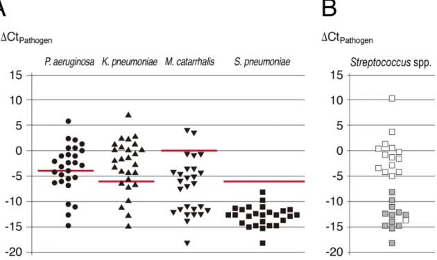
The cut-off values that discriminate the pathogens causing pneumonia were determined for P. aeruginosa, K. pneumoniae, M. catarrhalis, and S. pneumoniae in a previous HIRA-TAN study that investigated untreated pneumonia.[8] We adopted the same cut-off values in this study because it was considered to be a good starting point to preliminarily investigate the utility of the HIRA-TAN in progressive pneumonia without any preceding studies targeting the similar patient group. The 3 steps for the HIRA-TAN are usually completed in 6 h. The graph drawn for these 4 pathogens indicates the samples in which these pathogens are likely the cause of pneumonia (Figure 1A). The HIRA-TAN clearly discriminated S. pneumoniae (Figure 1A, rightmost plots) from Streptococcus spp. (Figure 1B). In many samples, the HIRA-TAN provided 2 pathogens suggestive of being the causative agent, which is also clear from the
12 HIRA-TAN results plotted for each sample (Figure 2). The bacterial culture definitely detected in nearly half of the samples. Some bacteria are difficult to culture because their natural milieu in the culture medium is difficult to simulate. Moreover, previous antibiotic history may also prohibit the growth of colonies. The results of the culture-positive samples and those obtained by the HIRA-TAN were in close agreement (Table 3). In addition, for samples with non-diagnostic culture, the HIRA-TAN provided the likely causative pathogens. No fluorescent signal was observed from other probes, indicating that the other bacteria do not exist in the sample.
Figure 1 The results of the HIRA-TAN
(A) ∆𝐶𝑡,&-./01' values for the 4 pathogens for which the cut-off value has been reported in a previous study.[7, 8] The cut-off values (shown as red lines) were −4 for P. aeruginosa, −6 for
K. pneumoniae, 0 for M. catarrhalis, and −6 for S. pneumoniae. (B) The ∆𝐶𝑡@-:1<-/=/==$? AB. for S. pneumoniae was obtained by the detection unit for both S. pneumoniae and Streptococcus spp. (gray boxes), whereas that for other bacteria belonging to Streptococcus spp. was detected only by the detection unit for Streptococcus spp. (white boxes).
13 Figure 2 ∆𝐶𝑡,&-./01'− 𝑐𝑢𝑡𝑜𝑓𝑓 for each sample
The ∆𝐶𝑡,&-./01'− 𝑐𝑢𝑡𝑜𝑓𝑓 value was graphed for each sample. The pathogens that show values above 0 (a red line) are likely to be those causing pneumonia. Samples are in the same order as in Table 3: samples 1–25 are sputum, whereas samples 26 and 27 are pleural effusion.
14 Table 3. Results of the culture and the HIRA-TAN
Sample name Previous antibiotics Culture HIRA-TAN
Pathogen 1 Pathogen 2 Sputum 1 CTRX Coagulase-negative Staphylococcus
Klebsiella pneumoniae Pseudomonas aeruginosa
Sputum 2 LVFX No growth Klebsiella pneumoniae
Sputum 3 LVFX No growth Negative
Sputum 4 CTRX No growth Klebsiella pneumoniae
Sputum 5 CTRX Enterobacter aerogenes Klebsiella pneumoniae Pseudomonas aeruginosa
Sputum 6 CTRX No growth Negative
Sputum 7 CTRX+AZM Klebsiella pneumoniae Klebsiella pneumoniae Pseudomonas aeruginosa
Sputum 8 CTRX Coagulase-negative Staphylococcus Klebsiella pneumoniae
Sputum 9 LVFX Coagulase-negative Staphylococcus Negative
Sputum 10 LVFX Pseudomonas aeruginosa Pseudomonas aeruginosa Klebsiella pneumoniae
Sputum 11 CTRX Pseudomonas aeruginosa Negative
Sputum 12 CTRX Moraxella catarrhalis Moraxella catarrhalis Klebsiella pneumoniae
Sputum 13 LVFX Moraxella catarrhalis Moraxella catarrhalis
Sputum 14 LVFX No growth Klebsiella pneumoniae Pseudomonas aeruginosa
Sputum 15 CTRX Pseudomonas aeruginosa Negative
Sputum 16 CTRX+AZM Pseudomonas aeruginosa Klebsiella pneumoniae Pseudomonas aeruginosa
Sputum 17 LVFX No growth Negative
Sputum 18 CTRX Pseudomonas aeruginosa Klebsiella pneumoniae Pseudomonas aeruginosa
Sputum 19 LVFX Klebsiella pneumoniae Klebsiella pneumoniae Pseudomonas aeruginosa
Sputum 20 LVFX No growth Klebsiella pneumoniae Pseudomonas aeruginosa
Sputum 21 CTRX Klebsiella pneumoniae Klebsiella pneumoniae Pseudomonas aeruginosa
15
Sputum 23 CTRX Klebsiella pneumoniae Klebsiella pneumoniae Pseudomonas aeruginosa
Sputum 24 CTRX+AZM No growth Klebsiella pneumoniae
Sputum 25 CTRX+AZM Klebsiella pneumoniae Klebsiella pneumoniae Pseudomonas aeruginosa
Effusion 1 CTRX No growth Klebsiella pneumoniae Pseudomonas aeruginosa
Effusion 2 CAZ No growth Klebsiella pneumoniae Pseudomonas aeruginosa
Abbreviations: CTRX, ceftriaxone; LVFX, levofloxacin; AZM, azithromycin; CAZ, ceftazidime. The pathogen 1 was that which gave the highest ∆𝐶𝑡,&-./01', while pathogen 2 was that which gave the second highest value.
4. Discussion
In the current study, we aimed to preliminarily evaluate the clinical utility of the HIRA-TAN for pneumonia with a progressive course after a failure of antibiotic treatment. In many cases, the HIRA-TAN identified pathogen(s) with numbers overwhelming those of inflammatory cells, thus judging them as likely causative agents of the pneumonia.
More sensitive and quicker results within 6 h were demonstrated by the HIRA-TAN method than the culture. Bacterial culture that was performed in parallel often failed to detect any pathogens. This disparity may be explained by the low performance of culture. We suspect that, in some cases, a trace amount of the previous antibiotics remaining may have prohibited the growth of colonies.[14, 15]
Detection of a bacterial genome by PCR does not necessarily indicate that the bacterium is viable in the sample, because PCR detects DNA in both living and dead cells. However, a report of the HIRA-TAN for CAP demonstrated that the copy number of bacterial DNA in a sample quickly decreased after effective antibiotic treatment.[7] Therefore, practically, when a pathogen(s) is detected as the causative pathogen by the HIRA-TAN, it should be considered viable and set as the treatment focus.
K. pneumoniae, P. aeruginosa, or both were detected in most patients. On the other
hand, S. pneumoniae, which is the most common pneumonia pathogen worldwide, was not detected. This suggests that the antibiotics used in the area investigated in the current study
16 may be effective for S. pneumoniae, pending confirmation. Our data suggests that K.
pneumoniae and P. aeruginosa survived the treatment and became the causative pathogen(s)
for the subsequent pneumonia. This finding is slightly different from that of a previous report, in which most of the causative pathogens in HAP and ventilator-associated pneumonia (VAP) were S. aureus, K. pneumoniae, or P. aeruginosa.[16] This difference may reflect hospital ward representation differences or geographical variance. However, such information is important for installing region-specific therapeutic strategies.
The overgrowth of K. pneumoniae and P. aeruginosa is important from the perspective of the development of drug resistance. The list of antibiotics previously administered (Table 2) shows the predominance of a few kinds of antibiotics in the hospitals studied. Some of these antibiotics are expected to be effective against K. pneumoniae and P. aeruginosa; however, these pathogens are detected.
There were discrepancies between HIRA-TAN and culture where both K. pneumoniae and P. aeruginosa were detected. Quantitative PCR has a wide dynamic range and it may explain some portion of the discrepancies. A positive result of HIRA-TAN indicates that a large number of bacterial cells exist in the sample and thus it necessitates serious consideration. A small sample size and a lack of a control group prohibited us to draw definite conclusion. A larger clinical trial is warranted to assess the sensitivity and specificity of HIRATAN in the same clinical setting.
The finding of HIRA-TAN can be utilized for selecting second line antibiotics in the absence of culture-positive result. However, the finding must be carefully considered according to clinical judgment. Clinical monitoring as respond to second line antibiotic must be analyzed. The HIRA-TAN may also be effective in identifying local tendencies of bacterial drug resistance and the biased use of specific antibiotics in the region. The detection of multiple pathogens by the HIRA-TAN is in clear contrast to the results of the HIRA-TAN in untreated
17 CAP, where only 1 pathogen was suggested in most of the samples.[8] Our result was consistent with that of a previous report, in which multiple pathogens were found in nearly half the HAP cases that occurred in patients with risk factors, including prior antibiotic use, older age, and existing comorbid disease.[17] The management of nosocomial infection is complex, and the treatment should be directed by the accurate knowledge of the pathogens involved.[18] The detection of multiple pathogens by the HIRA-TAN will help to control nosocomial infection, not in an empirical manner, but in a pathogen-oriented way.
18 5. Conclusion
We consider that the HIRA-TAN is useful as an alternative diagnostic test and can provide important information while waiting for confirmation from cultures. Early initiation of a pathogen-directed, second-line therapy will become possible by employing the HIRA-TAN as salvage microbiology. However, the study was limited by a small sample size within only one ward, pending confirmation by a larger clinical trial. We conclude that the HIRA-TAN provided valuable information for determining the second-line treatment for pneumonia that fails the initial round of antibiotic therapy.
19 6. References
1 Lozano R, Naghavi M, Foreman K, Lim S, Shibuya K, Aboyans V, Abraham J, Adair T, Aggarwal R, Ahn SY, Alvarado M, Anderson HR, Anderson LM, Andrews KG, Atkinson C, Baddour LM, Barker-Collo S, Bartels DH, Bell ML, Benjamin EJ, Bennett D, Bhalla K, Bikbov B, Bin Abdulhak A, Birbeck G, Blyth F, Bolliger I, Boufous S, Bucello C, Burch M, Burney P, Carapetis J, Chen H, Chou D, Chugh SS, Coffeng LE, Colan SD, Colquhoun S, Colson KE, Condon J, Connor MD, Cooper LT, Corriere M, Cortinovis M, de Vaccaro KC, Couser W, Cowie BC, Criqui MH, Cross M, Dabhadkar KC, Dahodwala N, De Leo D, Degenhardt L, Delossantos A, Denenberg J, Des Jarlais DC, Dharmaratne SD, Dorsey ER, Driscoll T, Duber H, Ebel B, Erwin PJ, Espindola P, Ezzati M, Feigin V, Flaxman AD, Forouzanfar MH, Fowkes FG, Franklin R, Fransen M, Freeman MK, Gabriel SE, Gakidou E, Gaspari F, Gillum RF, Gonzalez-Medina D, Halasa YA, Haring D, Harrison JE, Havmoeller R, Hay RJ, Hoen B, Hotez PJ, Hoy D, Jacobsen KH, James SL, Jasrasaria R, Jayaraman S, Johns N, Karthikeyan G, Kassebaum N, Keren A, Khoo JP, Knowlton LM, Kobusingye O, Koranteng A, Krishnamurthi R, Lipnick M, Lipshultz SE, Ohno SL, Mabweijano J,
MacIntyre MF, Mallinger L, March L, Marks GB, Marks R, Matsumori A, Matzopoulos R, Mayosi BM, McAnulty JH, McDermott MM, McGrath J, Mensah GA, Merriman TR, Michaud C, Miller M, Miller TR, Mock C, Mocumbi AO, Mokdad AA, Moran A,
Mulholland K, Nair MN, Naldi L, Narayan KM, Nasseri K, Norman P, O'Donnell M, Omer SB, Ortblad K, Osborne R, Ozgediz D, Pahari B, Pandian JD, Rivero AP, Padilla RP, Perez-Ruiz F, Perico N, Phillips D, Pierce K, Pope CA, 3rd, Porrini E, Pourmalek F, Raju M, Ranganathan D, Rehm JT, Rein DB, Remuzzi G, Rivara FP, Roberts T, De Leon FR, Rosenfeld LC, Rushton L, Sacco RL, Salomon JA, Sampson U, Sanman E, Schwebel DC, Segui-Gomez M, Shepard DS, Singh D, Singleton J, Sliwa K, Smith E, Steer A, Taylor JA, Thomas B, Tleyjeh IM, Towbin JA, Truelsen T, Undurraga EA, Venketasubramanian N, Vijayakumar L, Vos T, Wagner GR, Wang M, Wang W, Watt K, Weinstock MA, Weintraub R, Wilkinson JD, Woolf AD, Wulf S, Yeh PH, Yip P, Zabetian A, Zheng ZJ, Lopez AD, Murray CJ, AlMazroa MA, Memish ZA. Global and regional mortality from 235 causes of death for 20 age groups in 1990 and 2010: a systematic analysis for the Global Burden of Disease Study 2010. Lancet. 380: 2095-128; 2012.
2 Azmi S, Aljunid SM, Maimaiti N, Ali AA, Muhammad Nur A, De Rosas-Valera M, Encluna J, Mohamed R, Wibowo B, Komaryani K, Roberts C. Assessing the burden of pneumonia using administrative data from Malaysia, Indonesia, and the Philippines. Int J
Infect Dis. 49: 87-93; 2016.
3 Niederman MS. Use of broad-spectrum antimicrobials for the treatment of pneumonia in seriously ill patients: maximizing clinical outcomes and minimizing selection of resistant organisms. Clin Infect Dis. 42 Suppl 2: S72-81; 2006.
4 Leone M, Martin C. How to break the vicious circle of antibiotic resistances? Curr
Opin Crit Care. 14: 587-92; 2008.
5 Kerremans J, Verboom P, Stijnen T, Hakkaart-van Roijen L, Goessens W, Verbrugh H, Vos M. Rapid identification and antimicrobial susceptibility testing reduce antibiotic use and accelerate pathogen-directed antibiotic use. J Antimicrob Chemother. 61: 428-35; 2008. 6 Mackay IM. Real-time PCR in the microbiology laboratory. Clin Microbiol Infect. 10: 190-212; 2004.
7 Hirama T, Yamaguchi T, Miyazawa H, Tanaka T, Hashikita G, Kishi E, Tachi Y, Takahashi S, Kodama K, Egashira H, Yokote A, Kobayashi K, Nagata M, Ishii T, Nemoto M, Tanaka M, Fukunaga K, Morita S, Kanazawa M, Hagiwara K. Prediction of the pathogens that are the cause of pneumonia by the battlefield hypothesis. PLoS One. 6: e24474; 2011.
20 8 Hirama T, Minezaki S, Yamaguchi T, Kishi E, Kodama K, Egashira H, Kobayashi K, Nagata M, Ishii T, Nemoto M, Tanaka M, Fukunaga K, Kanazawa M, Hagiwara K. HIRA-TAN: a real-time PCR-based system for the rapid identification of causative agents in pneumonia. Respir Med. 108: 395-404; 2014.
9 Farrell JJ, Hujer AM, Sampath R, Bonomo RA. Salvage microbiology: opportunities and challenges in the detection of bacterial pathogens following initiation of antimicrobial treatment. Expert Rev Mol Diagn. 15: 349-60; 2015.
10 Kurniawan FD, Alia D, Priyanto H, Mahdani W, Hagiwara K. HIRA-TAN detects pathogens of pneumonia with a progressive course despite antibiotic treatment. Respir
Investig. 57: 337-44; 2019.
11 Miller DL. A study of techniques for the examination of sputum in a field survey of chronic bronchitis. Am Rev Respir Dis. 88: 473-83; 1963.
12 Chung DR, Song JH, Kim SH, Thamlikitkul V, Huang SG, Wang H, So TM, Yasin RM, Hsueh PR, Carlos CC, Hsu LY, Buntaran L, Lalitha MK, Kim MJ, Choi JY, Kim SI, Ko KS, Kang CI, Peck KR. High prevalence of multidrug-resistant nonfermenters in hospital-acquired pneumonia in Asia. Am J Respir Crit Care Med. 184: 1409-17; 2011.
13 Farida H, Gasem MH, Suryanto A, Keuter M, Zulkarnain N, Satoto B, van der Eijk AA, Djokomoeljanto R, Wahyono H, Verbrugh HA, Severin JA, van den Broek PJ. Viruses and Gram-negative bacilli dominate the etiology of community-acquired pneumonia in Indonesia, a cohort study. Int J Infect Dis. 38: 101-7; 2015.
14 Stewart EJ. Growing unculturable bacteria. J Bacteriol. 194: 4151-60; 2012. 15 Abers MS, Musher DM. The yield of sputum culture in bacteremic pneumococcal pneumonia after initiation of antibiotics. Clin Infect Dis. 58: 1782-3; 2014.
16 Jones RN. Microbial etiologies of hospital-acquired bacterial pneumonia and ventilator-associated bacterial pneumonia. Clin Infect Dis. 51 Suppl 1: S81-7; 2010. 17 Lynch JP, 3rd. Hospital-acquired pneumonia: risk factors, microbiology, and treatment. Chest. 119: 373S-84S; 2001.
18 Breathnach AS. Nosocomial infections and infection control. Medicine. 41: 649-53; 2013.