Hemodynamic and Cardiac Motion Analyses in an Experimental Model of Off‑pump Coronary Bypass Surgery
Effects of Stabilizer and Heart Positioner
Shinichi ISHII,Ryuichi NAGAHORI,Motohiro OSHIUMI,Hiroshi OKUYAMA, Yoshimasa SAKAMOTO,Kiyozo MORITA,and Kazuhiro HASHIMOTO
Department of Cardiovascular Surgery, The Jikei University School of Medicine
ABSTRACT
Objective:During off‑pump bypas s surgery,it is most important to prevent hemodynamic deterioration. However,the precise mechanism under lying hemodynamic changes has not been clarified and information regarding regional car diac wall motion is lacking.
Methods:Twelve Landrace pigs were used to study the effect of volume loading(Study 1,n=
6)and infusion of norepinephrine(Study 2,n=6). We evaluated various hemodynamic parameters and regional cardiac wall motion,while using a hear t stabilizer and exposing each target vessel with or without a heart positioner. A digital two‑di mensional sonomicrometer was used to evaluate regional cardiac motion.
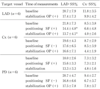
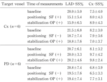
Results:Study 1(under volume loading):1)For the left anterior descending artery(LAD) anastomosis:No significant changes were noted in any of the parameters. 2)For the left circum- flex artery(Cx)anastomosis:Arterial pressure(AP)and right ventricular end‑diastolic volume (RVEDV)decreased compared with the baseline level(P<0.001〜0.05). 3)For the posterior descending artery(PD)anastomosis:AP decreas ed compared with the baseline value(P<0.01),but no significant change of RVEDV was recognized. Us e of a positioner ameliorated the decrease of AP at the Cx(P<0.05),but reduced segmental shor tening(%)in the LAD territory when the heart was made vertical for exposure of the Cx(P<0. 05)and the PD(P=0.06). Study 2(under volume loading with norepinephrine infusion). After the administration of norepinephrine,there were no significant changes of any parameters or regional wall motion in any territory.
Conclusion:1)Various hemodynamic parameters were useful for monitoring,but RVEDV seems to be an excellent parameter for monitori ng volume loading. 2)Adequate volume loading in combination with norepinephrine should be cons idered at appropriate times,before exposure of the Cx or PD. 3)A heart positioner reduced the decrease of AP,but its effect on regional wall motion was unsatisfactory. (Jikeikai Med J 2005;52:93‑101)
Key words:coronary artery bypass surgery,off‑pump coronary bypass surgery,hemodynamics
INTRODUCTION
Coronary bypass surgery under cardiopulmonary bypass(CPB)is a safe procedur e for the treatment of
ischemic heart disease. However,use of CPB is known to induce activati on of the proteolytic and inflammatory systems,as wel l as consumption of clotting factors and platel ets. Procedures that do
Jikeikai Med J 2005;52:93‑101
Received for publication,June 10,2005
石井 信一,長堀 隆一,鴛海 元博,奥山 浩,坂本 吉正,森田紀代造,橋本 和弘
Mailing address:Kazuhiro HASHIMOTO,Department of Cardiovascular Surgery,The Jikei University School of Medicine,3‑25‑8, Nishi‑shimbashi,Minato‑ku,Tokyo 105‑8461,Japan.
E‑mail:kaz‑hashi@jikei.ac.jp
93
not use CPB are likely to avoid these unwanted effects. Several off‑pump cor onary bypass surgery
(OPCAB)series,in which CPB was not used during coronary bypass surgery,have been r eported since early 1980s . Application of OPCAB r esulted in reduced cardiac enzyme rel ease,reduced use of blood products,and a slightly s horter hospital stay .
However,to achieve the full benefit of OPCAB,
hemodynamic stability is mandatory,particularly when vertical displacement of the heart is required to achieve optimal exposure of the circumflex artery
(Cx)and posterior descending artery(PD). To facili- tate access to the target vessels,numerous positioning techniques and devices have been developed,which include deep pericardial s utures(Lima sutures;Fig.
1),sur ical ads,cardiac ositionin devices(Fi.2),
Fig.2 Fig.1
and various types of cardiac stabilizers (Fig.2). These devices have expanded the performance of OPCAB , with excellent r esults. However, a detailed hemodynamic study has not been performed during OPCAB and uncer tainties about the hemodynamic changes stil l exist.
Vertical displacement causes significant hemodynamic instability,whi ch manifests as a reduc-
tion of stroke volume and arterial blood pressure. In order to prevent these det eriorations,several investi-
gations have been performed . However,it has not been well establi shed how to prevent hemodynamic deteriorat ion. Furthermore, little information is available r egarding the change of regional cardiac wall moti on.
In the experimental setting, we evaluated hemodynamic changes dur ing OPCAB by monitoring various parameters, incl uding continuous right ventricular end‑diastolic vol ume,while using an Octo-
pus stabilizer(Medtronic,Fig.2)and exposing each target vessel with or wi thout the Starfish heart positioner(Medtronic,Fig.2) . In addition,a digital two‑dimensional sonomicr ometer(Sono Metrics)was used to evaluate regional car diac wall motion.
SUBJECT AND METHODS
Twelve Landrace pigs(weight:45‑55 kg)were used in this study. The ani mals were divided into two experimental groups.
Operative procedure and experimental setup
After fasting for 24 hours,general anesthesia was induced with 10 mg/kg of ket amine intramuscularly.
Volume‑controlled ventilation was started with a 1:1 mixture of oxygen and room ai r,as well as inhalation of 3% sevoflurane after endot racheal intubation.
The external electrocardiogram was recorded contin- uously to monitor the heart rate(HR)and rhythm.
Under general anesthesia,each pig was placed in the supine position. A Swan‑Ganz catheter(continuous end‑diastolic volume t hermodilution catheter 774HF75;Edwards Life Sci ences)was introduced via the right internal jugular vei n for measurement of the mean central venous pressur e(CVP),mean pulmonary
artery pressure (PAP),cardiac output(CO),right ventricular end‑diastolic vol ume (RVEDV),stroke volume(SV),right ventricul ar ejection fraction(EF),
and mixed venous blood oxygen saturation(SVO ). Principle of measuring of RVEDV,SV and EF was introduced in the previous l iterature . For measure-
ment of mean arterial blood pressure(AP),an 8 Fr sheath was inserted into t he right femoral artery.
After median sternotomy,the pericardium was opened and the heart was s uspended. To prevent the onset of ventricular arrhyt hmia,1 mg/kg of lidocaine hydrochloride was injected i ntravenously on opening of the pericardial sac,and cont inuous infusion was performed at the rate of 1 mg/kg/hour . To evaluate regional cardiac motion wi th a digital two‑dimen-
sional sonomicrometer,four ultrasonic crystals were fixed on the surface of the heart(one pair of crystals in the right and left side of t he left anterior descending artery(LAD)and another pai r of crystals near the Cx.
This system allowed us to calculate segmental shortening (SS%)to asses s regional cardiac wall motion. For calculation of SS%,we used the follow-
ing equation:SS%=(SL(ED)(−SL(ES))/SL(ED), where SL is segment length,ED is end‑diastolic length, and ES is end‑s ystolic length. After hemodynamic stability was at tained,baseline data were collected,including HR,AP,CVP,PAP,CO,
RVEDV,SV,EF,SVO ,and SS%.
Then the heart was positioned to expose each target vessel with or without using the Starfish(Fig.
2). The LAD was exposed by placing a sponge under the left side of the heart. When the Starfish was not used to position the heart ver tically,deep pericardial sutures(Lima sutures,Fig.1)wer e used for exposure of both Cx and PD. One s uture was placed just below the left lower pulmonary vei n,and two other sutures were placed on the diaphr agmatic surface of the pericardium near the infer ior vena cava and halfway between the previous t wo sutures. After hemodynamics became st able,data were collected.
Then the Octopus stabilizer(Fig.2)was positioned around a target vessel cor responding to#7,#4 or
#12(or 14)to obtain the optimum setting for sham coronary anastomosis and t he same measurements were repeated. In both St udy 1 and Study 2,the
Hemodynamics in Off‑pump Bypass
December,2005 95
target vessels were not occluded during measurement and anastomosis was not actually carried out. An adequate interval(at leas t 10 minutes)was allowed between measurements in var ious settings to mini-
mize the effect of the previous setting.
Study 1:Effect of simple volume loading:After adequate volume loading wi th one liter of saline fol-
lowing induction of anesthesia,infusion of normal saline at 10 ml/kg/hour was continued intravenously from the start of the operat ion. In study 1,inotropic agents were not used for t he treatment of a decrease in arterial pressure or other hemodynamic changes.
Based on the assumption that adequate volume load- ing had been performed,no dditional volume loading was done,even when hemodynami c deterioration was observed.
Study 2:Effect of norepinephrine under volume loading:Norepinephrine ( 0.05μg/kg/min) was in-
fused during exposure of the Cx or the PD under volume loading by the same pr otocol as in study 1.
This experiment was designed to evaluate the preven- tive effect of norepinephrine on hemodynamic deterio- ration in the presence of adequate volume loading.
All animals received humane care in compliance with the“Guide for the Car e Use of Laboratory Animals” published by t he National Institutes of Health(NIH publication 85‑25,r evised in 1985).
Statistical analysis
Continuous variables are expressed as the mean±
standard deviation. The paired t‑test(LAD terri- tory)and repeated measures ANOVA with Fisherʼs post‑hoc test(Cx and PD t erritories)were used to assess the statistical si gnificance of differences between baseline values and dat a obtained during vertical displacement and s tabilization of the heart.
In addition,the unpaired t‑test was used for compari- son between two groups at baseline. In all analyses, P<0.05 was considered significant.
RESULTS
Study 1 (Effect of simple volume loading) (N=6)
Hemodynamics changes are summarized in Table 1.
1) For LAD anastomosis.(stabilization with the Octopus in placing a sponge under the left side of the heart)
No significant changes were noted in any of the parameters(HR,AP,PAP,CVP,CO,RVEDV,SV,
EF,and SVO ).
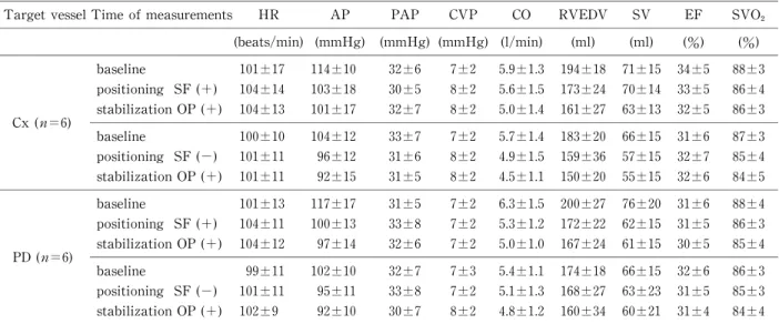
2) For Cx anastomosis
(I) Positioning with the Starfish and stabilization with the Octopus
a) Pos itioning:AP decreased from 87±9 mmHg
(baseline)to 64±6 mmHg(positioning)(P<0.001)and PAP decreased from 25±3 mmHg(baseline)to 21±2 mmHg (positioning)(P<0. 05). However,the other parameters did not show any significant changes.
b) Addition of stabilization:When the Octopus was set,there were no s ignificant changes of the parameters,except that RVEDV s howed a significant decrease(P<0.05)from 163±15 ml(baseline)to 140±
15 ml after stabilization. The deterioration of AP and PAP (P<0.05‑0.001)was cons istently observed compared with the baseline values.
(II) Positioning with deep pericardial stitches(with- out the Starfish)and stabilization with the Octopus
a) Positioning:AP decreased f rom 80±5 (baseline)mmHg to 60±5 mmHg(positioning)(P< 0.001)and CO decreased from 4.3±0.7 l/min(baseline) to 3.5±0.7 l/min(positioning)(P<0.05),but the other parameters did not show s ignificant changes.
b) Addition of stabilization:Only AP decreased further from 60±5 mmHg ( positioning) to 52±6 mmHg(stabilization)(P<0. 05)after the addition of stabilization. Compared wi th the data obtained at baseline,deterioration of CO,SV,and SVO became obvious(P<0.05‑0.001),and there was a decrease of RVEDV from 166±35 ml(bas eline)to 136±16 ml(sta-
bilization)(P<0.05). 3) For PD anastomosis
(I) Positioning with the Starfish and stabilization with the Octopus
a) Pos itioning:After positioning,only AP de-
creased from 87±7 mmHg(baseline)to 73±9 mmHg (positioning)(P<0.01),while the other parameters showed no significant changes .
b) Addition of stabilization:Although significant changes of AP were stil l detected compared with