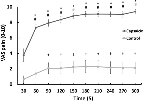
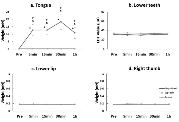
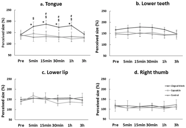
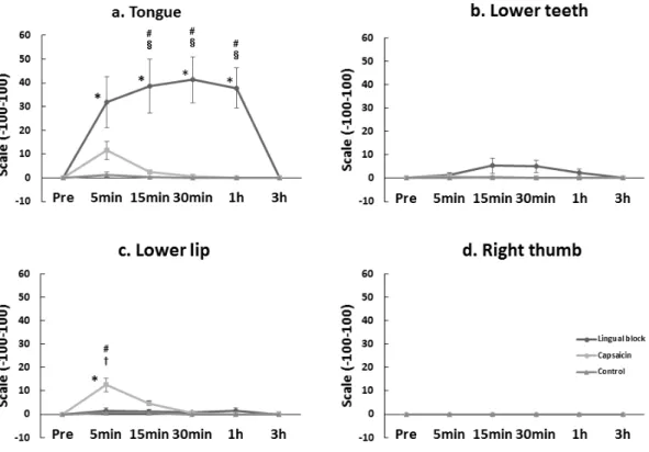
Influence of somatosensory changes in tongue to somatosensory function and perceptual distortion of tongue in healthy participants
(舌の体性感覚の変化が舌の感覚機能および知覚の歪みに及ぼす影響)
Mika Honda
Nihon University Graduate School of Dentistry at Matsudo Oral Function and Rehabilitation
(Director: Prof. Misao Kawara)
日本大学大学院松戸歯学研究科 顎口腔機能治療学専攻 本田 実加
(指導:川良 美佐雄 教授)