Contents lists available atScienceDirect
Pathology - Research and Practice
journal homepage:www.elsevier.com/locate/prp
Clinical signi fi cance of M2 macrophages expressing heme oxygenase-1 in malignant transformation of ovarian endometrioma
Yuki Yamada
a,⁎, Tomoko Uchiyama
b, Fuminori Ito
c, Naoki Kawahara
a, Kenji Ogawa
a, Chiho Obayashi
b, Hiroshi Kobayashi
aaDepartment of Obstetrics and Gynecology, Nara Medical University, 840 Shijocho, Kashihara, Nara, Japan
bDepartment of Diagnostic Pathology, Nara Medical University, 840 Shijocho, Kashihara, Nara, Japan
cDepartment of Obstetrics and Gynecology, Nara Prefecture General Medical Center, 2-897-5, Shichijonishimachi, Nara, Japan
A R T I C L E I N F O
Keywords:
Ovarian endometrioma
Endometriosis-associated ovarian cancer Macrophage
Phenotype Heme oxygenase-1
A B S T R A C T
Malignant transformation of endometriosis is a rare and still poorly understood event, but is associated with the distortion of the pro-oxidant and anti-oxidant balance. The aim of the present study was to quantify the numbers of macrophages polarized as M1 or M2 phenotypes and the expression of heme oxygenase (HO)-1 in tissue sections from patients with benign ovarian endometrioma (OE) and its malignant transformation (endometriosis- associated ovarian cancer, EAOC). We performed a retrospective study at the Department of Gynecology, Nara Medical University hospital from December 2012 to March 2015. This study included 53 patients with OE (n = 33) and EAOC (n = 20), and we evaluated polarized functional status of macrophages by im- munohistochemical staining of CD68, CD11c, CD163 and HO-1. The number of the M1 phenotype (CD11c+, p = 0.001) and the M2 phenotype (CD163+, p = 0.009) was significantly lower in EAOC patients than in OE patients. Analyzing the correlations between the studied markers, the expression of CD68, CD11c, and CD163 proteins significantly correlated with each other (p < 0.001). The number of M2 phenotypes expressing HO-1 was significantly decreased in the EAOC group, compared with the OE group (P < 0.001), demonstrating sustained downregulation of an antioxidant marker, HO-1, in EAOC. In conclusion, reduced number of M2 macrophages expressing HO-1 may have an important role in promoting malignant transformation of OE.
1. Introduction
Endometriosis is defined as the presence of endometrial glands and stroma outside the uterus, most often in the pelvic peritoneum and ovaries. This disorder affects an estimated 10% of women in the re- productive age group and is basically an estrogen-dependent benign gynecological disease. Repeated episodes of retrograde menstruation or ovarian hemorrhage occur in the peritoneal cavity or ovarian en- dometrioma (OE), respectively [1].
Endometriosis results in a local accumulation of hemoglobin, heme and iron species, which causes severe oxidative stress and antioxidants depletion, leading to distortion in the redox balance [2]. Altered homeostatic redox balance of the environment may support chronic inflammation, uncontrolled proliferation and then malignant transfor- mation [2]. Actually, endometriosis increases the subsequent risk of developing endometriosis-associated ovarian cancer (EAOC) [3,4].
Endometriotic lesions are highly infiltrated with various leukocytes, including macrophages that secrete antioxidants and
immunosuppressive factors [1,5–7]. Endometriosis infiltrating macro- phages might adapt to these stressful environmental conditions by se- creting antioxidants that control excess oxidative stress [1].
Oxidative stress and inflammation in the surrounding environment contribute to several aspects of macrophage functions including re- cruitment, activation and the shift in cell polarity. Macrophage polar- ization may have a distinct role in the inflammatory, immune and neoplastic diseases [8,9]. Macrophages are classified into the pro-in- flammatory, classically activated M1 macrophages that possess anti- tumor activity and the alternatively activated M2 macrophages that support tumor progression and malignancy [8,9]. For the phenotypical characterization of infiltrating macrophages, immunohistochemistry employing selected literature-based prototype-antibodies against CD11c, CD163 and CD68 was evaluated in this study. Although CD11c is a specific marker in macrophages and dendritic cells [10], CD11c- based immunohistochemistry is used as a M1 phenotype specific marker [11,12].
CD11c, a member of integrin family, induces tissue injury and the
https://doi.org/10.1016/j.prp.2018.12.017
Received 10 October 2018; Received in revised form 24 November 2018; Accepted 11 December 2018
⁎Corresponding author at: Department of Obstetrics and Gynecology, Nara Medical University, 840 Shijo-cho, Kashihara, 634-8522, Japan.
E-mail address:[email protected](Y. Yamada).
Pathology - Research and Practice 215 (2019) 639–643
0344-0338/ © 2018 Elsevier GmbH. All rights reserved.
T
inflammatory response [13]. The protein expression of M2 phenotype specific marker CD163 is tested via immunohistochemistry. CD163 is a hemoglobin/haptoglobin scavenger receptor and acts to protect tissues from oxidative damage [5]. CD68 is specifically expressed by tissue macrophages and used as a pan-macrophages marker [14].
When the microenvironment is altered in endometriosis by an ex- cess oxidative stress and inflammatory insult, knowledge on how macrophages respond to its changes is limited. An antioxidant enzyme, heme oxygenase-1 (HO-1), is a key mediator that allows the resolution of inflammatory processes [15,16]. HO-1 is responsible for the cata- bolism of heme to carbon monoxide (CO), biliverdin, and iron. HO-1 was poorly expressed in peritoneal mesothelium and macrophages, but highly expressed in endometriotic lesions [17].
However, HO-1 expression in the microenvironment of OE and its malignant transformation remain to be investigated.
The aim of the present study was to quantify the numbers of mac- rophages polarized as M1 or M2 and the expression of HO-1 in tissue sections from patients with OE and EAOC.
2. Materials and methods 2.1. Tissue samples
The data of patient demographic features and clinicopathologic characteristics were collected from a database containing medical re- cords and pathology reports. The samples consist of surgically resected tissues from hospitalized patients. A total of 53 formalin-fixed, paraffin- embedded tissue specimens, including 33 cases of pathologically con- firmed benign OE and 20 cases of EAOC, were retrospectively collected from Department of Gynecology at Nara Medical University hospital, Japan, between December 2012 to March 2015. In EAOC cases, we confirmed morphologic documentation of the continuous transition from benign endometriotic epithelial cells to atypical endometriosis andfinally to invasive carcinoma within the same specimen. No pa- tients underwent hormonal therapy or chemotherapy prior to the sur- gery. The protocols were approved by the ethics committee of Nara Medical University(Approved number 1570/2017). This study is a retrospective observational study, carried out by the opt-out method of our hospital website.
2.2. Immunohistochemical staining for CD68, CD11, CD163, and HO-1 Paraffin sections of serial 2μm thickness were taken from each original block, one section was stained with hematoxylin and eosin for diagnostic confirmation and the other sections were immunostained for CD68, CD11c, CD163 and HO-1. Immunohistochemically staining for tissue sections was performed using anti-Human CD11c monoclonal antibody (1:100 dilution; Leica, Newcastle, UK), anti-Human CD68 monoclonal antibody (1:200 dilution; Dako, Denmark), anti-Human CD163 monoclonal antibody (1:200 dilution, Leica, Newcastle, UK), and anti-HO-1 monoclonal antibody (1:100 dilution; Abcam, Cambridge, UK). Immunohistochemistry was performed using the Leica Bond Max systems (Mitsubishi Chemical Medicine Co., Tokyo, Japan) following the manufacture’s instructions. The primary antibodies were replaced with PBS for negative control experiments. The slides were counterstained with hematoxylin, and mounted under coverslips.
Light microscopy was used to evaluate the intensity and localization of the staining. In each tissue section, round cells with a well-re- presented cytoplasm positive for CD68, CD11c, CD163, HO-1 were identified by screening the entire area in a low-powerfield (100×field) andfive visualfields showing the highest infiltration rate of positive cells were selected (400×field) as described previously [14]. Results were expressed as the number of positive cell staining with brown in cytoplasm or cell membrane. Most of the staining intensity were moderate to strong brown particles. Cell counting results were obtained by 2 independent observers (TU and YY). If discrepant results were
obtained by two observers, these slides were re-evaluated using a dual- vision microscope, in order to achieve a consensus.
2.3. Statistical analysis
Analyses were performed by SPSS version 21.0 (IBM SPSS, Armonk, NY, USA). Numerical variables, including age, were presented as the mean ± SD. Variables that did not present normal distribution were expressed by median, minimum and maximum values. Categorical data were analyzed by the Chi-square test or the Fisher’s 2-tailed test. To determine the level of significance in differences in positive cell num- bers between the two groups, Mann-Whitney U test and t-test were applied. The Pearson, Spearman’s correlation coefficient analysis was implemented as descriptive statistics. Two-sided P < 0.05 was con- sidered to indicate a statistically significant difference.
3. Results
3.1. Study population
The major clinical and pathological characteristics of the two groups of patients are listed inTable 1. There was a significant difference be- tween the OE and EAOC group in median values of variables such as age (p < 0.001), tumor size (p < 0.001), menopausal status (p < 0.001) and CA125 levels (p < 0.001). There were no statistical differences for parity status between the two study groups (P > 0.05).
3.2. The number and phenotypes of macrophages in the OE and EAOC group
We identify and quantify the amounts of accumulated cells in tissue samples by immunostaining. Sample immunohistochemical images are shown in Fig. 1 CD68-, CD11c- and CD163-positive cell staining is mainly seen in the cytoplasm and/or membrane of macrophages.
Generally, CD163+cells and CD68+cells were ampler than CD11c+ cells in both tissues. CD marker-positive cells revealed a randomly dispersed distribution, but "hot-spot" areas were identified in the OE and EAOC tissue specimens. A greater number of CD68+cells, CD11c+
Table 1
Demographic and clinical characteristics of the study population.
Baseline characteristics of the two groups
The OE group The EAOC group p-value
Number 33 20
Benign ovarian endometrioma
Clear cell (n=13) Endometrioid (n=3) Serous (n=1) Mucinous (n=1) Other (n=2)
FIGO stage – IA(n=5)
IC(n=13) II(n=2) Age at diagnosis,
mean ± SD 37.2 ± 8.5 51.1 ± 11.7 <0.001
median (range) 38 (26–65) 50 (36–69)
Nulliparous
n (%) 10 (30.3%) 10 (50.0%) 0.152
Premenopause
n (%) 32 (97.0%) 11 (55.0%) <0.001
A maximum diameter of the cyst
mean ± SD 72.0 ± 24.4 131.6 ± 47.0 <0.001
median (range) 70 (25-137) 120 (38-230) CA125
mean ± SD 73.9 ± 47.1 687.7 ± 1428.6 <0.001
median (range) 61(17-235) 110(6-6272)
OE ovariana endometrioma, EAOC endometriosis-associated ovarian cancer, FIGO The International Federation of Gynecology and Obstetrics.
cells, or CD163+cells was observed in the stroma of both tissues. The number of CD68+cells (Fig. 1B and G), CD11c+cells (Fig. 1C and H) and CD163+ cells (Fig. 1D and I) were lower in the EAOC group, compared with the OE group. Our immunohistochemical results also showed that CD68, CD11c and CD163 expression was downregulated in the majority of EAOC samples compared with corresponding adjacent noncancerous endometriotic tissues (Fig.2).
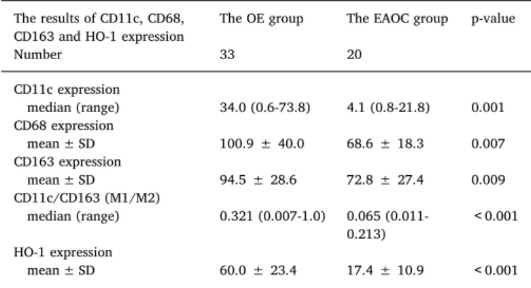
The number of macrophage infiltration according to CD68, CD11c and CD163 counts are summarized in Table 2. The mean CD68+, CD11c+ and CD163+ counts in the EAOC group were significantly lower than those in the OE group. We further analyzed M1-like cells/
M2-like cell ratio (M1/M2) based on cell staining with CD11c+cells and CD163+cells in the consecutive sections. The M1/M2 ratio was significantly lower in the EAOC group, compared with the OE group (p < 0.001).
Table 3shows comparison of the correlations between the studied markers, including CD68, CD11c, and CD163. Analyzing the correla- tions between the studied markers, the expression of CD68, CD11c, and CD163 proteins significantly correlated with each other (p < 0.001)
Fig. 1.Immunohistochemical analysis of CD68, CD11c, CD163 and HO-1 expression in OE and EAOC tissues. To evaluate the changes in the macrophage phenotypes, consecutive tissue sections were immunohistochemically analyzed for the presence of CD68, CD11c, CD163 and HO-1 using avidin-biotin complex method. These slides show polarized macro- phage infiltration. Representative images of hematoxylin and eosin stains (AandF), as well as CD68 (BandG), CD11c (CandH), CD163 (D and I) and HO-1 (E and J) im- munohistochemical staining in OE (A, B, C, D andE) and EAOC tissue (F, G, H, I andJ) (original magnification x100). CD68, CD11c and CD163 were localized to the cytoplasm and cell membranes. HO-1 was predominantly expressed in the cytoplasm of macrophages.
Hematoxylin was used to stain the nuclei.
Fig. 2.CD68, CD11c and CD163 expression in EAOC tissues and matched adjacent nontumor tissue samples. Our immunohistochemical re- sults showed that CD11c, CD163, and CD68 expression was downregulated in the majority of EAOC samples (black arrow) compared with corresponding adjacent noncancerous en- dometriotic tissues (white arrow).
Table 2
Distribution of cells expressing CD11c, CD68, CD163 and HO-1 in OE and EAOC tissues. OE ovariana endometrioma, EAOC endometriosis-associated ovarian cancer, HO-1 heme oxygenase-1.
The results of CD11c, CD68, CD163 and HO-1 expression
The OE group The EAOC group p-value
Number 33 20
CD11c expression
median (range) 34.0 (0.6-73.8) 4.1 (0.8-21.8) 0.001 CD68 expression
mean ± SD 100.9 ± 40.0 68.6 ± 18.3 0.007
CD163 expression
mean ± SD 94.5 ± 28.6 72.8 ± 27.4 0.009
CD11c/CD163 (M1/M2)
median (range) 0.321 (0.007-1.0) 0.065 (0.011- 0.213)
<0.001 HO-1 expression
mean ± SD 60.0 ± 23.4 17.4 ± 10.9 <0.001
OE ovariana endometrioma, EAOC endometriosis-associated ovarian cancer, HO-1 heme oxygenase-1.
3.3. The HO-1 expression in OE and EAOC tissues
Next, the HO-1 expression was evaluated using im- munohistochemistry in the OE and EAOC group (Fig. 1E and J). The ectopic endometrial cells and epithelial cancer cells were generally unstained. HO-1 staining was strongly observed in the stroma. The number of HO-1-positive cells were significantly lower in the EAOC tissues, compared with the OE tissues (P<0.001) (Table 2). Since HO-1 acts as a target for M2 macrophages, we further investigated the co- expression pattern of HO-1 and CD163 in OE and EAOC tissues. Indeed, examination of consecutive sections of OE tissue revealed that HO-1 is expressed in the majority of the cells expressing CD163, suggesting that HO-1 is co-expressed in a major subset of M2 macrophages (Fig. 1D and E). This co-expression was observed in all specimens, regardless of age (data not shown).
Fig. 3shows comparison of the association between the numbers of CD163+ cells and HO-1+ cells. We observed statistically significant correlations between CD163 and HO-1 (r = 0.201, p = 0.003).
4. Discussion
To our knowledge, this is the first study that investigates the number and phenotypes of macrophages in malignant transformation of endometriosis. Here we evaluated the immunohistochemical expression of CD68 (pan-macrophage marker), CD11c (M1 marker), CD163 (M2 marker) and the influence of HO-1 in a benign OE and its malignant transformation, EAOC. A higher number of macrophages were observed in patients with OE compared to those with EAOC, and most had the M2 phenotype expressing HO-1. This study demonstrates sustained down- regulation of an antioxidant marker, HO-1 in EAOC, suggesting that reduced number of M2 macrophages expressing HO-1 may have an important role in promoting malignant transformation of OE.
First, we found that CD163+M2-like cells were outnumbered by
CD11c+M1-like cells in the two groups. M1 macrophages secrete in- flammatory cytokines and contribute to the adaptive immune response through Th1 responses [8,18]. In contrast, anti-inflammatory M2 macrophages induce Th 2 responses and tissue repair [8,18]. Environ- mental stimuli shape macrophage plasticity in the OE and EAOC groups, and modify macrophages phenotypes from an anti-tumor M1 type to a pro-tumor M2 type [8,18–20]. It has been established that CD163 is classified into the marker of alternatively activated M2 phe- notype and acts as a hemoglobin scavenger receptor on macrophage [5]. The possible role of macrophages in carcinogenesis has not been directly examined between the OE and its malignant transformation.
However, our data support thefinding that endometriosis environment itself has a propensity to develop into ovarian cancer.
Second, although HO-1 expression in the stromal macrophages has been seen in the other cancer tissues [21], the impact of HO-1 ex- pression on EAOC progression has not been explored. We have found that the CD163+ M2-like cells were HO-1+ in OE, but malignant transformation may be associated with, and at least partly due to, re- duced number of M2 phenotypes expressing HO-1. Endometriotic cyst fluid contains much higher levels of iron-related compounds, such as hemoglobin species, heme and free iron, compared with EAOC sample [22]. Repeated episodes of hemorrhage in OE induce excess oxidative stress and trigger DNA damage, mutations and genome instability, de- monstrating the dichotomy between cytotoxicity and proliferation in endometriotic cells [1,2,19,23]. Thus, stimuli in environment create increased cellular susceptibility to oxidant-mediated cell killing or carcinogenesis. In benign OE, autoxidation and Fenton reaction of he- moglobin from the ferrous Fe2+(oxyhemoglobin) state to the ferric Fe3+ (methemoglobin) state lead to production of excess reactive oxygen species (ROS), such as O2−and∙OH, that are major sources of DNA damage and mutation [24]. HO-1 is strongly and rapidly induced in response to oxidative cytotoxic conditions, such as excessive pro- duction of ROS. The ferric Fe3+rich in OE can induce a high expression of HO-1 [25], supporting thefinding that the number of HO-1+cells was significantly higher in the OE group than in the EAOC group. In contrast, EAOC showed lower oxidant activity than OE, due to the de- creased expression of ferric Fe3+(methemoglobin) and increased ex- pression of ferrous Fe2+ (oxyhemoglobin) [2]. EAOC appears to be associated with an antioxidant environment and decreased response to oxidative stress. Ourfindings support that M2 phenotypes no longer produce HO-1 in the EAOC microenvironment. EAOC macrophages demonstrate sustained downregulation of an antioxidant marker, HO-1, possibly due to decreased oxidative stress. Taken together, the pattern of redox balance supports that reduced oxidative stress may be involved in the pathogenesis of malignant transformation.
Third, the expression of HO-1 in macrophages was decreased in some cancers such as lung cancer [26]. Theoretically, HO-1 down- regulation leads to the increase of ROS and DNA damage in cells, which may promote the initiation of carcinogenesis [27]. Conversely, HO-1 has been detected in tumor-infiltrating macrophages and shows the impact on cancer progression, aggressiveness, invasion, metastasis, and poor prognosis [27,28]. At late phase of tumorigenesis, HO-1 over- expression may promote cancer progression through inducing the ex- pression of angiogenic factors, such as vascular endothelial growth factor (VEGF) [29]. Several lines of evidence have highlighted the role of HO-1 in cancer progression through modulating tumor micro- environment [27]. Thus, the function of HO-1 in the pathogenesis of cancer progression remains controversial.
In conclusion, the aberrant microenvironment in endometriotic milieus can induce alterations in macrophage recruitment and its po- larization phenotype, which significantly induces the shift to M2 phe- notype expressing HO-1. Reduced number of M2 macrophages expres- sing HO-1 may have an important role in promoting malignant transformation of endometriosis. Although the exact reasons for EAOC carcinogenesis are unclear at the present time, this study supports the homeostatic redox balance hypothesis that there are at least two phases Table 3
Comparison of the correlations between the studied markers. Correlations be- tween the distribution of CD68+, CD11c+, and CD163+ cells of each case are shown.
CD11c CD163
CD68 y=0.484x-19.370
rs**=0.524, p < 0.001
y=0.610+33.011 r*=0.552, p < 0.001
CD163 y=0.423x-14.328
rs**=0.270, p < 0.001
* Pearson’s product moment correlation coefficient.
** Spearman’s rank correlation coefficient.
Fig. 3.Comparison of the association between CD163 and HO-1. The Pearson’s correlation coefficient analysis for the correlation of CD163+cells with HO-1+ cells. Plot of the number of HO-1+cells against that of CD163+cells.
of EAOC tumorigenesis: the initial wave of the reduced infiltration of M2 macrophages would be followed by the second wave of subsequent increase of ROS and DNA damage in endometriotic cells, and then the final big wave of EAOC carcinogenesis. The specific molecular me- chanisms by which reduced HO-1 expression favors the promotion of malignant transformation of endometriosis still require further in- vestigation.
Funding
This study was not funded.
Conflict of interest
The authors declare that the research was conducted in the absence of any commercial orfinancial relationships that could be construed as a potential conflict of interest.
Ethical approval
All procedures performed in studies involving human participants were in accordance with the ethical standards of the institutional re- search committee and with the 1964 Helsinki declaration and its later amendments or comparable ethical standards. The protocols were ap- proved by the ethics committee of Nara Medical University(Approved number 1570/2017). This study is a retrospective observational study, carried out by the opt-out method of our hospital website.
Acknowledgements
The present study was supported by grant-in-aid for Scientific Research from the Ministry of Education, Science, and Culture of Japan to the Department of Obstetrics and Gynecology, Nara Medical University (to H.K.).
References
[1] H. Kobayashi, Potential scenarios leading to ovarian cancer arising from en- dometriosis, Redox. Rep. 21 (3) (2016) 119–126.
[2] T. Iwabuchi, C. Yoshimoto, H. Shigetomi, H. Kobayashi, Oxidative stress and an- tioxidant defense in endometriosis and its malignant transformation, Oxid. Med.
Cell Longev. (2015),https://doi.org/10.1155/2015/848595.
[3] H. Kobayashi, K. Sumimoto, N. Moniwa, M. Imai, K. Takakura, T. Kuromaki, E. Morioka, K. Arisawa, T. Terao, Risk of developing ovarian cancer among women with ovarian endometrioma: a cohort study in Shizuoka, Japan, Int. J. Gynecol.
Cancer 17 (1) (2007) 37–43.
[4] H. Kobayashi, Ovarian cancer in endometriosis: epidemiology, natural history, and clinical diagnosis, Int. J. Clin. Oncol. 14 (5) (2009) 378–382.
[5] M. Bacci, A. Capobianco, A. Monno, L. Cottone, F. Di Puppo, B. Camisa, M. Mariani, C. Brignole, M. Ponzoni, S. Ferrari, P. Panina-Bordignon, A.A. Manfredi, P. Rovere- Querini, Macrophages are alternatively activated in patients with endometriosis and required for growth and vascularization of lesions in a mouse model of disease, Am. J. Pathol. 175 (2) (2009) 547–556.
[6] F. Itoh, Y. Komohara, K. Takaishi, R. Honda, H. Tashiro, S. Kyo, H. Katabuchi, M. Takeya, Possible involvement of signal transducer and activator of transcription- 3 in cell-cell interactions of peritoneal macrophages and endometrial stromal cells in human endometriosis, Fertil. Steril. 99 (6) (2013) 1705–1713.
[7] K.A. Smith, C.B. Pearson, A.M. Hachey, D.L. Xia, L.M. Wachtman, Alternative ac- tivation of macrophages in rhesus macaques (Macaca mulatta) with endometriosis, Comp. Med. 62 (4) (2012) 303–310.
[8] Y. Wang, Y. Fu, S. Xue, A. Ai, H. Chen, Q. Lyu, Y. Kuang, The M2 polarization of macrophage induced by fractalkine in the endometriotic milieu enhances inva- siveness of endometrial stromal cells, Int. J. Clin. Exp. Pathol. 15 (1) (2013) 194–203.
[9] F. Porcheray, S. Viaud, A.C. Rimaniol, C. Léone, B. Samah, N. Dereuddre-Bosquet, D. Dormont, G. Gras, Macrophage activation switching: an asset for the resolution of inflammation, Clin. Exp. Immunol. 142 (3) (2005) 481–489.
[10] J.A. Villadangos, P. Schnorrer, Intrinsic and cooperative antigen-presenting func- tions of dendritic-cell subsets in vivo, Nat. Rev. Immunol. 7 (7) (2007) 543–555.
[11] M.E. Shaul, G. Bennett, K.J. Strissel, A.S. Greenberg, M.S. Obin, Dynamic, M2-like remodeling phenotypes of CD11c+ adipose tissue macrophages during high-fat diet–induced obesity in mice, Diabetes 59 (5) (2010) 1171–1181.
[12] M. Weber, C. Iliopoulos, P. Moebius, M. Büttner-Herold, K. Amann, J. Ries, R. Preidl, F.W. Neukam, F. Wehrhan, Prognostic significance of macrophage po- larization in early stage oral squamous cell carcinomas, Oral Oncol. 52 (2016) 75–84.
[13] E.F. Plow, T.A. Haas, L. Zhang, J. Loftus, J.W. Smith, Ligand binding to integrins, J.
Biol. Chem. 275 (29) (2000) 21785–21788.
[14] J.X. Song, Z.J. Dian, Y. Wen, F. Mei, R.W. Li, Y.L. Sa, Assessment of the number and phenotype of macrophages in the human BMB samples of CML, Biomed. Res. Int.
(2016),https://doi.org/10.1155/2016/8086398.
[15] T.H. Tu, Y. Joe, H.S. Choi, H.T. Chung, R. Yu, Induction of heme oxygenase-1 with hemin reduces obesity-induced adipose tissue inflammation via adipose macro- phage phenotype switching, Mediat. Inflamm. (2014),https://doi.org/10.1155/
2014/290708.
[16] E. Sierra-Filardi, M.A. Vega, P. Sánchez-Mateos, A.L. Corbí, A. Puig-Kröger, Heme oxygenase-1 expression in M-CSF-polarized M2 macrophages contributes to LPS- induced IL-10 release, Immunobiology 215 (9-10) (2010) 788–795.
[17] A. Van Langendonckt, F. Casanas-Roux, M.M. Dolmans, J. Donnez, Potential in- volvement of hemoglobin and heme in the pathogenesis of peritoneal en- dometriosis, Fertil. Steril. 77 (3) (2002) 561–570.
[18] F.O. Martinez, A. Sica, A. Mantovani, M. Locati, Macrophage activation and po- larization, Front. Biosci. 1 (13) (2008) 453–461.
[19] S.Y. Ko, A. Ladanyi, E. Lengyel, H. Naora, Expression of the homeobox gene HOXA9 in ovarian cancer induces peritoneal macrophages to acquire an M2 tumor-pro- moting phenotype, Am. J. Pathol. 184 (1) (2014) 271–281.
[20] Y.F. He, M.Y. Zhang, X. Wu, X.J. Sun, T. Xu, Q.Z. He, W. Di, High MUC2 expression in ovarian cancer is inversely associated with the M1/M2 ratio of tumor-associated macrophages and patient survival time, PLoS One (2013),https://doi.org/10.1371/
journal.pone.0079769.
[21] T. Matsuo, Y. Miyata, K. Mitsunari, T. Yasuda, K. Ohba, H. Sakai, Pathological significance and prognostic implications of heme oxygenase 1 expression in non- muscle-invasive bladder cancer: correlation with cell proliferation, angiogenesis, lymphangiogenesis and expression of VEGFs and COX-2, Oncol. Lett. 13 (1) (2017) 275–280.
[22] C. Yoshimoto, T. Iwabuchi, H. Shigetomi, H. Kobayashi, Cystfluid iron-related compounds as useful markers to distinguish malignant transformation from benign endometriotic cysts, Cancer Biomark. 15 (4) (2015) 493–499.
[23] M. Zhang, Y. He, X. Sun, Q. Li, W. Wang, A. Zhao, W. Di, A high M1/M2 ratio of tumor-associated macrophages is associated with extended survival in ovarian cancer patients, J. Ovarian Res. (2014),https://doi.org/10.1186/1757-2215-7-19.
[24] K.A. Bauckman, E. Haller, I. Flores, M. Nanjundan, Iron modulates cell survival in a Ras- and MAPK-dependent manner in ovarian cells, Cell Death Dis. (2013),https://
doi.org/10.1038/cddis.2013.87.
[25] T. Kassa, S. Jana, F. Meng, A.I. Alayash, Differential heme release from various hemoglobin redox states and the upregulation of cellular heme oxygenase-1, FEBS Open Bio. 8;6 (9) (2016) 876–884.
[26] P. Boschetto, E. Zeni, L. Mazzetti, D. Miotto, N. Lo Cascio, P. Maestrelli, E. Marian, P. Querzoli, M. Pedriali, B. Murer, E. De Rosa, L.M. Fabbri, C.E. Mapp, Decreased heme-oxygenase (HO)-1 in the macrophages of non-small cell lung cancer, Lung Cancer 59 (2) (2008) 192–197.
[27] L.Y. Chau, Heme oxygenase-1: emerging target of cancer therapy, J. Biomed. Sci.
(2015),https://doi.org/10.1186/s12929-015-0128-0.
[28] Y.S. Liu, H.S. Li, D.F. Qi, J. Zhang, X.C. Jiang, K. Shi, X.J. Zhang, X.H. Zhang, Zinc protoporphyrin IX enhances chemotherapeutic response of hepatoma cells to cis- platin, World J. Gastroenterol. 20 (26) (2014) 8572–8582.
[29] H. Was, T. Cichon, R. Smolarczyk, D. Rudnicka, M. Stopa, C. Chevalier, J.J. Leger, B. Lackowska, A. Grochot, K. Bojkowska, A. Ratajska, C. Kieda, S. Szala, J. Dulak, A. Jozkowicz, Overexpression of heme oxygenase-1 in murine melanoma: increased proliferation and viability of tumor cells, decreased survival of mice, Am. J. Pathol.
169 (6) (2006) 2181–2198.