ORIGINAL ARTICLE
EFFECTS OF HALOTHANE ANESTHESIA ON ELECTROCARDIOGRAM PARAMETERS IN MICE
Shuhei Nozaki1),Yoshiki Ogata1),Manabu Yonekura2),Chong Han1),Hidetoshi Niwa3), Tetsuya Kushikata3),Kazuyoshi Hirota3),Yasushi Matsuzaki4),Hirofumi Tomita2), Tadaatsu Imaizumi5),Shirou Itagaki6),Daisuke Sawamura4),and Manabu Murakami1)
Abstract We analyzed the effects of halothane as an inhalational anesthetic agent on electrocardiogram (ECG)
parameters in C57/BL6 mice. Following induction of anesthesia using 2% halothane, the ECGs showed a regular pattern and the heart rate (HR) was within an acceptable range (~500 bpm). The HR decreased with increasing halothane concentration in a concentration-dependent fashion.
The frequency domain analysis showed that the high-frequency (HF) component decreased and the low- frequency (LF) component increased in a concentration-dependent fashion. Therefore, the LF/HF ratio increased with increasing halothane concentration, suggesting effects on the autonomic nervous system.
We analyzed the pharmacological response to sympathetic blockade with propranolol, a typical adrenergic ȕ-blocker, under halothane anesthesia. Propranolol administration resulted in a decreased HR; interestingly, intraperitoneal injection of propranolol (120 ȝg/kg body weight) resulted in arrhythmia (sick sinus syndrome)
during anesthesia with 3% halothane.
Our results indicate the importance of selecting a suitable anesthetic agent for C57/BL6 mice in pharmacological studies.
Hirosaki Med.J. 67:129―135,2017
Key words: Inhalation halothane anesthesia; barbiturate; mouse; ECG.
1) Department of Pharmacology, Hirosaki University Graduate School of Medicine
2) Department of Cardiology and Nephrology, Hirosaki University Graduate School of Medicine
3) Department of Anesthesiology, Hirosaki University Graduate School of Medicine
4) Department of Dermatology, Hirosaki University Graduate School of Medicine
5) Department of Vascular Biology, Institute of Brain Science, Hirosaki University Graduate School of Medicine
6) Department of Pharmacy, Hirosaki University, Graduate School of Medicine
Correspondence: M. Murakami Recieved for publication, May 30, 2016 Accepted for publication, June 28, 2016
Introduction
In pharmacological animal studies, reliable methods of inducing general anesthesia are important. Transgenic mice are used in research on inherited human cardiac diseases, and electrophysiological studies have been performed in transgenic mice to characterize the electrical phenotype of the heart. However, studies of mouse electrocardiogram (ECG) and heart rate variability (HRV) under inhalational anesthesia,
especially with halothane, are lacking 1-6).
Halothane is a volatile anesthetic agent with a minimum alveolar concentration (MAC)
of 0.74%, which allows attainment of various depths of anesthesia. Its blood-gas partition coefficient is 2.4, which enables rapid induction and recovery. In addition, halothane is included in the WHO Model List of Essential Medicines and was formerly considered to be one of the most important medicines. However, it has been replaced by other anesthetic agents̶such as
component (LF; 0.15‒1.5 Hz) and high-frequency component (HF; 1.5‒5 Hz) were evaluated by means of ECG stimulation (ML846 Power Lab System, AD Instruments, Dunedin, New Zealand).
The ECG and heart rate variability (HRV)
were analyzed with HRV program with the original tools (AD Instruments). The LF component is considered to be an indicator of sympathetic nerve tonus, and the HF component an indicator of para-sympathetic nerve tonus
(cardiac vagal control)2, 3). Therefore, the LF/
HF ratio reflects autonomic nerve tonus.
After achieving a stable condition, the h a l o t h a n e c o n c e n t r a t i o n w a s i n c r e a s e d incrementally to 2.5, 3, 3.5, and 4%, and the effect of each concentration was evaluated. For sympathetic blockade, propranolol (60, 120, 240, or 480 ȝg/kg body weight) was administered intraperitoneally to mice anesthetized with 2 or 3% halothane. Each group contains at least 8 mice.
Statistical Analysis
The results are expressed as means ± standard error (S.E.). Statistical significance was determined using an analysis of variance
(ANOVA) followed by Dunnettʼs t-test, and p values < 0.05 were considered to indicate significant differences.
Results
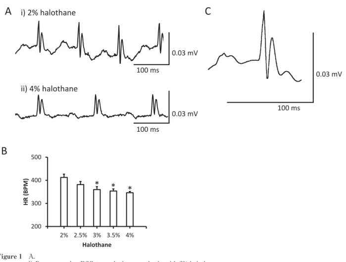
Concentration-dependent changes in the ECG Changes in the ECGs of mice during anesthesia with 2 and 4% halothane are shown in Fig. 1A, and the statistical analysis is shown in Fig. 1B. The HR decreased significantly in a concentration-dependent manner (412 ± 14, 381 ± 13, 360 ± 13, 354 ± 9, and 346 ± 6 bpm with 2, 2.5, 3, 3.5, and 4% halothane, respectively;
n = 8). An averaging view of the ECGs showed conserved P, Q, R, S, and T waves during sevoflurane, isoflurane, and desflurane̶due to
its hepatotoxicity. Trifluoroacetic acid, a dead- end metabolite of halothane, plays a critical role in the development of acute liver injury.
According to a previous report, ~1 in 10,000‒
30,000 exposures results in halothane hepatitis.
Since this side effect rarely develops in infants, halothane is used mainly in pediatric patients. In addition, halothane elevates the catecholamine sensitivity of the myocardium, which can lead to cardiac events, such as arrhythmia1).
In the present study, we analyzed the effects of halothane on the ECGs of mice. Our purpose was to determine optimal halothane concentration for pharmacological experiments with mouse.
Moreover, we evaluated the pharmacological effect of a sympathetic ȕ-blocker (propranolol)
during halothane anesthesia.
Materials and Methods
This study was performed in accordance with the institutional guidelines of Hirosaki University (Hirosaki, Japan) and was approved by its Animal Care and Use Committee. C57BL6 mice, which are commonly used in Japan, were purchased from Japan SLC, Inc. (Hamamatsu, Japan) and were housed under standard laboratory conditions. Ten male C57BL6 mice
(12‒16 weeks old) weighing 32 ± 1 g were used.
Protocol
The mice were placed in a chamber (15 × 15
× 7 cm) containing 4% enflurane for anesthesia induction. After achieving sedation, the forefeet were immobilized using needles. The anesthetic agent in the chamber was then changed to 2%
halothane.
Electrocardiograms (ECGs) were recorded by placing needles in the forelimbs of mice as lead I. The HR, R-R interval, low-frequency
anesthesia with 2% halothane (Fig. 1C). The ECG parameters of eight mice during anesthesia with 2% halothane were analyzed statistically;
the results were as follows: PR interval (30.3
± 3.5 ms), P duration (20.4 ± 1.1 ms), QRS interval (13.5 ± 1.1 ms), QT interval (26.0 ± 3.3 ms), and rate-corrected QT (QTc) interval (69.3
± 8.6).
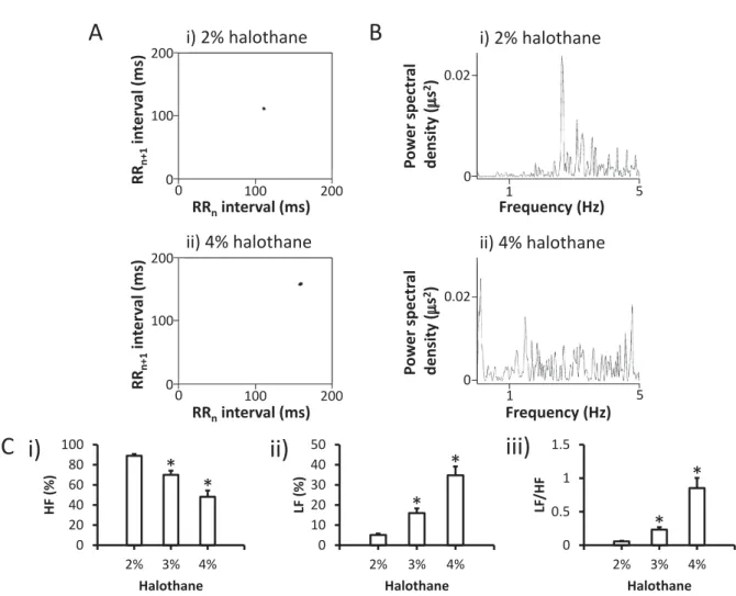
Changes in HRV
We analyzed the HRV during halothane anesthesia. Fig. 2A shows Poincaré plots (RRn vs. RRn + 1) of beat-to-beat dynamics during anesthesia with 2 and 4% halothane. Anesthesia with 4% halothane resulted in an elongated and
fluctuating R-R interval.
In the frequency domain analysis, the LF
(0.15‒1.5 Hz) and HF (1.5‒5 Hz) components were resolved in power spectral density (Fig.
2B). Statistical analysis of the HF and LF components indicated a significant decrease and increase, respectively, during halothane a n e s t h e s i a . T h e r e f o r e , t h e L F / H F r a t i o increased significantly in a concentration- dependent manner, suggesting decreased para- sympathetic nerve tonus (Fig. 2B and C).
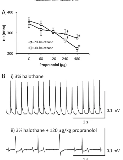
Sympathetic blockade
Intraperitoneal administration of propranolol to halothane-anesthetized mice resulted in a
Figure 1 A.
i) Representative ECG trace during anesthesia with 2% halothane.
ii) Representative ECG trace during anesthesia with 4% halothane.
B. Changes in heart rate according to halothane concentration. *P < 0.05 between anesthesia with 2% halothane and other concentrations (n = 8).
C. Averaging view of ECG with 2% halothane (from the mouse of Ai).
A
100ms 100ms i)2%halothane
ii)4%halothane
0.03mV 0.03mV
100ms
0.03mV
C
Fig.1
200 300 400 500
2% 2.5% 3% 3.5% 4%
,Z;WDͿ
,ĂůŽƚŚĂŶĞ
B
* * *
decrease in HR. Mice anesthetized with 3%
halothane showed greater changes in HR than those anesthetized with 2% halothane (Fig.
3A, at 240 and 480 ȝg/kg BW). On the other hand, under lower dose administration (120 ȝg/kg BW) of propranolol, it seems that mice anesthetized with 2% halothane showed greater change in HR compared to 3% halothane (Fig.
3A). It may be referred from the different effects of halothane for adrenoceptor at low or high concentrations. At low concentrations, Į-adrenergic effects inhibit phase 4 diastolic depolarization, slowing automatic rate, whereas
at higher concentrations, ȕ-adrenergic effects enhance phase 4 diastolic depolarization and increase the rate. Therefore, HR decreased with 2% halothane more greatly than 3% halothane, and it considered that HR change is the results of complex adrenergic effects. In addition, some mice showed atrioventricular block and sick sinus syndrome upon propranolol administration during 3% halothane anesthesia (Fig. 3B).
Therefore, we decided not to examine the effect of propranolol during anesthesia with 4%
halothane.
Figure 2 Heart rate variability (HRV)
HRV during anesthesia with 2% (i) and 4% (ii) halothane (A and B).
A. Poincaré plots (RRn vs. RRn + 1) of consecutive pairs of RR intervals during the control period, together with the nth + 1 RR interval plotted against the nth RR period. Note the elongated RR interval during anesthesia with 4% halothane.
B. Representative power spectral densities during anesthesia with 2% (i) and 4% (ii) halothane.
C. Statistical comparison of LF (i), HF (ii), and the LF/HF ratio (iii). *P < 0.05 between anesthesia with 2%
halothane and other concentrations (n = 8).
B A
200 100
0 100 0 200
ZZŶŝŶƚĞƌǀĂů;ŵƐͿ ZZŶнϭŝŶƚĞƌǀĂů;ŵƐͿ
200
200 100
0 100 0
ZZŶŝŶƚĞƌǀĂů;ŵƐͿ ZZŶнϭŝŶƚĞƌǀĂů;ŵƐͿ
i)2%halothane
ii)4%halothane
0 0.02
1 5
&ƌĞƋƵĞŶĐLJ;,njͿ WŽǁĞƌƐƉĞĐƚƌĂů ĚĞŶƐŝƚLJ;PƐϮͿ
1 5 0 0.02
&ƌĞƋƵĞŶĐLJ;,njͿ WŽǁĞƌƐƉĞĐƚƌĂů ĚĞŶƐŝƚLJ;PƐϮͿ
i)2%halothane
ii)4%halothane
Fig. 2
0 0.5 1 1.5
2% 3% 4%
>&ͬ,&
,ĂůŽƚŚĂŶĞ 0
10 20 30 40 50
2% 3% 4%
>&;йͿ
,ĂůŽƚŚĂŶĞ 0
20 40 60 80 100
2% 3% 4%
,&;йͿ
,ĂůŽƚŚĂŶĞ
C i) ii) iii)
* *
* *
*
*
Discussion
We studied changes in basal ECG and HRV during halothane anesthesia in mice. Inhalation anesthesia using 2% halothane resulted in a conserved response to a sympathetic ȕ-blocker, indicating that halothane might be useful for studies involving small rodents. However, halothane anesthesia resulted in a concentration- dependent decrease in HR, emphasizing the importance of using an appropriate concentration. In a previous study, we reported that both isoflurane and halothane functioned as negative chronotropic agents4). In this study, sick sinus syndrome developed in some
mice administered 4% isoflurane, but no such arrhythmia occurred in mice that received 4%
halothane.
Halothane anesthesia also resulted in a decreased HF component and increased LF component in a concentration-dependent fashion, which suggests that even a slight change in halothane concentration could affect electrophysiological analysis. The LF/HF ratio reflects autonomic nerve tonus4). A decreased HF and LF/HF ratio are thought to be related to decreased para-sympathetic nerve tonus, and an increased LF/HF ratio to increased sympathetic nerve tonus4). Although the LF/
Figure 3 A. Changes in heart rate according to propranolol concentration. *P < 0.05 between anesthesia with 2% and 3%
halothane (n = 8).
B. Typical trace of a mouse ECG during anesthesia with 3% halothane (i). Intraperitoneal injection of the sympathetic ȕ-blocker, propranolol (120 ȝg/kg body weight), resulted in sick sinus syndrome-type arrhythmia during anesthesia with 3% halothane (ii).
1s
1s i)3%halothane
ii)3%halothane+120Pg/kgpropranolol
B
0.1mV
0.1mV 200
300 400
C 60 120 240 480
,Z;WDͿ
WƌŽƉƌĂŶŽůŽů;PŐͿ 2%halothane 3%halothane
A
*
*
* *
HF ratio was significantly increased, inhalation anesthesia usually affects both sympathetic and para-sympathetic nerves. The significant decrease in HF suggests that the increased LF/HF ratio was likely due to decreased para- sympathetic nerve tonus. Anesthesia using 3 % halothane concentrations resulted in sick sinus arrhythmia in response to propranolol (120 ȝg/kg body weight), for which halothane and propranolol were responsible.
In the present study, we first determined t h e o p t i m u m h a l o t h a n e c o n c e n t r a t i o n . Halothane anesthesia decreased the HR in a concentration-dependent fashion (Fig. 1A, B).
We also examined 1.5% halothane because of its low MAC (0.74%). Appropriate anesthesia, in terms of loss of the righting reflex, could not be achieved using 1.5% halothane. In contrast, 3% halothane sometimes resulted in arrhythmia upon administration of a high dose of propranolol (Fig. 3B). Thus, 2% halothane inhalation anesthesia was used in subsequent experiments.
Szczesny et al. reported that isoflurane anesthesia was useful for experimental studies on mice because of its simple administration, rapid induction, and control of the depth of anesthesia5), while inhalation anesthesia required a specialized apparatus. Since halothane has been widely used since 1959, we examined its effect on ECG and whether it could be used for studies involving transgenic mouse. Mouse inhalation anesthesia has been performed in a limited number of laboratories.
In our previous study, isoflurane anesthesia also conserved the HR (494 ± 3.7 bpm, n = 146). The data obtained with 2% halothane anesthesia were comparable with those of isoflurane and sufficient for interpreting the results of pharmacological manipulation using an adrenergic ȕ-blocker (propranolol). In contrast, Shintaku et al. reported that isoflurane anesthesia resulted in sick sinus arrhythmia6),
while halothane did not. This suggests halothane to be more suitable than isoflurane for rodent research.
Halothane decreases the action potential duration (APD) of both Purkinje and ventricular muscle fibers by reducing the rate of phase-0 depolarization (Vmax), the action potential amplitude, and the overshoot7). This effect could be mediated by depression of the peak inward Na+ current during the upstroke of Purkinje fiber action potential in ventricular muscle fibers8). Additionally, at higher concentrations, ȕ-adrenergic effects enhance phase-4 diastolic depolarization and increase the rate9-11). In the present study, the induction of arrhythmias by halothane might have been due to its electrophysiological or ȕ-adrenergic effect, which should be analyzed in future studies.
In summary, we examined the changes in ECGs during halothane anesthesia in mice.
Anesthesia with 2% halothane resulted in a desirable HR and a relatively good response to sympathetic blockade with propranolol. We also evaluated the standard HRV during halothane anesthesia. Our results indicate the importance o f s e l e c t i n g a n a n e s t h e s i a m e t h o d o l o g y according to the aim of the pharmacological experiment.
References
1)Kristensen J, Helbo-Hansen HS, Toft P, Hole P.
The cardiovascular effects of vecuronium during halothane anaesthesia. Acta Anaesthesiol Scand.
1989;33:494-7.
2)Routledge HC, Chowdhary S, Townend JN. Heart rate variability̶a therapeutic target? J Clin Pharm Ther. 2002;27:85-92.
3)Murakami M, Niwa H, Kushikata T, Watanabe H, Hirota K, Ono K, et al. Inhalation anesthesia is preferable for recording rat cardiac function using an electrocardiogram. Biol Pharm Bull.
2014;37:834-9.
4)Shintaku T, Ohba T, Niwa H, Kushikata T, Hirota K, Ono K, Matsuzaki Y, et al. Effects of isoflurane inhalation anesthesia on mouse ECG. Hirosaki Med J. 2015;66:1-7.
5)Szczesny G, Veihelmann A, Massberg S, Nolte D, Messmer K. Long-term anaesthesia using inhalatory isoflurane in different strains of mice- the haemodynamic effects. Lab Anim. 2004;38:64-9.
6)Shintaku T, Ohba T, Niwa H, Kushikata T, Hirota K, Ono K, Matsuzaki Y, et al. Effects of propofol on electrocardiogram measures in mice. J Pharmacol Sci. 2014;126:351-8.
7)Hauswirth O. Effects of halothane on single atrial, ventricular, and Purkinje fibers. Circ Res.
1969;24:745-50.
8)Ikemoto Y, Yatani A, Imoto Y, Arimura H.
Reduction in the myocardial sodium current by halothane and thiamylal. Jpn J Physiol.
1986;36:107-21.
9)Rosen MR, Hordof AJ, Ilvento JP, Danilo P.Jr.
Effects of adrenergic amines on electrophysiological properties and automaticity of neonatal and adult canine Purkinje fibers: evidence for alpha- and beta-adrenergic actions. Circ Res. 1977;40:390-400.
10)Rosen MR, Weiss RM, Danilo P.Jr. Effect of alpha adrenergic agonists and blockers on Purkinje fiber transmembrane potentials and automaticity in the dog. J Pharmacol Exp Ther. 1984;231:566-71.
11)Bosnjak ZJ, Turner LA. Halothane, catecholamines, and cardiac conduction: anything new? Anesth Analg. 1991;72:1-4.