Hirosaki Med.J.
64(Suppl.):S81―S85,2013FORMATION AND DEGRADATION OF MICROSCOPIC GLOBULAR BODY: A PROTEINACEOUS SPHERON ORIGINATED FROM DENDRITE
Saori Odagiri
1,2),Fumiaki Mori
1),Kunikazu Tanji
1),Naohito Kuroda
3)and Koichi Wakabayashi
1)Abstract Microscopic globular bodies (MGBs) are eosinophilic spherical inclusions, 1-10 μm in diameter and are located in dendrites. Although MGBs are distributed widely in the gray matter, they are most frequent and numerous in the cerebral neocortex and hippocampus. They are found in the human subjects with and without neurological diseases, ranging in age from 1 to 99 years. Previous histochemical studies have shown that MGBs consist of a proteinaceous component. Recently, we immunohistochemically examined the hippocampus from five control subjects. The marginal portion of MGBs was positive for lysosomal proteases (cathepsins B, D and L), and markers of dendrite (MAP2) and dendritic spine (drebrin). They were negative for ubiquitin, ubiquitin-proteasome system (p62, NUB1 and EDD1), autophagosome (LC3), cytoskeletal proteins (neurofilament, actin, tubulin and cytokeratin), tau, α-synuclein and TDP-43. These findings suggest that MGBs are sequestered by lysosome-protease system, but not by ubiquitin-proteasome system or autophagosome.
Hirosaki Med.J. 64, Supplement:S81―S85,2013 Key words: cathepsin; dendrite; immunohistochemistry; lysosome-protease system;
microscopic globular body
What is microscopic globular body?
Microscopic globular bodies (MGBs) were first described in the neuropil of normal human brain by Hara.
1, 2)MGBs are brilliantly and homogenously eosinophilic spherical inclusions, 1-10
μm in diameter (Figure 1). Several investigators have referred to these structures as eosinophilic globular body,
1)granular osmiophilic body,
3)dense microsphere
4-6)and spheron.
7, 8)They are mainly distributed in the frontal and temporal neocortex and hippocampus in the human subjects with and without neurological diseases, ranging in age from 1 to 99 years.
2, 7)Such structures are also seen in the cerebral cortex of monkeys, dogs, cows, pigs, raccoons, deer, goats, sheep, horses and bears.
7, 9)Ultrastructurally, they are composed of electron-dense granular material surrounded by single unit membrane and are located
Departments of 1)Neuropathology, 2)Neuroanatomy, Cell Biology and Histology, and 3)Forensic Medicine, Hirosaki University Graduate School of Medicine,
Hirosaki, Japan Fax: +81-172-39-5006,
E-mail: [email protected]
in cytoplasmic processes, contained a few mitochondria and other organelles.
2 - 4)In addition, synaptic junctions were often present on the cytoplasmic process which contained MGBs.
2, 4)These morphological features suggest that MGBs are located in dendrites.
Composition of MGB
Previous histochemical studies have shown that MGBs consist of a proteinaceous component, but without a significant amount of carbohydrate or neutral glycoprotein.
2, 4, 6, 9)Furuoka et al.
9)reported that MGBs in the horse brain is not ubiquitinated. However, immunohistochemical profile of MGBs has not been thoroughly investigated. Recently, we immunohistochemically examined the hippocampus from five control subjects ranged from 25 to 76 years.
10)MGBs were found in the hippocampus
in all five cases on HE-stained sections.
Immunohistochemically, MGBs were labeled with antibodies against cathepsins B, D and L
(Figure 2a-c). They were also immunolabeled with markers of dendrite (MAP2) (Figure 2d), dendritic spine (drebrin), endoplasmic reticulum
(IP
3R) and mitochondria (Tom20). The MGBs were classified into two types by cathepsin D immunohistochemistry. The common type (type 1) showed positive staining in the marginal portion (Figure 2a, b). The other type (type 2)
was entirely immunostained with anti-cathepsin
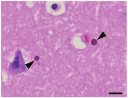
Figure 1 Light micrograph of microscopic globular bodies (arrowheads) in the hippocampus, stained with HE. Bar = 10 μm.
Figure 2 Light micrographs of microscopic globular bodies (MGBs) (arrowheads) in the hippocampus, immunostained with cathepsin D and MAP2. a-c: Catepsin D immunoreactivity in the marginal portion (a, b) or entire MGBs (c). d:
Immunoreactivity of MAP2 in the marginal portion of MGBs. Bars = 10 μm.
D (Figure 2c). On the other hand, marginal portion of MGBs were positive for cathepsin B, cathepsin L, MAP2, drebrin, IP
3R and Tom20. However, they were not immunolabeled with monoclonal and polyclonal anti-ubiquitin antibodies. They were also negative for proteins implicated in the ubiquitin-proteasome system
(p62, NUB1 and EDD1), cathepsins A and S, autophagosome (LC3), early endosome
(Rab5), cytoskeletal proteins (neurofilament, vimentin, cytokeratin, GFAP, actin and tubulin), structural elements of abnormal inclusions in neurodegenerative disorders (phosphorylated
α-synuclein, phosphorylated tau, β-amyloid, TDP-43 and FUS), a marker of Golgi-endoplasmic reticulum complex (cystatin C and
β-COP), amarker of endoplasmic reticulum (ryanodine receptor and TxNDC4), a marker of mitochondria
(COX I V), and a marker of presynapse
(SNAP25).
The proportion of immunolabeled MGBs relative to the total number of inclusions was as follows: cathepsin B, 12.1%; cathepsin D, 27.6%;
cathepsin L, 7.1%, MAP2, 19.4%; drebrin, 6.3%;
IP
3R, 7.0%; and Tom20, 9.5%.
Formation and degradation of MGB
As described above, MGBs are immunopositive for cathepsins B, D and L. Cathepsins B and L are cysteine proteases. Cathepsin D is an aspartate protease, which activates pro-enzyme forms of cathepsins B, D, H and L. Among the cathepsins we examined, MGBs were most frequently immunolabeled with anti-cathepsin D. The association of neurodegeneration with the endosome-lysosome protease system has been reported in various neurological disorders including Alzheimer’s disease (AD), motor neuron disease, demyelinating disease and ischemia.
11-18)Cathepsin B immunoreactivity is i ncrea sed i n neu rof ibr i l la r y t a ngle - containing neurons and dystrophic neurites
of senile plaques in AD
12)as well as in motor neurons in amyotrophic lateral sclerosis.
17)Immunoreactivity of cathepsins B and D is increased in the degenerative regions in the early stage of AD.
13)These findings suggest that cathepsins B and D are associated with neuronal cell death. Felbor et al.
19)reported that cathepsin B/cathepsin L double knockout mice showed brain atrophy, neuronal loss and lysosomal inclusion bodies in large cortical neurons, suggesting that cathepsins B and L have a role in maintenance of the brain. Considering that MGBs are immunopositive for cathepsins B, D and L, they are degraded, at least in part, by the lysosome-protease system. This is supported by the finding that MGBs are not ubiquitinated and are negative for proteins implicated in the ubiquitin-proteasome system and a marker of autophagosome.
MGBs are also immunolabeled with markers of dendrite and dendritic spine. This is consistent with previous ultrastructural studies that MGBs are located in the dendrite.
2-4)The marginal portion of MGBs is also positive for markers of the endoplasmic reticulum and mitochondria.
This is explained by the facts that the normal dendrites contain a few mitochondria and endoplasmic reticulum and that MGBs are surrounded by the cytoplasm of the dendrites.
2, 9)However, the major component of MGBs is still uncertain.
MGBs are negative for proteins implicated in the cytoskeletal proteins and structural elements of abnormal inclusions in neurodegenerative disorders. It is unlikely that MGBs are composed of cytoskeletal proteins.
MGB and aging
Previous studies have shown that MGBs are
not seen in newborns but are regularly present
by the age of one year.
7)They are found in
the brain of normal individuals ranged in age
from 1 to 99 years.
2, 7)The number of MGBs in the temporal neocortex is stable across age groups,
7)whereas the number of MGBs in the whole hemisphere at the level of mammillary body is highest in the fourth decade.
2)Thus, the occurrence of MGBs does not simply reflect aging phenomenon. Moreover, the density of MGBs in the temporal neocortex is significantly decreased in AD compared to age-matched normal controls.
7)Immunoreactivity of cathepsins B and D is increased in the degenerative regions of AD.
12, 13)In addition, the levels of cathepsin B in plasma and cathepsin D in cerebrospinal fluid are elevated in patients with AD.
20, 21)It is possible to consider that the degradation of MGBs is accelerated in AD.
In conclusion, MGBs are dendritic in origin and are sequestered by lysosome-protease system, but not by ubiquitin-proteasome system or autophagosome.
Acknowledgements
This work was supported in part by Grants- in-Aid for Scientific Research from the Ministry of Education, Culture, Sports, Science, and Technology, Japan (F.M., K.T. and K.W.) and a Grant for Hirosaki University Institutional Research (K.W.). The authors wish to express their gratitude to M. Nakata for her technical assistance.
References
1)Hara M. Eosinophilic globular body in the cerebral grey matter. Neurol Med (Tokyo) 1981;14:76-9.
2)Hara M. Microscopic globular bodies in the human brain. J Neuropathol Exp Neurol 1986;45:169-78.
3)Rees S. A quantitative electron microscopic study of atypical structures in normal human cerebral cortex. Anat Embryol (Berl) 1975;148:303-31.
4)Averback P. Dense microspheres in normal
human brain. Acta Neuropathol (Berl) 1983;61:
148-52.
5)Averback P. Immunofluorescent staining of dense microspheres in human brain. Arch Pathol Lab Med 1982;106:394-6.
6)George DH, Munoz DG and McConnell T. Dense microspheres in the human hippocampus. Acta Neuropathol 1993;86:86-9.
7)Averback P, Morse D and Ghanbari H. Bursting dense microspheres (spherons) in Alzheimer’s disease. J A1zheimers Dis 1998;1:1-34.
8)Averback P, Fitzpatrick J, Iversen L, Kaufman D and Morse D. Spherons and Alzheimer’s disease:
biochemical, immunological and cell biological studies. Alzheimer’s Reports 2000;3:177-84.
9)Furuoka H, Yamada M, Miyazawa K, Taniyama H and Matsui T. Dense microspheres in normal horse brain. Acta Neuropathol 1996;91:440-3.
10)Odagiri S, Mori F, Tanji K, Kuroda N and Wakabayashi K. Immunohistochemical study of microscopic globular bodies of normal human brain. Biomed Res 2011;32:337-42.
11)Nakamura Y, Takeda M, Suzuki H et al. Abnormal distribution of cathepsins in the brain of patients with Alzheimer’s disease. Neurosci Lett 1991;130:195- 8.
12)Ii K, Ito H, Kominami E and Hirano A. Abnormal distribution of cathepsin proteases and endogenous inhibitors (cystatins) in the hippocampus of patients with Alzheimer’s disease, parkinsonism- dementia complex on Guam, and senile dementia and in the aged. Virchows Arch A Pathol Anat Histopathol 1993;423:185-94.
13)Cataldo AM, Hamilton DJ and Nixon RA. Lyso- somal abnormalities in degenerating neurons link neuronal compromise to senile plaque develop- ment in Alzheimer disease. Brain Res 1994;640:
68-80.
14)Bever CT Jr and Garver DW. Increased cathepsin B activity in multiple sclerosis brain. J Neurol Sci 1995;131:71-3.
15)Hill IE, Preston E, Monette R and MacManus JP. A comparison of cathepsin B processing
and distribution during neuronal death in rats following global ischemia or decapitation necrosis.
Brain Res 1997;751:206-16.
16)Ginsberg SD, Hemby SE, Lee VM, Eberwine JH and Trojanowski JQ. Expression profile of transcripts in Alzheimer’s disease tangle-bearing CA1 neurons. Ann Neurol 2000;48:77-87.
17)Kikuchi H, Yamada T, Furuya H et al. Involvement of cathepsin B in the motor neuron degeneration of amyotrophic lateral sclerosis. Acta Neuropathol 2003;105:462-8.
18)Nakanishi H. Neuronal and microglial cathepsins in aging and age-related diseases. Aging Res Rev
2003;2:367-81.
19)Felbor U, Kessler B, Mothes W et al. Neuronal loss and brain atrophy in mice lacking cathepsin B and L. Proc Natl Acad Sci USA 2002;99:7883-8.
20)Schwageri AL, Mohan PS, Cataldo AM, Vonsattel JP, Kowall NW and Nixon RA. Elevated levels of the endosomal-lysosomal proteinase cathepsin D in cerebrospinal fluid in Alzheimer disease. J Neurochem 1995;64:443-6.
21)Sundelöf J, Sundström J, Hansson O et al. Higher cathepsin B levels in plasma in Alzheimer’s disease compared to healthy controls. J Alzheimers Dis 2010;22:1223-30.