Title of Thesis
“Studies on the facts of the loss of MMP3 on tumoroid formation and the integrity of extracellular vesicles”
“MMP3の欠損が腫瘍形成と細胞外小胞の完全性に
及ぼす影響に関する研究”
September 2020
Eman Ahmed Mohamed Mohamed Taha
Graduate School of Natural Science and Technology Okayama University
Doctoral Program OKAYAMA UNIVERSITY
Acknowledgment
Acknowledgment
First, I would like to express my sincere gratitude to my supervisor Dr. Ayano Satoh for the valuable support, understanding, and thoughtful comments and recommendations on this dissertation.
Thank you for supporting me to find a friendly atmosphere where I can learn and grow. I would like to express my deep gratitude to Dr. Takanori Eguchi for his valuable and constructive suggestions during the planning and development of this research work. He helped me a lot in editing and revising the manuscript as well as teaching me how to use and represent my data. Besides, he allowed me to improve my academic writing skills, thanks to him I was able to build such a good publication record.
My grateful thanks are also extended to Dr. Chiharu Sogawa for her advice and assistance in designing the experimental protocols, teaching me how to use different instruments and programs. Also, for her help in visualizing the data, editing and revising the manuscript, and paying the publication fees. I appreciate your kindness, support, and personal approach. Words cannot express how grateful I am for the kindness and generosity of Prof. Dr. Kuniaki Okamoto. Thank you for your valuable support, insightful discussions, and accepting me as a member of your lab.
Besides my advisors, I would like to thank the rest of my thesis committee: Prof. Dr. Takashi Otsuki and Prof. Dr. Hiroshi Tokumitsu for their insightful comments, and valuable suggestions.
My special thanks are extended to Prof. Dr. Masaharu Seno for allowing me to study in Japan and to Dr. Maram Hussein for her warm welcome as well as to Prof. Dr. Heiichiro Udono for being considerate and kind towards me. Also, for allowing me to visit his lab and his explanation of various immunological techniques.
Additionally, I am grateful to the help provided by Ms. Sarah Chikako, and Dr. Sabina Mahmoud as well as Dr. Mariko Muzuka.
Acknowledgment
I am particularly grateful for the assistance given by Dr. Yuka Okusha, Dr. Hotaka Kawai, Prof. Dr. Satoshi Kubota, Prof. Dr.
Masaharu Takigawa, Prof. Dr. Hitoshi Nagatsuka, Dr. Eriko Aoyama, Dr. Abdellatif Elseoudi, Mr. Haruo Urata, Ms. Kazuko Kobayashi, May Wathone Oo, my lab mates; Dr. Kisho Ono, Yanyin Lu, Feng Yunxia, Penggong Wei, and Toshiki Nara for their valuable assistance and contribution to my research project.
Besides, the scholarship provided by Egypt-Japan Education Partnership (EJEP) agency, took away the financial burden, allowing me to focus on my study. A special appreciation is extended to Prof.
Dr. Hany A. El-Shemy; Cultural Counselor, and Dr. Hanem Ahmed; Cultural Attache, Mr. Fathy Dawoud and Ms.
Keiko Morishita from the Culture, Education, and Science Bureau in Tokyo, as well as to the members of the Ministry of Higher Education and Scientific Research in Cairo for their support and help.
I am grateful to all professors and colleagues from the Biochemistry department, Faculty of Science in Cairo for their support and encouragement. Especially, I am grateful to Prof. Dr.
Ibrahim Kamal, Prof. Dr. Mohamed Ragaa, Prof. Dr. Nadia Abdallah, Prof. Dr. Magda Kamal, Prof. Dr. Tahany Abd El- Moneam (who passed away before I visited Japan), Prof. Dr.
Fawzya Ashoor, Dr. Rania Hassan, Dr. Elham Abdel-Badiea, Dr.
Walaa Elabd, Dr. Hayam Rushdy for their encouragement and valuable support.
Besides, I would like to express my very great appreciation to Dr.
Hamdy Aly and his wife Mrs. Basma Fakery, for being so kind, very supportive, and helping me out when I needed it.
Finally, I must express my very profound gratitude to my parents, sisters, and brothers for providing me with unfailing support and continuous encouragement. Thank you so much for being there for me and for believing in me. This accomplishment would not have been possible without your sincere prayers during these difficult times. Thank you.
Eman A. Taha
Table of Contents
Content Page
Abbreviations I
List of Figures and Tables III
1. Introduction 1
1.1. Classification of extracellular vesicles (EVs) 1 1.2. Biogenesis and characteristics of EVs 2 1.3. EVs as modulators of the tumor microenvironment 5
1.4. Fluorescent labeling of EVs 7
1.5. Structure of MMPs 8
1.6. Complex roles of MMPs in tumorigenesis 10
1.7. MMPs in EVs 13
1.8. The two-dimensional and three- dimensional culture systems
15
2. Aim and objective 17
3. Materials and Methods 18
3.1. Cells 18
3.2. Tumoroid culture 18
3.3. 2D re-seeding assay 19
3.4. Preparation of EVs and conditioned media 19 3.5. Transmission electron microscopy 20 3.6. Particle diameter distribution 20
3.7. Western blotting 21
3.8. Coomassie blue staining 22
3.9. EV-driven in vitro tumorigenesis 22
3.10 Palm fluorescent cells 22
3.11. EVs exchange assay 23
3.12. 2D confocal laser-scanning microscopy 24 3.13 Immunofluorescence of tumoroids 25 3.14. Hematoxylin and eosin staining 26
3.15. Tracing EV-uptake in vitro 27
3.16. Statistical analysis 27
4. Results 28
4.1. MMP3 knockout reduces the release of CD9 and CD63 within extracellular vesicles
28 4.2. MMP3 knockout impacts the physical and biological
integrities of extracellular vesicles
31 4.3. Loss of Mmp3 reduces the in vitro tumorigenicity 33 4.4. The addition of MMP3-rich EVs accelerated the in
vitro tumorigenesis of MMP3-KO cells
36
Table of Contents
4.5. Establishment of fluorescent-labeled LuM1 and MMP3-null cells
40 4.6. Penetration of MMP3-rich EVs into organoids 43 4.7. The knockout of the Mmp3 gene significantly
decreased the transmissive potential of tumoroid- derived EVs
47
4.8. MMP3-rich EVs and CM rescue the cell proliferation of MMP3-KO tumoroids
49
5. Discussion 52
5.1. Summary 52
5.2. Potential mechanism of how MMP3 promotes tumorigenesis
52 5.3. Potential role of MMP3 in cryoprotection 56 5.4. Release of L-EVs and s-EVs from 3D tumoroids 58
5.5. Fluorescent labeling of EVs 59
5.6. Inter MMPs regulation 60
6. Conclusion 61
7. Supplementary Figures 62
8. References 67
Abbreviations
I
Abbreviations EVs Extracellular vesicles
MV Microvesicles
MVBs Multivesicular endosomes
MHC Major histocompatibility complex proteins ILVs Intraluminal vesicles
Rab The Rab family is part of the Ras superfamily of small GTPases.
ESCRT The endosomal sorting complex required for transport
TSG101 Tumor Susceptibility 101 HSP Heat shock proteins TME Tumor microenvironment CAFs Cancer-associated fibroblasts
VEGFA Vascular endothelial growth factor-A CXCL C-X-C motif chemokine
IL-6 Interleukin 6
ECM Extracellular matrix
TGF-β1 Transforming growth factor-beta CRC Colorectal cancer
EGFR Epidermal growth factor receptor MET Tyrosine-protein kinase Met
tdTomato Palmitoylation signal-fused fluorescent proteins such as tandem dimer Tomato
EGFP Enhanced green fluorescence protein MMPs Metalloproteinases
PEX A hemopexin-like repeat domain
IGFBP Insulin-like growth factor-binding protein IGFR Insulin-like growth factor 1receptor
Abbreviations
II
HB-EGF Heparin-binding epidermal growth factor-like growth factor
EGFR/ERBB Epidermal growth factor receptor FGF-2 Fibroblast growth factor- 2 EMT Epithelial-mesenchymal transition MET Mesenchymal-epithelial transition
ZEB Zinc finger E-box-binding homeobox protein SNAIL Zinc finger transcriptional repressor
ESCC Esophageal squamous cell carcinoma CCN2/CTGF Connective tissue growth factor IFN-α Interferon-alpha
NFKIBA Nuclear factor-κB inhibitor alpha NLS Nuclear localization signaling sequence PARP Poly (ADP-ribose) polymerase
FADD FAS-associated death domain protein Bax BCL2 associated X
Bcl-xs B-cell lymphoma-shorter form
XRCC1 X-ray repair cross-complementing protein RANKL Receptor activator of nuclear factor kappa B
ligand
VEGFR-1TK Vascular endothelial growth factor receptor 1 tyrosine kinase
2D Two-dimensional
3D Three-dimensional NCP NanoCulture plates ULA Ultra-low attachment
List of Figures &Tables
III List of Figures
No. Figure Legend Page
1. Schematic representation of exosome biogenesis and release.
3
2. The domain structure of MMPs. 9
3. The Pivotal role of MMPs in cancer progression. 13 4. MMP3 knockout reduces the release of CD9 and
CD63 within extracellular vesicles.
30 5. MMP3 knockout impacts the physical integrities of
extracellular vesicles.
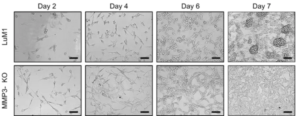
33 6. Loss of Mmp3 reduces the in vitro tumorigenicity. 34 7. Representative images of re-cultured LuM1 and
MMP3-KO cells in 2D culture.
36 8. The addition of MMP3-rich EVs accelerated the in
vitro tumorigenesis of MMP3-KO cells.
37 9. Tumoroid size was altered by the addition of LuM1-
EVs versus MMP3-KO-EVs.
39 10. Establishment of fluorescent-labeled LuM1 and
MMP3-null cells.
42 11. The EV-mediated deep transfer of MMP3 into
tumoroids.
45 12. Treatment with LuM1-derived EVs and CM
recovered CD9 in MMP3-null tumoroids.
46 13. The knockout of the MMP3 significantly decreased
the transmissive potential of tumoroid-derived EVs.
48 14. MMP3-knockout resulted in necrotic cell death in
tumoroids.
50 15. MMP3 enriched-EVs and CM rescue the proliferation
of MMP3-KO tumoroids.
51 16. Graphical abstracts summarizing the role of MMP3
on tumorigenesis in vitro.
53 S1. Full images of Western blotting. 62
List of Figures &Tables
IV
S2. Images of GAPDH Western blotting and CBB staining of the SDS-PAGE.
63 S3. Column scatters plotting of the size of LuM1
tumoroids versus MMP3-KO tumoroids cultured for 7 days or 14 days in serum-containing or stemness media.
64
S4. Evaluation of the effect of three different concentrations of LuM1-EVs on the MMP3-KO tumoroids growth.
65
S5. The specificity of the MMP3 antibody. 66
List of Tables
No. Table title Page
1 Comparison of particle size distributions between LuM1-EVs and MMP3-KO-EVs.
32
2 Necrotic areas in the LuM1 tumoroid versus MMP3-KO tumoroid
49
Introduction
1 1. Introduction
1.1. Classification of extracellular vesicles (EVs)
Communications of cancer cells with each other or with neighboring cells or cells at distant sites are crucial for tumor proliferation and dissemination (Jakhar and Crasta 2019).
Extracellular vesicles (EVs) are lipid bi-layered vesicles that are released from almost all cells under physiological and pathological conditions. EVs play a crucial role in intercellular communications at local and distant sites (Yáñez-Mó et al. 2015). These small vesicles carry various molecular cargo such as; nucleic acids (DNA, RNA), proteins, lipids, and metabolites that could be transferred into the recipient cells leading to genetic alterations and reprogramming of these cells (Raposo and Stoorvogel 2013; Colombo, Raposo, and Théry 2014; Yáñez-Mó et al. 2015; Eman A Taha, Ono, and Eguchi 2019).
According to the vesicle size, EVs are mainly classified into three categories; exosomes (50-200 nm), ectosomes are also known as microvesicles (MV) (100-500 nm), and apoptotic bodies (1-10 μm) (Andreola et al. 2002; Janowska-Wieczorek et al. 2005; Lawson et al.
2016). In addition to this heterogenous population, other vesicles have been reported including., oncosomes (Al-Nedawi et al. 2008; Rak 2013; Choi et al. 2019), large oncosomes (1-10 μm) (Di Vizio et al.
2012; Vagner et al. 2018), matrix vesicles (Mebarek et al. 2013; Chen et al. 2016; Schmidt et al. 2016), migrasomes (50 nm to 3 μm) (Ma et al. 2015; Huang et al. 2019), exopheres (~4 μm), exomeres (~35 nm), and bacterial outer membrane vesicles (OMV) (Raposo and
Introduction
2
Stoorvogel 2013; Kim et al. 2017; Van Niel, D’Angelo, and Raposo 2018; Coelho et al. 2019).
Notably, EVs are heterogeneous populations, so there is no unanimous consensus on the nomenclature of them. General terms such as “exosomes” and “microvesicles” have been broadly used. The International Society for Extracellular Vesicles (ISEV) proposed to use the term EVs in general to describe vesicles naturally released from the cells and surrounded by a lipid bilayer unless authors can establish specific markers of subcellular origin with a description based of physical characteristics, such as size (Théry et al. 2018).
Thus, I will use the general term EVs, and classify it into small and large EVs based on the size of vesicles.
1.2. Biogenesis and characteristics of EVs
Exosomes are vesicles of endosomal origin. Their biogenesis starts with the inward budding of the cellular plasma membrane, internalization of extracellular ligands from the cell surface (e.g., growth factor receptors) or from the Golgi apparatus (e.g., MHC class-II molecules) forming early endosomes (Babst 2005; Trajkovic et al. 2008; Fader and Colombo 2009; Babst 2011; Colombo et al.
2013; Jakhar and Crasta 2019; Eman A Taha, Ono, and Eguchi 2019).
Early endosomes mature into late endosomes. After that, the endosomal membrane undergoes a second inward (intraluminal) budding to generate smaller vesicles within the late endosome lumen to form multivesicular bodies (MVBs), which are carrying various bioactive molecules such as proteins, lipids, and nucleic acids of the parent cell. Finally, MVBs either fuse with lysosomes to be degraded or fuse with the plasma membrane thereby releasing the intraluminal
Introduction
3
vesicles, termed exosomes, into the extracellular space (Figure 1 ) (Trajkovic et al. 2008; Fader and Colombo 2009; Babst 2011;
Colombo et al. 2013; Jakhar and Crasta 2019; Eman A Taha, Ono, and Eguchi 2019). Once generated within the MVB, the release exosomes into the extracellular space are mediated by small transport GTPases molecules such as; Rab27A, Rab11, and Rab31, which can collaborate with SNARE (a soluble N-ethylmaleimide sensitive factor attachment protein receptor) proteins to fuse the MVB membrane with their target membrane (Bobrie et al. 2011).
Figure 1. Schematic representation of exosome biogenesis and release (Jakhar and Crasta 2019).
It is worth noting that the formation of exosomes within the MVBs occurs by the endosomal sorting complex required for transport (ESCRT)-dependent machinery and ESCRT-independent mechanisms. Four distinct ESCRT protein complexes have been
Introduction
4
identified (ESCRT-0, -I, -II, and -III). The ESCRT-dependent biogenesis starts with the inward budding of the cell membrane with aid from ESCRT-0 to produce early endosomes. The other ESCRT complexes contributing to the packaging of exosome contents into late endosomes. Whereas, the ESCRT-independent biogenesis involves the packaging of proteins from the Golgi into exosomes within MVBs and discharged into the extracellular milieu in the absence of ESCRT machinery (Babst 2011; Jakhar and Crasta 2019).
In general, EVs are characterized by their cup-shaped lipid bilayers structure under the electron microscope (Szatanek et al.
2017). Despite the absence of specific protein markers to distinguish between the different subtypes of EVs, the protein profiles of MVs, exosomes, and apoptotic bodies are different due to their different routes of formation (F. T. Borges, Reis, and Schor 2013; Yáñez-Mó et al. 2015; Mikołaj P Zaborowski et al. 2015). For instance, the membrane of Exosome contains cholesterol, sphingomyelin, phosphatidylinositol, ceramide, and lipid rafts (Théry, Ostrowski, and Segura 2009; Ciardiello et al. 2016; Tamkovich, Tutanov, and Laktionov 2016).
Besides, exosomes protein markers including tetraspanin family proteins (CD63, CD9, CD81, and CD82), members of ESCRT complex (TSG101, Alix), and heat shock proteins (HSP60, HSP70, HSPA5, CCT2, and HSP90) (Théry, Ostrowski, and Segura 2009;
Simpson et al. 2009; Yoshioka et al. 2013; Fernanda T. Borges et al.
2013; Yokoi, Yoshioka, and Ochiya 2015; Ciardiello et al. 2016; Ha, Yang, and Nadithe 2016). While, microvesicles membrane are enriched with cholesterol, diacylglycerol, and phosphatidylserine (Colombo, Raposo, and Théry 2014); and integrins, selectins, and
Introduction
5
CD40 are the main protein markers for this category of EVs (Colombo, Raposo, and Théry 2014).
Furthermore, apoptotic bodies are distinguished from the other two major EV groups by the presence of fragmented DNA and cell organelles from their host cell (Mathivanan, Ji, and Simpson 2010;
Akers et al. 2013; Boukouris and Mathivanan 2015). Moreover, apoptotic bodies have exposed phosphatidylserine on their membranes, and their major protein markers include histones, thrombospondin (TSP), and complement protein C3b (Théry et al.
2001).
1.3. EVs as modulators of the tumor microenvironment
Tumor-derived EVs have been recently emerged as putative biological mediators in cancer (Rak and Guha 2012). EVs are highly specialized molecules in cellular communication, as they carry several oncogenic proteins, nucleic acids, and signaling molecules that can be transferred horizontally to the target cells and modulate the tumor microenvironment (TME) for supporting tumor growth, invasion, and metastasis (Higginbotham et al. 2011; Rak and Guha 2012; Tovar-Camargo, Toden, and Goel 2016). The role of EVs in cancer progression and metastasis is described in detail below.
The tumor microenvironment does not only consist of cancer cells but also a heterogeneous population of fibroblasts, endothelial cells, immune cells, cytokines, extracellular vesicles, and extracellular matrix, adipocytes, and vasculature (Balkwill, Capasso, and Hagemann 2012). The crosstalk between cancer cells and their surrounding environment plays a pivotal role in tumor development and progression (Balkwill, Capasso, and Hagemann 2012).
Introduction
6
Cancer-associated fibroblasts (CAFs) are one of the most important members within the TME that represent the largest proportion of stroma cells by secreting extracellular matrix components (Xing, Saidou, and Watabe 2010). CAFs can promote the tumor invasion and metastasis, via the secretion of many cytokines such as vascular endothelial growth factor A (VEGFA), C-X-C motif chemokine 12 (CXCL12), Interleukin 6 (IL-6), as well as remodeling of the extracellular matrix (ECM) (Alkasalias et al. 2018).
It was reported that ovarian cancer-derived EVs are capable of modulating fibroblast's behavior towards a CAF-like state. The secretome of these CAFs stimulates the surrounding cells to promote the proliferation, motility, and invasion of the tumor and endothelial cells (Giusti et al. 2018). Moreover, it has been shown that transforming growth factor-beta TGF-β1-associated EVs secreted from prostate cancer can trigger the differentiation of fibroblast into a myofibroblast phenotype resembling stromal cells isolated from cancerous prostate tissue; promoting in vitro angiogenesis and accelerating in vivo tumor growth (Webber et al. 2015).
Abdouh et al. demonstrated that colorectal cancer (CRC) - derived EVs were able to induce the transformation of fibroblasts into colon carcinoma cells in vitro (Abdouh et al. 2019). They showed that fibroblasts treated with CRC-derived EVs mediated the transfer of DNA that was actively transcribed in the fibroblasts after the EVs exposure (Abdouh et al. 2019).
The groups also observed that a definite set of miRNA molecules was transferred from the CRC-derived EVs to the fibroblasts;
activating cell cycle progression and cell survival pathways.
Introduction
7
Furthermore, the injection of CRC-derived EVs in the tail vein of NOD-SCID mice prompted malignant transformation and metastases in the lungs of the mice (Abdouh et al. 2019).
1.4. Fluorescent labeling of EVs
Several methods have been developed to monitor the biogenesis, transmission, distribution, and subcellular localization of EVs, such as lipid-based fluorescence labelings (Yoshimura et al. 2016; Namba et al. 2018), such as the transmembrane protein CD63 fused with a green fluorescent protein (GFP) and red fluorescent protein (RFP) (CD63-GFP/RFP fusion) (Piao, Kim, and Moon 2019; Mikołaj Piotr Zaborowski et al. 2019), and membrane lipid-binding palmitoylation signal-fused fluorescent proteins such as tandem dimer Tomato (tdTomato) or enhanced green fluorescence protein (EGFP) as I abbreviate as palmGFP (palmG) and palmtdTomato (palmT) (Lai et al. 2015).
Protein S-acylation is a lipid modification that enables the covalent attachment of long-chain palmitic fatty acids to thiol groups of cysteine residues through a thioester linkage (Xu 2011; Verpelli et al. 2012). This type of protein modification is commonly known as S- palmitoylation (S-PALM) allows the association of proteins with cellular membranes (Triola, Waldmann, and Hedberg 2012). The fusion of the fluorescent proteins with palmitoylation sequence to the cell membranes, enabling the whole-cell labeling (Zuber, Strittmatter, and Fishman 1989; Zacharias et al. 2002). As EVs are derived from the plasma membrane (Raposo and Stoorvogel 2013), I assumed that tagging the plasma membrane with fluorescent proteins would enable the labeling of multiple EVs types.
Introduction
8 1.5. Structure of MMPs
Metalloproteinases (MMPs) constitute a large family of zinc- calcium dependent endopeptidases and they are considered as the main players in ECM remodeling (Berg, Barchuk, and Miksztowicz 2019). Due to their ability to degrade numerous components of ECM, nucleus matrix, and non-ECM proteins, such as adhesion molecules, cytokines, protease inhibitors, and membrane receptors (Berg, Barchuk, and Miksztowicz 2019).
MMPs play crucial roles in wound healing, angiogenesis, tissue remodeling, as well as in pathological processes, including wound healing (Nagaset and Woessner 1999; Ravanti and Kähäri 2000; Visse and Nagase 2003), inflammation (Y, H, and Jr 1987), and cancer (Coussens and Werb 1996; Curran and Murray 1999; Sternlicht and Werb 2001; Kessenbrock, Plaks, and Werb 2010). So far, the MMP family consists of about 28 members that share similarities in their structure, regulation, and function (Berg, Barchuk, and Miksztowicz 2019).
Based on structure and substrate specificity, MMPs can be further divided into six major subfamilies including collagenases, gelatinases, stromelysins, matrilysins, membrane-type MMPs, and other MMPs (Peng et al. 2012). All MMPs have three principal domains; (1) a pro-domain that functions as an intramolecular inhibitor to maintain the enzyme in an inactive state, (2) a catalytic domain that promotes the proteolytic activity, and (3) a hemopexin- like repeat domain (PEX), which determines the substrate specificity (Figure 2) (Radisky and Radisky 2015).
Introduction
9
Figure 2. The domain structure of MMPs. S, signal peptide; Pro, pro-peptide; CAT, catalytic domain; F, fibronectin type-II repeats;
PEX, hemopexin domain; TM, transmembrane domain; GPI, glycophosphatidylinositol membrane anchor; C, cytoplasmic domain;
CA, cysteine array; Ig, immunoglobulin-like domain. Adapted from (Radisky and Radisky 2015).
The PEX domain is found in all MMPs except MMP-7 and MMP-26, thereby they are the smallest MMPs members that having only the pro-peptide and catalytic domains (Murphy et al. 1994;
Steffensen, Wallon, and Overall 1995; Shipley et al. 1996;
Mikhailova et al. 2012). A more specialized domain including three fibronectin type II repeats, present in MMP-2 and MMP-9 and assist in recognizing elastin and denatured collagen as extracellular matrix
Introduction
10
substrates (Murphy et al. 1994; Steffensen, Wallon, and Overall 1995;
Shipley et al. 1996; Mikhailova et al. 2012).
Additionally, while most MMPs are soluble extracellular proteins, MMPs-14, -15, -16, and -24 are type I membrane proteins that directly anchored through C-terminal transmembrane domains, MMP-17 and -25 is membrane localized via C-terminal glycophosphatidylinositol (GPI) anchors, and MMP-23 via an N- terminal type II transmembrane domain (Rangaraju et al. 2010).
Furthermore, MMP-23 possesses a unique cysteine array that modulates the ion channel activity and an adjacent immunoglobulin- like domain, that similar to the PEX domain of other MMPs (Rangaraju et al. 2010). This array mediates the protein-protein interactions involved in localization or substrate recognition (Rangaraju et al. 2010; Galea et al. 2014).
1.6. Complex roles of MMPs in tumorigenesis
The extracellular matrix serves as a niche for tumor cells to survive and proliferate. On the other hand, it acts as a barrier that suppresses the spreading of tumor cells. Degradation of ECM is one of the first steps in tumor invasion and metastasis (Lu et al. 2011;
Venning, Wullkopf, and Erler 2015). ECM remodeling is tightly controlled to maintain tissue homeostasis, integrity, and functions.
However, uncontrolled ECM dynamics causes deregulated cell proliferation, invasion, resistance to cell death, and can lead to the development of congenital defects and pathological diseases such as tissue fibrosis and cancer. Moreover, the ECM can act as a barrier against the immune cells or the anticancer drugs, e.g., blocking the penetration of immune cells into the tumor, or creating a high
Introduction
11
interstitial fluid pressure (IFP) to prevent the drugs perfusion, thus facilitating cancer immune-escaping and chemoresistance (Lu et al.
2011; Venning, Wullkopf, and Erler 2015).
MMPs were found to promote cell invasion and motility by pericellular ECM degradation. For instance, the expression and activity MMP-2 and MMP-9 are strongly upregulated in human cancers and correlated with the tumor stage, metastasis, and poor prognosis (Lubbe et al. 2006). Besides, MT1-MMP, plays a crucial role in invasion and metastasis, by activating proMMP-2 and directing the cleavage of collagen types I, II, and III (Poincioux, Lizárraga, and Chavrier 2009).
Degradation of the ECM structures by MMPs not only breaks the barrier that prevents the metastatic spread of tumor cells but also produces bioactive molecules that foster tumor growth, proliferation, invasion, and metastasis. For example, cleavage of laminin-5 by MMP-2 and MT1-MMP generates epidermal growth factor EGF-like motifs containing fragments that trigger the epidermal growth factor receptor (EGFR) signaling and other larger fragments that engage the integrin signaling, thereby inducing the tumor cell migration (Koshikawa et al. 2005; Sadowski et al. 2005).
Additionally, osteopontin cleavage by MMP-9 produces a 5-kDa fragment that facilitates tumor cell invasion (Takafuji et al. 2007).
What is more, MMP-7 and MMP-9 have been shown to cleave insulin-like growth factor-binding protein (IGFBP), as a result, enhancing the insulin-like growth factor (IGF) bioavailability and activation of insulin-like growth factor receptor (IGFR) signaling (Mañes et al. 1999; 1997; Rorive et al. 2008).
Introduction
12
Another study has reported that MT1-MMP cleaves heparin- binding EGF-like growth factor (HB-EGF) and removes the 20 amino acids from the amino (NH2)-terminal region, that are necessary for heparin-binding. The truncated HB-EGF form was found to stimulate the EGFR/ERBB signaling (Rorive et al. 2008; Koshikawa et al.
2010). Moreover, MT1-MMP degrades the protein-tyrosine kinase-7 (PTK7), an inhibitor of cell invasion, thus stimulating cell invasion and migration (Golubkov et al. 2010).
MMPs are capable of modulating cancer progression by promoting invasion, metastasis, and angiogenesis. Both tumor cells and neighboring stromal cells can secret MMPs which degrade the physical barriers and facilitate cancer cells angiogenesis as well as invasion and metastasis (Gialeli, Theocharis, and Karamanos 2011).
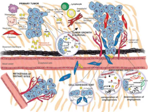
Furthermore, MMPs support tumor growth and angiogenesis via increasing the availability of signaling molecules, such as growth factors and cytokines, by liberating them from the ECM (IGF, bFGF, and VEGF) or by increasing their shedding by from the cell surface (EGF, TGF‐α, HB‐EGF). Besides, MMPs induce angiogenic switch through the downregulation of angiogenic inhibitors and upregulation of angiogenic stimulators factors. Moreover, MMPs can modulate the cell-cell interactions and provoke the ECM through the processing of E‐cadherin and integrins, respectively, thereby, increasing cell migration (Figure 3) (Gialeli, Theocharis, and Karamanos 2011).
Introduction
13
Figure 3. The Pivotal role of MMPs in cancer progression (Gialeli, Theocharis, and Karamanos 2011).
1.7. MMPs in EVs
Several studies have reported that several MMP family members were packaged in EVs from body fluids or various types of cell lines (Dolo et al. 1999; Taraboletti et al. 2002; Belhocine et al. 2010;
Shimoda and Khokha 2013; Reiner et al. 2017; Okusha et al. 2020).
For instance, prostate cancer-derived oncosomes were shown to contain bioactive MMP2, MMP9 molecules that are involved in local invasion, and correlated with tumor progression (Di Vizio et al. 2012).
Another study revealed that vesicles shed from the cultured human umbilical vein endothelial cells are containing active and proenzyme forms of gelatinases, MMP-2 and MMP-9 as well as the MT1-MMP proenzyme that was located on the external side of
Introduction
14
the vesicle membrane, all these proteases initiated the proteolysis necessary for tumor invasion and angiogenesis (Taraboletti et al.
2002). Furthermore, MMP13-containing exosomes were found to facilitate the metastasis of nasopharyngeal cancer cells (an endemic type of head and neck cancer associated with a high rate of cervical lymph node metastasis) through the induction of EMT (You et al.
2015).
Also Hendrix et al. reported that Rab27b-mediated exocytic release of HSP90 exosomes from metastatic breast cancer cells can activate MMP2 resulting in the degradation of ECM components and release of growth factors, promotion of cancer cell invasion (Hendrix et al. 2010). Additionally, San-chez et al. have recently demonstrated that prostate cancer stem cells secreted exosomes that are enriched with miRNAs such as miR-100-5p, miR21-5p, and miR-139-5p.
These exosomal miRNA increased the expression of MMP2, MMP9, MMP13, and RANKL, also enhanced the fibroblasts migration, thereby contributing to local invasion and pre-metastatic niche formation (Sánchez et al. 2016).
Moreover, Hiratsuka et al. have shown that MMP9 induced by primary tumors in lung endothelial cells and macrophages significantly promoted the lung metastasis, the induction of MMP9 was dependent on the tyrosine kinase VEGFR-1 (Hiratsuka et al.
2002). Blocking of the MMP9 induction via deletion of either VEGFR-1TK or MMP9 markedly diminished the lung metastasis in mice models (Hiratsuka et al. 2002).
Introduction
15
1.8. The two-dimensional and three- dimensional culture systems
The two-dimensional (2D) cell culture system has been frequently used for cancer research and drug screening (Yoshii et al.
2011). In conventional 2D culture systems, cells are cultured as monolayers on flat surfaces of plates which allow each cell to access the same amount of growth factors and nutrients present in the medium, resulting in homogenous growth and proliferation (Edmondson et al. 2014). Besides, the strong physical interaction present between cells and 2D culture substrates resulted in alteration in the tumor cell behaviors that differ from those of tumors growing in vivo (Yoshii et al. 2011). Thus, the 2D culture model fails to correctly mimic the proper tissue architecture and complex microenvironment in vivo (Lv et al. 2017).
To overcome the limitations of the 2D culture system, a three- dimensional (3D) cell culture model (aka a spheroid or organoid culture) have been developed to better mimic in vivo tissue microenvironments (Lv et al. 2017; Duval et al. 2017). The 3D culture model maintains the interactions between cells and their ECM, create gradient access of oxygen and nutrient, and buildup a combination of tissue-specific scaffolding cells (Griffith and Swartz 2006).
Similar to human cancers, proliferating, quiescent, and dying cells are coexisting in normoxic, hypoxic, or necrotic zones within tumor organoids (Hirschhaeuser et al. 2010; Eguchi et al. 2018;
Namba et al. 2018). Thus, the 3D tumor models reflect more closely the in vivo human tumors, which prompted us to define tumor organoids as “tumoroids”. Among several methodologies of tumoroid models, we have adopted gel-free tumoroid models cultured on
Introduction
16
NanoCulture Plates (NCP) and ultra-low attachment (ULA) plates (Arai et al. 2016; Eguchi et al. 2018; Namba et al. 2018; Sogawa et al. 2019; 2020).
A great advantage of the gel-free tumoroid model is the collectability of the secretome including EVs from their culture supernatants. NCP is a nanopatterned gel-free scaffold for 3D cell culture (Elsayed and Merkel 2014). The mogul field structure on NCPs restricts cells to sprawl on the base and enable tumor cells to migrate from a scaffold to another scaffold more actively than cells cultured on the 2D plate.
The increased migration and lesser attachment of cancer cells on the NCPs enable tumor cells to form 3D tumoroids (Arai et al. 2016;
Eguchi et al. 2018; Namba et al. 2018; Sogawa et al. 2019; 2020).
ULA plates have been also useful for the collection of secretome including EVs. Cells do not rapidly migrate on ULA plates compared to NCPs. We have examined a few types of culture media such as serum-containing media versus serum-free stemness-enhancing media in combination with the 3D culture systems. In vitro culture of tumoroids in such a 3D nano-environment combined with a defined stem cell medium enabled the cells to grow slowly and form large organoids that expressed multiple stem cell markers and intercellular adhesion molecules (Eguchi et al. 2018; Namba et al. 2018).
Aim & Objective
17 2. Aim and objective
Here, I explored (i) the tumorigenic role of MMP3 on the in vitro tumoroid formation under the 3D culture system and on their EVs integrity, (ii) whether MMP3-rich or MMP3-null EVs could alter tumoroid formation, and examined (iii) the EVs-mediated molecular transfer of MMP3 into the MMP3-KO tumoroids under the 3D culture system.
Material & Methods
18 3. Materials and methods
3.1. Cells
A rapidly metastatic murine cancer cell line LuM1 (Sakata et al.
1996; Namba et al. 2018; Sogawa et al. 2020) and MMP3-KO cells line (Okusha et al. 2020) were maintained in RPMI-1640 with 10%
fetal bovine serum (FBS) and penicillin, streptomycin, and amphotericin B. MMP3-KO cells were established using the CRISPR/Cas9 system from the LuM1 cell line (Okusha et al. 2020).
Briefly, Cas9 protein and guide RNA that targets Mmp3 exon 1 were co-transfected into LuM1 and stable MMP3-KO clones with frame- shifting deletion were obtained.
3.2. Tumoroid culture
Tumoroids were formed in the 3D culture systems using NanoCulture Plate (NCP) (Medical & Biological Laboratories, Nagoya, Japan) or ultra-low attachment (ULA) culture plates/dishes (Greiner, Kremsmunster, Austria) within mTeSR1 stem-cell medium (Stemcell Technologies, Vancouver, Canada) or the above-mentioned serum-containing medium as described previously (Eguchi et al.
2018; Namba et al. 2018; Sogawa et al. 2019; 2020).
For quantification of tumoroids size and number, cells were seeded in a 96 well NCP for 14 days at a concentration of 5.0 x 10³ cells in 200 μL mTeSR1 or RPMI-1640 media with 10% FBS.
Tumoroid maturation was monitored every day and photographed using the Floid cell imaging station (Thermo Fisher, Waltham, MA, USA) from day 1 until day 7 and a BZ-X microscope (Keyence, Osaka, Japan) starting from day 10 until the end of the experiment
Material & Methods
19
day 14. The tumoroid size was measured using Image J software (NIH, Bethesda, MD, USA).
3.3. 2D re-seeding assay
Tumoroids were cultured in the 3D and stem-cell medium condition for 14 days and detached by trypsin/EDTA. The detached cells were re-seeded in a 24-well 2D culture plate at a concentration of 1.5 × 10⁴ cells/well in RPMI-1640 with 10% FBS. The cell images were taken by using the Floid cell imaging station (Thermo Fisher, Waltham, MA, USA) on days 2, 4, 6, and 7 after the seeding.
3.4. Preparation of EVs and conditioned media
Tumoroid-derived EVs were used for tumoroid formation assays.
Otherwise, 2D cultured cells-derived CM was used for 2D experiments. EVs were prepared from culture supernatants of tumoroids using a modified polymer-based precipitation method (Fujiwara et al. 2018; Ono et al. 2018; Eguchi et al. 2020). Briefly, cells were seeded on a 10-cm ULA dish at a density of 1.0 × 10⁶ cells/8 mL mTeSR1 medium and cultured for 6 days. The formed tumoroids were washed with PBS (-), and then further cultured in serum-free medium (4 mL per dish) for 2 days. Cell culture supernatant was collected and centrifuged at 2,000 × g for 30 min at 4°C to remove detached cells. The supernatant was then centrifuged at 10,000 × g for 30 min at 4°C to remove cell debris. The supernatant (8 mL) was concentrated to less than 1 mL by using an Amicon Ultra- 15 Centrifugal Filter Devices for M.W. 100k (Merck Millipore, Burlington, MA). The concentrate was applied to the Total EVs Isolation System (Thermo Fisher, Waltham, MA, USA). The pass-
Material & Methods
20
through was concentrated using an ultrafiltration device for molecular weight 10 kD and used as a non-EV fraction. The EV fraction was suspended in 100 µL PBS (-) and used 3D-tumoroid-EVs. Protein concentration was measured using a micro BCA protein assay kit (Thermo Fisher, Waltham, MA, USA).
For immunofluorescence in the 2D culture system, culture supernatants were collected from serum-free media of 2D-cultured donor cells during the exponential growth phase (70% confluence).
The culture supernatants were centrifuged at 2,000 x g for 15 min to get rid of cells and debris, followed by diluting in a ratio 1:1 with a fresh culture medium. The CM was stored at -80°C. Recipient cells were treated with the CM for 48 h.
3.5. Transmission electron microscopy
As described previously (Eguchi et al. 2018; 2020), a 400-mesh copper grid coated with formvar/carbon films was hydrophilically treated. The EVs suspension (5-10 µL) was placed on Parafilm, and the grid was visualized at 5,000, 10,000, or 20,000 times magnification with an H-7650 transmission electron microscope (TEM) (Hitachi, Tokyo, Japan) at the Central Research Laboratory, Okayama University Medical School.
3.6. Particle diameter distribution
As described previously (Fujiwara et al. 2018; Eguchi et al. 2020), 40 µL of EV fraction within PBS (-) was used. Particle diameters of the EV fractions in a range between 0 and 6,000 nano-diameters were analyzed in Zetasizer nano ZSP (Malvern Panalytical, Malvern, UK).
Material & Methods
21 3.7. Western blotting
Western blotting was performed as described (Ono et al. 2018;
Eguchi et al. 2020). Cells were cultured for 6 days on a 6 well ULA plate at a density of 3.0 x 10⁵ cells/3 mL mTeSR1 medium in a well.
Cells were further cultured in serum-free media for 2 days. On day 8, the supernatants and tumoroids were collected, centrifuged at 2000 x g, 4˚C for 5 min. The supernatants were used for EV preparations as mentioned above.
To prepare whole cell lysate (WCL), tumoroids were lysed in a RIPA buffer (1% NP-40, 0.1% SDS, and 0.5% deoxycholate, and EDTA-free protease inhibitor cocktail in PBS) using 25-gauge syringes. The cell lysate was incubated for 30 min on ice and then centrifuged at 12,000 g for 20 min at 4˚C. The preparation method of EV and non-EV fraction was described above. The supernatant was used as WCL. The same protein amounts for each lane were subjected to sodium dodecyl sulfate-polyacrylamide gel electrophoresis (SDS- PAGE), followed by transfer to a polyvinylidene fluoride (PVDF) membrane using a semi-dry transfer system.
The membranes were blocked in 5% skim milk in Tris-buffered saline containing 0.05% Tween 20 for 60 min at room temperature (RT) and then incubated overnight with rabbit monoclonal antibodies;
anti-MMP3 (EP1186Y, ab52915, Abcam, Cambridge, UK) or anti- CD9 (EPR2949, ab92726, Abcam, Cambridge, UK) or anti-CD63 (EXOAB-CD63A-1, System Biosciences). For CD63, blocking was performed in 10% overnight and the primary antibody was reacted for 2 days. The membranes were incubated with horseradish peroxidase (HRP)-conjugated secondary antibodies. For GAPDH, the HRP- conjugated anti-GAPDH mouse monoclonal antibody (HRP-60004,
Material & Methods
22
Proteintech, Rosemont, IL, USA) was used. For Actin, anti-Actin rabbit antibody (A2066, Sigma Aldrich, St. Louis, MO, USA) was utilized. Blots were visualized with the ECL substrate (Merck Millipore, Burlington, MA, USA).
3.8. Coomassie blue staining (CBS)
Protein samples (1 µg each) were loaded on the SDS-PAGE.
After the electrophoresis run, the gel was stained with Coomassie Brilliant Blue R-250 solution (1610436, Bio-Rad, Hercules, CA, USA) for 30 min with gentle agitation followed by washing with the destaining solution (50% methanol, 10% glacial acetic acid) for 2 h until the background became less dark.
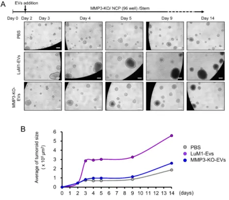
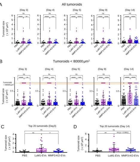
3.9. EV-driven in vitro tumorigenesis
MMP3-KO cells were seeded at 5.0 x 10³ cells/200 μL mTeSR1 medium in a well of 96-well NCP. After two days, EVs derived from 3D-tumoroids (LuM1 or MMP3-KO) were added to MMP3-null tumoroids at a final concentration of 5 μg/mL. Then the plate was centrifuged at 1,800 × g for 1 h at 4˚C to increase the internalization of EVs into the tumoroids (Lai et al. 2015). The MMP3-KO tumoroids maturation was monitored over 14 days using a microscope FSX100 (Olympus Life Science, Tokyo, Japan). Then tumoroid size was measured using Image J.
3. 10. Palm fluorescent cells
The lentiviral reporter constructs of CSCW-palmitoylation signal-tandem dimer Tomato (palmT) and CSCW-palmitoylation signal-EGFP (palmG) were kindly gifted from Dr. Charles P. Lai (Lai
Material & Methods
23
et al. 2015). For virus production, HEK293T cells at 70-80%
confluence were transfected with PalmT or PalmG constructs, psPAX2 packaging plasmid, and pMD2.G envelope plasmid using PEI max (Polysciences).
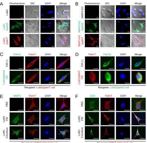
LuM1 or MMP3-KO cells were infected by using the spinfection method with the viral solution. Infected/transduced stable cells were selected using puromycin. Isolation of single clones was carried out by limiting dilution method. We established palmtdTomato-expressed LuM1 cells (designated LuM1/palmT), palmGFP-expressed LuM1 cells (designated LuM1/palmG), palmGFP-expressed MMP3-KO cells (designated MMP3-KO/palmT), and palmGFP-expressed MMP3-KO cells (designated MMP3-KO/palmG).To confirm fluorescent labeling, the palm fluorescent cells were seeded on a type I collagen-coated coverslip in a 24-well plate at a density of 1 × 10⁴ cells/well in a serum-containing culture media and cultured for 48 h.
3.11. EVs exchange assay
Two different colored fluorescent cells (LuM1/palmG and LuM1/palmT cells) were used as donor cells or recipient cells with each other in the 2D culture system. The donor cells were seeded at 1
× 106 cells in a 60 cm dish and cultured overnight in a serum- containing culture media. The grown cells of 70-80% confluence were washed twice with PBS, then the culture media was replaced with a serum-free medium and cultured for a further 2 days. The culture supernatant was collected and centrifuged at 2,000 × g for 15 min at 4°C to remove detached cells and the supernatants were diluted in a ratio 1:1 with a fresh culture medium and used as CM. Recipient cells were seeded on a type I collagen-coated coverslip inserted in a
Material & Methods
24
24-well plate at a density of 1 × 10⁴ cells/well and cultured for 24 h in a serum-containing culture media. The recipient cells were treated by donor cells-derived CM for 48 h. For coculturing, recipient MMP3- KO cells were seeded on coverslips. LuM1-donor cells (1 × 10⁴ cells/well) were seeded on a culture insert with a 0.45-μm pore (Greiner, Kremsmunster, Austria) in a 24 well plate. The insert with donor cells was placed on the well containing the recipient cells and cocultured for 48 h.
3.12. 2D confocal laser-scanning microscopy
Cells were fixed in 4% paraformaldehyde (PFA) for 10 min at RT and permeabilized with 0.5% Tween-20 for 10 min. For blocking the non-specific reaction of primary antibodies, cells were blocked in 10% normal goat serum solution (Dako, Tokyo, Japan) for 30 min, then incubated overnight at 4℃ with rabbit anti-MMP3 antibody (EP1186Y, ab52915, Abcam, Cambridge, UK) or rabbit anti-CD9 antibody (EPR2949, ab92726, Abcam, Cambridge, UK), for overnight at 4˚C and subsequently with a secondary antibody, goat anti-rabbit IgG Alexa Fluor 488 (A-11034, Thermo Fisher, Waltham, MA, USA) for 1 h at RT.
As a negative control, the same protocol was performed without primary antibody staining. Washes after antibody reactions were done with PBS, three times for 3 min each, on a shaker at RT. The mounting and DNA staining was performed by using Immunoselect Antifading Mounting Medium DAPI (SCR-038448, Dianova, Germany).
Fluorescent images were taken using a confocal laser scanning microscopy LSM780 (Carl Zeiss, Oberkochen, Germany) at Central Research Laboratory, Okayama University Medical School.
Material & Methods
25 3.13. Immunofluorescence of tumoroids
For tumoroid formation, cells were cultured for 6 days at a density of 3.0 x 10⁵/3 mL mTeSR1 in a well of a 6-well ULA plate.
Then tumoroids were treated with PBS, the 3D-tumoroid LuM1-EVs at a final concentration of 5 μg/mL or the 3D-tumoroid LuM1-CM (diluted 1:1 with fresh mTeSR1) for 24 h. Then the plate was centrifuged at 1,800 × g for 1 h at 4˚C to increase the internalization of EVs into the tumoroids (Lai et al. 2015). Tumoroids were washed with PBS and fixed in 4% PFA for 10 min.
Tumoroids were additionally washed with PBS for 5 min 3 times and embedded in paraffin. Tumoroids sections (5 µm thickness) were deparaffinized and hydrated through xylenes and graded alcohol series. Antigen retrieval was performed by heating the specimens in Tris/EDTA buffer, pH 9.0 (Dako target retrieval solution S2367, DAKO, Carpenteria, CA) using a microwave for 3 min for CD9 or by autoclaving in 0.01M citrate buffer pH 6.0 (sodium citrate dihydrate, citric acid; Sigma Aldrich, USA) in a pressure cooker for 8 min for MMP3 and Ki-67.
Sections were treated with blocking solution (Dako) for 30 min at RT, then incubated with primary antibodies; rabbit anti-CD9 (EPR2949, ab92726, Abcam, Cambridge, UK), rabbit anti-MMP3 (EP1186Y, ab52915, Abcam, Cambridge, UK), or rat anti-Ki-67 antibody (TEC-3, M7249, Dako); individually at 4°C overnight.
Then, sections were subsequently stained with a secondary antibody goat anti-rabbit IgG, Alexa Fluor 488 (A-11034, Thermo Fisher, Waltham, MA, USA) for 1 h at RT. Then samples were counterstained with 1 mg/mL of DAPI (Dojindo Laboratories, Kumamoto, Japan). Fluorescent images were taken using a confocal
Material & Methods
26
laser scanning microscopy LSM780 (Carl Zeiss, Oberkochen, Germany) at Central Research Laboratory, Okayama University Medical School.
For IHC staining of Ki67, a biotinylated secondary antibody was applied for 30 min (Vector Lab, Burlingame, CA) and the color was developed with 3, 3’-diaminobenzidine (DAB) (Histofine DAB substrate; Nichirei, Tokyo, Japan). Then, samples were counterstained with Myer’s hematoxylin and images were taken using an optical microscope BX53 (Olympus).
To calculate the Ki-67 labeling index (%), we counted approximately 100 Ki67-positive cells were counted in random five fields under the 40× objective. Areas with severe necrosis were avoided. The Ki-67 labeling index (%) was calculated by dividing the total Ki-67 positive cells by the total numbers of cells multiplied by 100. The total tumoroid areas, as well as the area of necrotic regions, were measured using Image J. The percentage of necrosis was calculated by dividing the total necrotic area by the total tumoroid area.
3.14. Hematoxylin and eosin staining
For tumoroid formation, LuM1 or MMP3-KO cells were cultured for 8 days on 6 well ULA plates at a density of 3.0 x 10⁵/3 mL mTeSR1/well. Then tumoroids were washed with PBS, fixed in 4%
PFA for 10 min, and embedded in paraffin. Tumoroids sections (5 µm thickness) were deparaffinized in a series of xylene for 15 min, rehydrated in graded ethanol solutions, and washed well in distilled water. Then sections were incubated in Harris hematoxylin solution for 10 min and rinsed in tap water until the water was colorless.
Material & Methods
27
Finally, after sequential treatment with hydrogen chloride and 80%
ethanol solution, sections were incubated in eosin for 7 min.
3.15. Tracing EV-uptake in vitro
Ten micrograms of tumoroid-derived EVs were incubated with 0.25 μM BODIPY TR Ceramide (Thermo Fisher, Waltham, MA, USA) for 20 min at 37°C. Excessive BODIPY TR Ceramide was removed with Exosome Spin Columns (MW 3000) (Thermo Fisher, Waltham, MA, USA) (Namba et al. 2018). Cells were seeded at a concentration of 5.0 x 10³ cells/200 μL mTeSR1 in a well of 96-well NCP. The next day, EVs were added at a final concentration of 5 μg/mL. The EVs- uptake was monitored over 24 h using the ArrayScan High Content Screening (HCS) system (Thermo Fisher, Waltham, MA, USA). The fluorescence intensity of each cell was determined using a filter set (485/594) for (GFP/ BODIPY TR). The average fluorescence intensity of the PBS treatment group at time point 0 h was evaluated as background and subtracted from raw values.
3.16. Statistical analysis
Statistical significance was calculated using GraphPad Prism and Microsoft Excel. The difference between the sets of data was analyzed using ANOVA Tukey’s multiple comparisons test and all data are expressed as the mean ± standard deviation unless otherwise indicated.
Results
28 4. Results
4.1. MMP3 knockout reduces the release of CD9 and CD63 within extracellular vesicles
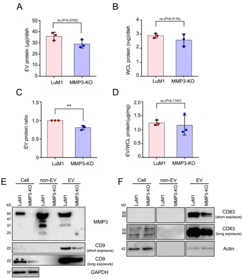
To explore the role of MMP3 on cellular communication in cancer, our research group has generated MMP3-KO cells by the CRISPR/Cas9 genome editing system from a rapidly metastatic murine cancer cell line LuM1 with a parental cancer cell line Colon26 (aka CT26) (Okusha et al. 2020). The release of EV proteins from MMP3-KO-tumoroids tended to decrease compared to LuM1- tumoroids (Figure 4A-D). However, there was no statistically significant difference in the protein concentration of the whole cell lysate (WCL) between LuM1 and MMP3-KO-tumoroids.
MMP3 was markedly detected in the cellular, non-EV, and EV fractions of the LuM1, while the complete loss of MMP3 was confirmed in MMP3-knockout LuM1 cells, non-EV (including soluble proteins), and EV fractions (Figure 4E, top row; Figure S1A), suggesting a successful knockout of the Mmp3 gene.
Next, I examined CD9 (a category-1 EV marker protein) expression pattern. Interestingly, the CD9 content was significantly down-regulated in both cellular and EV fractions of the MMP3-null cells compared to their counterpart (Figure 4E, second and third rows;
Figure S1B). Moreover, CD63 was reduced in the EV fraction of the MMP3-null cells (Figure 4E, fourth and fifth rows; Figure S1C).
Recently, we have shown that GAPDH and β-actin were released in the EV and non-EV soluble fractions upon membrane-damaging cell stress (Eguchi et al. 2020). Therefore, I examined the expression
Results
29
levels of β-actin and GAPDH not only as a loading control but also to investigate whether they were released within EVs or non-EV soluble proteins. Our results revealed that both β-actin and GAPDH were detected in the EVs derived from both cell lines, but not in the non- EV fractions (Figure 4E, bottom lane; Figure S1D, E; Figure S2A).
Notably, β-actin levels were considered as a loading control for WCL and EV fractions, whereas GAPDH was not (Figure S1D, E).
Furthermore, the SDS-polyacrylamide gel was stained with Coomassie brilliant blue (CBB) after the electrophoretic separation (Figure S2B).
These findings demonstrate that MMP3 controls the secretion of CD9/CD63-contained EVs.
Results
30
Figure 4. MMP3 knockout reduces the release of CD9 and CD63 within extracellular vesicles. Tumoroids were formed in 10-cm ultra-low attachment (ULA) plates for 6 days.
Extracellular vesicle (EV) and non-EV fractions were collected from the culture supernatants. (A, B) The total protein concentration in the (A) EV and (B) whole cell lysate (WCL) fractions of LuM1-tumoroids and MMP3-KO tumoroids. (C) Relative EV protein ratio comparing two cell lines. (D) EV protein concentration per the WCL proteins. ** p < 0.01; ns, not significant. (E) Western blotting showing MMP3, CD9, CD63, and β-actin in tumoroids, non-EV, and EV fractions. The 54-kD
Results
31
bands indicate the full-length MMP3, the 47-kD bands represent the active form consists of the catalytic, hinge, and PEX domains, the 37-kD represents the catalytic domain, and the 25-kD shows the PEX domain of MMP3. The expression level of β-actin was examined as a loading control as well as to check if it was released from cells. The protein amounts loaded from WCL and EV fractions were 10 µg per lane, while 5 µg per lane was loaded from the non-EV fractions. The experiments were repeated twice.
For full images of Western blotting, see Figure S1.
4.2. MMP3 knockout impacts physical integrities of extracellular vesicles
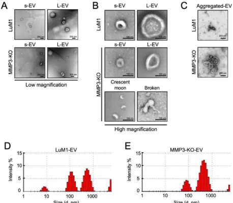
Further, I examined the morphology and size of EVs secreted from “tumoroids” by transmission electron microscopy (TEM) and Zetasizer, respectively. Both LuM1- and MMP3-KO tumoroids released two types of EVs small EVs (s-EVs) ranged approximately 50-200 nm and large EVs (L-EVs) more broadly ranged between 200 and 1000 nm (Figure 5A, B, Table 1). According to their size, the s- EVs were supposed to be exosomes, while the L-EVs were supposed to be microvesicles.
Meanwhile, crescent moon-like shaped and broken EVs were particularly seen in the MMP3-KO EV fraction released by MMP3- KO tumoroids (Figure 5A, B). Additionally, I observed large aggregates (500-800 nm) of EVs derived from MMP3-KO compared to their counterparts (Figure 5C).
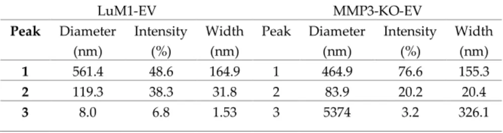
Particle diameter distribution analysis using Zetasizer revealed that the size of both s-EVs (peaked at 84 nm) and L-EVs (peaked at 465 nm) released from MMP3-KO tumoroids were smaller than those (peaked at 119 nm and 561 nm, respectively) of LuM1 tumoroids
Results
32
(Figure 5D, E). The small particles detected at 5-10 nm in the LuM1- EV fraction might be damaged membrane vesicles or lipoprotein particles e.g. HDL (5–12 nm) which have similar size ranges as EVs (Schumaker 1994).
These findings demonstrate that MMP3 is important for maintaining the physical integrities of EVs, and the loss of MMP3 resulted in disorganizing the EVs structures.
Table 1. Comparison of particle size distributions between LuM1- EVs and MMP3-KO-EVs.
LuM1-EV MMP3-KO-EV
Peak Diameter (nm)
Intensity (%)
Width (nm)
Peak Diameter (nm)
Intensity (%)
Width (nm)
1 561.4 48.6 164.9 1 464.9 76.6 155.3
2 119.3 38.3 31.8 2 83.9 20.2 20.4
3 8.0 6.8 1.53 3 5374 3.2 326.1
Results
33
Figure 5. MMP3 knockout impacts the physical integrities of extracellular vesicles. (A-C) TEM images of EV fractions derived from the LuM1 and MMP3-KO tumoroids. at (A) low magnification, (B) high magnification, and of (C) aggregated EVs. s-EVs, small EVs; L-EVs, large EVs. Scale bars, 200 nm (in low magnification), and 100 nm (in high magnification). (D, E) Representative histograms showing the particle diameter distributions of EVs derived from (D) LuM1 tumoroids and (E) MMP3-KO tumoroids. The experiments were repeated twice.
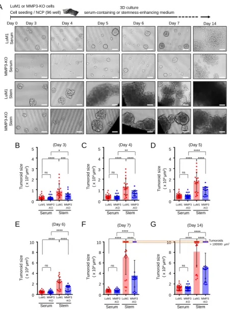
4.3 Loss of the Mmp3 gene reduces the in vitro tumorigenicity Next, I examined the consequences of Mmp3 loss on the in vitro tumoroid formation. The LuM1 and MMP3-KO cells were cultured in the 3D culture system either under serum-containing or mTeSR1 stemness-enhancing conditions for 14 days. Larger tumoroids were formed in the stemness-enhancing medium compared to smaller
tumoroids seen in the serum-containing medium (Figure 6A-G).