緒 言
くも膜下出血発症の解離性椎骨動脈瘤は,早期に再出 血をきたす頻度が高く,迅速かつ確実な再出血予防処置 が求められる.開頭手術による椎骨動脈遮断術に代わっ て,最近ではdetachable coilを用いた塞栓術が行われ,
良好な成績が報告されているが,解離部に後下小脳動脈
(PICA)の起始部が含まれる,PICA-involved typeの解 離性動脈瘤では,PICAの閉塞を生じるため急性期の血 管内治療による根治的治療は困難であると考えられてき た.しかし今回我々は,急性期の血管内治療にて解離腔
へのentryおよび破裂部に限局した,部分的trappingを行 うことによりPICA起始部を温存し,再出血を防止し得 た症例を経験した.PICA-involved typeの解離性動脈瘤 に対する急性期治療の選択肢の一つになる可能性がある と考えられるため,報告する.
症例呈示
症 例:!"歳 男性
主 訴:突然の後頭部痛,嘔吐,意識障害 既往歴・家族歴:高血圧症(治療中)
現病歴:#""$年%%月%#日,突然の後頭部痛,嘔吐に続き
症例報告
PICA!involved typeの破裂解離性椎骨動脈瘤に 対する急性期血管内治療の " 例
─ PICA を温存した部分的 trapping ─
今岡 充%) 長谷川真作#) 尾崎 聡&) 小櫃久仁彦&) 谷井雅人&) 吉澤 卓&) 伊藤建次郎&)
Endovascular treatment for ruptured PICA-involved vertebral artery dissecting aneurysm: partial trapping preserving the PICA: a case report
Takashi IMAOKA1) Shinsaku HASEGAWA2) Satoshi OZAKI3) Kunihiko KOBITSU3) Masahito TANII3) Takashi YOSHIZAWA3) Kenjiro ITOH3)
1) Department of Neurosurgery, Nishijima Hospital 2) Department of Neurosurgery, Kawasaki Saiwai Hospital
3) Department of Neurosurgery, Yokohama Shintoshi Neurosurgical Hospital
●Abstract●
Objective: There is an ongoing debate as to the proper management of a ruptured vertebral artery dissecting aneurysm (VADA) involving the origin of the posterior inferior cerebellar artery (PICA). This article describes a case of ruptured VADA successfully treated in the acute stage with endovascular intervention.
Clinical Presentation: A 50-year-old male presented with subarachnoid hemorrhage and was admitted to our institution. Preoperative right vertebral angiogram showed a VADA in which the PICA arose from the wall of the aneurysm and a sufficient interval between the rupture point and the origin of PICA. The aneurysm was treated on the day of rupture while preserving the origin of PICA with limited internal trapping of the vertebral artery at the rupture point and entry into the pseudolumen (partial trapping). Postoperative MR angiography demonstrated rapid shrinkage and almost complete disappearance of the residual aneurysm within 2 months while maintaining the patency of PICA. The patient recovered completely without any episodes of recurrent hemorrhage.
Conclusion: Partial trapping preserving the origin of PICA offers potential as a treatment for this complex lesion in a select group of patients.
●Key Words●
dissecting aneurysm, partial trapping, PICA-involved type
(Recieved January #', #""':Accepted May $, #""')
%! 西島病院 脳神経外科
#! 川崎幸病院 脳神経外科
&! 横浜新都市脳神経外科病院 脳神経外科
<連絡先:〒(%""""## 静岡県沼津市大岡#'&!"$ E-mail: [email protected]>
JNET 2:38-44, 2008
意識障害が出現し,救急搬送された.
入院時現症:GCS %#(E&+V(+M!)で,WFNS Grade (.バイタルサイン(血圧%#)/!"mmHg,体温&*.'℃,脈 拍'*/分・整)には異常なく,髄膜刺激症状を含め,そ の他,神経学的異常所見は認められなかった.
神経放射線学的所見:頭部CT検査にて後頭蓋窩に強い くも膜下出血を認めた.MRAの+D再構成画像では右側 椎骨動脈に,約&"mm長の連続した不整な紡錘状変化が 認められ,解離性動脈瘤と考えられた(Fig. 1A).右側 PICAは 動 脈 瘤 の 中 央 よ り や や 心 臓 側 か ら 分 岐 し,
PICA起始部より遠位側には特に不整な瘤様拡張部が認 められた(Fig. 1A, B).また椎骨動脈は右側優位で健 常側は低形成であり,後交通動脈は両側とも明らかな描 出が認められなかった(Fig. 1B).
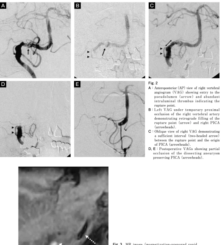
入院後経過:入院後直ちに,局所麻酔下,両側大腿動脈 アプローチにて脳血管撮影および右側椎骨動脈閉塞試験 を行った.脳血管撮影では,MRA所見と同様に右側 PICAは動脈瘤の中央部からやや心臓側で起始してお り,,mm程度の距離を置いた遠位側にはintimal flap,
および血栓形成等の所見が限局して認められ,破裂部位 であることが強く示唆された(Fig. 2A).右側椎骨動脈 V-遠位部に.Frのバルーン付ガイディングカテーテル
(PATLIVE:Clinical Supply,岐阜)を誘導し,右側 椎骨動脈遮断下に側副血行動態を評価すると,低形成で
ある左側椎骨動脈から脳底動脈先端部までの十分な血流 が明らかとなり,両側後大脳動脈も両側後交通動脈を介 して内頸動脈系から良好に描出されることが判明した.
意識状態,神経症状にも変化を認めないため,右側椎骨 動脈は安全に閉塞可能と判断した.引き続き血管内治療 を行ったが,左側椎骨動脈から逆行性に動脈瘤破裂部,
右側PICAまで明瞭に描出されるため,右側椎骨動脈近 位部閉塞では再出血防止には不十分であると考えられた
(Fig. 2B).しかし斜位像にて再評価すると,破裂部お よびentryとPICA起始部との間に十分な距離的安全域を 確保できるため(Fig. 2C),破裂部およびentryを十分 に塞栓してもPICA起始部は温存可能と判断し,同部の 部分的trappingを試みることとした.PATLIVEによる 右側椎骨動脈の一時的遮断下に,マイクロカテーテルを 右 側 椎 骨 動 脈 の 遠 位 ま で 誘 導 し,GDC %"(Boston Scientific, Natick, MA, USA)を用いて破裂部および entryを含めて椎骨動脈を閉塞した.右側PICA起始部直 後まで詰め戻ったところで右側椎骨動脈の遮断を解除し 造影したところ,PICA起始部より近位側に約%"mm長の 膨隆が残存していたが,同部分には解離腔へのentryお よび破裂を示す所見が認められず,非出血性解離部であ ると判断できたため,それ以上の塞栓は行わず,PICA 起始部を温存した部分的trappingで手術を終了した(Fig.
2D, E).
A B
Fig. 1 &-dimensional rendering image of MR angiography #A: anteroposterior #AP! view, B: posteroanteror #PA! view!
showing fusiform transformation of the right vertebral artery, indicating a dissecting aneurysm with particularly irregular dilatation #arrowheads! at the distal portion, and the PICA #arrow! originating from the wall of the aneurysm.
Imaoka T, et al
術後経過:術後/日目のMRI(magnetization-prepared rapid gradient echo:MPRAGE)では,PICA起始部よ り近位側の椎骨動脈に壁在血腫を示唆する所見が認めら れた(Fig. 3).術後-日目のMRAで右側椎骨動脈径の細
小化と残存動脈瘤の著明な縮小が認められ(Fig. 4B),
術後%$日目にはPICAの開通性を維持したまま動脈瘤は 殆ど消失していた(Fig. 4C).術後の再出血,症候性脳 血管攣縮,水頭症等は出現せず,第&&病日にmodified
A B C
D E Fig. 2
A: Anteroposterior #AP! view of right vertebral angiogram #VAG! showing entry to the pseudolumen #arrow! and abundant intraluminal thrombus indicating the rupture point.
B: Left VAG under temporary proximal occlusion of the right vertebral artery demonstrating retrograde filling of the rupture point #arrow! and right PICA
#arrowheads!.
C: Oblique view of right VAG demonstrating a sufficient interval #two-headed arrow!
between the rupture point and the origin of PICA #arrowheads!.
D, E:Postoperative VAGs showing partial occlusion of the dissecting aneurysm preserving PICA #arrowheads!.
Fig. 3 MR image #magnetization-prepared rapid gradient echo: MPRAGE! obtained ' days after the treatment showing a high intensity area adjacent to the vertebral artery corresponding to intramural hematoma #arrowheads!. Arrow indicates the proximal vertebral artery and dotted arrow indicates the basilar artery.
Rankin Scale0にて独歩退院した.術後-ヵ月目のMRA でも動脈瘤の再発傾向はみられなかった(Fig. 4D).
考 察
くも膜下出血で発症する解離性椎骨動脈瘤は急性期の 再出血率が高く,再出血は予後を悪化させる因子の一つ である.その多くは発症#(時間以内に生じるが,特に1
〜-時間以内の,病院到着直後から手術室に至るまでの 超早期に生じることが多いとの報告もあり%%),近年では
手術に時間を要する開頭手術に代わって,血管内治療の 有効性が多く報告されている$,%(,%!).
術式に関して,従来は近位部母血管閉塞が標準的術式 であったが,健側椎骨動脈からの逆行性血流による再出 血が治療の確実性の観点から問題視され%)),最近では解 離部を含めて椎骨動脈を閉塞するinternal trappingが行 われるようになった.しかし解離部にPICA起始部が含 まれるPICA-involved typeの場合,internal trappingで はPICA閉塞による虚血性合併症の発生が危惧される.
A B
C D
Fig. 4 Serial MR angiography demonstrating rapid shrinkage and almost complete disappearance of the residual aneurysm within # months, while maintaining the patency of PICA. A : pretreatment. B : Day #. C : Day %$. D : # months.
Imaoka T, et al
PICA-involved typeの頻度は%("#$%(,!,',%*)程度で高くは ないが,治療に際しては迅速性,確実性に加え,PICA 領域の虚血回避を加えた+要件を満たすものが求めら れ,治療戦略に困難を強いられている.
PICA-involved typeの解離性動脈瘤の治療方針につい て,PICA起始部を含めたtrappingによる良好な治療成 績が報告されている(,#").これは,PICA領域には側副血 行路が発達しており,PICAの遮断は問題にならない場 合があるとの理由によるが,一方でPICAを犠牲にする 術式(trapping,近位部母血管閉塞+PICA clipping)で は!"%に小脳梗塞を生じ,手術morbidityの主原因になる との報告もあることから%%),虚血性合併症を確実に回避 するためにはPICAの血行再建または温存が欠かせない と思われる.
治療の確実性,PICA領域の虚血回避の観点から,開 頭手術にて一期的にtrappingとPICAの血行再建を行う 報告が散見される%,#,&,%*).しかし,本症は超早期の再出 血の頻度が高いため発症直後の緊急手術が必要であるこ と,手術にかなりの習熟を要すること,下位脳神経障害 等の手術合併症などを考慮すると*),最良の治療法であ るとは言い難い.また,急性期に血管内治療にて近位部 母血管閉塞のみを行って再出血を軽減し,慢性期に PICA閉塞試験を施行して,必要があればPICAの血行 再建を併用してtrappingするという,段階的治療での良 好な治療成績も報告されている().しかしこの方法では,
急性期に健側の椎骨動脈より病側PICAへの血流があり 再出血の危険が残るため,不確実であると思われる&,#"). 血管内治療によるinternal trappingは迅速性と確実性 が利点であるが,当然ながらPICAの血行再建は不可能 である.解離性動脈瘤の治療目標は罹患部位を血流から 完全に排除することであるが,実際の術式では,破裂部 である瘤様拡張部から詰め始めて解離腔へのentryまで をカバーする方法%')や,瘤様拡張部のみをtightに塞栓 し壁在血腫による解離性狭窄部は塞栓しない方法%!)な ど,破裂部とentryに重点を置いたtrappingが標準的に行 われており,解離部全長のtrappingを行わずとも良好な 治療成績が得られている%#,%!,%'!.また病理学的検討によ ると,動脈瘤のpseudolumenへのentryは通常破裂部の直 下にあり,解離腔の端の方では内弾性板の連続性が保た れていることから),%"),PICAを温存して動脈瘤部分の椎 骨動脈を遮断する方法の可能性が示唆されている%%).す なわち,再出血防止には破裂部とentryの双方を血流か ら除外することが最重要であり,それ以外の非出血性解
離部へのtrapping範囲の拡大は,再出血防止の観点から は必ずしも必要でないと思われる.PICA-involved type のように解離部全長のtrappingでは虚血性合併症のリス クが高い場合であっても,破裂部とentryを確実に診断 し得る症例であれば,同部に限定的なtrappingを行うこ とで,重要な血管を温存したままの再出血防止が成立し 得ると考えられる.しかしながら,今回のようにMRA での不整形拡張所見や脳血管撮影でのintimal flap,血栓 形成等の所見から,破裂部とentryを明瞭に同定し得る 症例は,むしろ例外的であるのかもしれない.
また今回の症例は病変が長い上,脳血管撮影(Fig.
2C)ではPICA起始部直後に非膨隆部を認め病変全体が dumbbell様にもみえること等から,病変全体が同一病変 であるのかどうか疑問が残る.つまり以前からの解離そ の他の原因によりPICA起始部より近位側が無症候性に 膨隆していた椎骨動脈に,今回PICA起始部より遠位側 に新たな解離が起こり出血した可能性が考えられる.解 離性動脈瘤の病理学的検討に関する報告は少ないが,
Mizutaniらは2症例の臨床的,病理学的所見に基づき解 離性および紡錘状動脈瘤を,Type %:classic dissecting aneurysm,Type #:segmental ectasia,Type &:
dolichoectatic dissecting aneurysm,Type (:saccular aneurysm,arising from arterial trunk,の3型に分類し
た),%").Type % は急激に内弾性板が断裂して解離腔を生
じ,解離腔内に血栓を伴うもので血管撮影上は不整な形 態を示すが,Type # は慢性的な血行力学的負荷により 内弾性板の菲薄化や断裂を生じ,血管撮影上は滑らかな 紡錘状で血栓を伴わず無症候性であることが多いとして おり,純粋な解離性動脈瘤はtype % であるとしている.
また各Typeの判別には脳血管撮影に加えてMRI所見が 有用であると述べている),%").今回の症例は,MRA(Fig.
1)にみられる病変の全長にわたる連続した不整形所見 や,MRI(Fig. 3)でPICA起始部より近位側の膨隆部 に壁在血腫の存在が示唆される所見等から,Type %,
Type # の合併ではなく,病変全体がType % の病変であ ったと考えられる.また椎骨動脈解離性動脈瘤の大部分 は血管腔からのentryのみを有しreentryを有しないone entry typeであると考えられているが),%"),今回の症例は 病変が長くintimal flapの方向が末梢側から中枢側へ向 かっている所見等から,reentryを有するentry-reentry typeであった可能性がある.しかし解離腔やentryの部 分的閉塞がしばしばみられるため,病理診断以外で-つ のtypeを判別するのは困難であるとされている),%").
今回の部分的trappingにより残存した動脈瘤の術後経 過は興味深い.術後早期から動脈瘤の縮小が始まり,術 後%$日目には殆ど消失し,以後も再発傾向はみられてい ない.椎骨動脈の血流がPICAのみに集束する形となる ために,血流量低下による椎骨動脈径の細小化が起こり,
それに伴って残存動脈瘤も縮小したものと思われる.解 離性動脈瘤の形態変化については一定の見解をみていな
いが%&,#%),一部の例外を除き,解離の進行や解離腔の血
栓化などの病巣変化はかなり早期に急激に生じ,初期変 化はおおむね+ヵ月から半年程度で安定すると考えられ
ている*,%&,%$).今回の症例も早期に治癒傾向となったも
のの,今後も継続的な経過観察が必要であり,動脈瘤の 明らかな増大を認めた場合には,PICAの血行再建を含 めたtrapping追加を検討する必要があろう.
結 論
PICA-involved typeの破裂解離性椎骨動脈瘤で,画像 所見から破裂部およびentryと思われる部位の診断が可 能であった症例に,急性期の血管内治療にてPICAを温 存した部分的なtrappingを行うことで,迅速な再出血防 止と残存部の改善が得られた.しかし本法はPICA- involved typeの解離性動脈瘤に対する普遍的な治療法で はなく,破裂部とentryを確実に同定でき,PICA起始部 との間に距離的安全域が確保できる症例に限られるた め,脳血管撮影やMRI,MRA等を詳細に検討した上で,
慎重に適応を判断せねばならない.
文 献
1) Hamada J, Kai Y, Morioka M, et al: Multimodal treatment of ruptured dissecting aneurysms of the vertebral artery during the acute stage. J Neurosurg )):)*"")**, #""&.
-) 濱田潤一郎 , 森岡基浩 , 甲斐 豊 , 他:後下小脳動脈を 含んだ出血発症解離性動脈瘤に対する浅側頭動脈移植を 用いた血行再建術 . 脳卒中の外科 &&:&)'"("%, #""!.
+) 堀 内 哲 吉 , 新 田 純 平 , 北 沢 和 夫 , 他:PICA-involved dissecting aneurysm に対する OA"PICA anastomosis を 用いた急性期直達手術 . 脳卒中の外科 &&:("#"("!, #""!.
3) Iihara K, Sakai N, Murao K, et al: Dissecting aneurysms of the vertebral artery: a management strategy. J Neurosurg )$:#!)"#*$, #""#.
,) Kim CH, Son YJ, Paek SH, et al: Clinical analysis of vertebrobasilar dissection. Acta Neurochir #Wien!
%(':&)!"("(, #""*.
4) Kitanaka C, Sasaki T, Eguchi T, et al: Intracranial vertebral artery dissections: clinical, radiological features, and surgical considerations. Neurosurgery &(:*#""*#$,
%))(.
.) Kurata A, Ohmoto T, Miyasaka Y, et al: Coil embolization for the treatment of ruptured dissecting vertebral aneurysms.
AJNR ##:%%"%', #""%.
/) Lee JW, Jung JY, Kim YB, et al: Spontaneous dissecting aneurysm of the intracranial vertebral artery: management strategies. Yonsei Med J (':(#!"(&#, #""$.
2) Mizutani T, Miki Y, Kojima H, et al: Proposed classification of nonatherosclerotic cerebral fusiform and dissecting aneurysms. Neurosurgery (!:#!&"#*", %))).
%") Mizutani T, Kojima H, Asamoto S, et al: Pathological mechanism and three-dimensional structure of cerebral dissecting aneurysms. J Neurosurg )(:$%#"$%$, #""%.
%%) 水谷 徹:解離性脳動脈瘤に対する手術適応と術式 . 脳 卒中の外科 &":(#("(#', #""#.
%#) 中村 貢 , 藤田敦史 , 甲村英二 , 他:椎骨動脈解離性病 変に対する瘤様拡張部塞栓術 . 脳卒中の外科 &&:%*$"%$&,
#""&.
%&) 小野純一 , 平井伸治 , 芹澤 徹 , 他:椎骨脳底動脈系の 非出血性解離性病変の治療方針 . 脳卒中の外科 &&:#""#!,
#""!.
%() Raymond J, Roy D: Safety and efficacy of endovascular treatment of acutely ruptured aneurysms. Neurosurgery (%:%#&!"%#(*, %))$.
%!) 杉生憲志 , 徳永浩司 , 伊達 勲:破裂急性期椎骨動脈解 離 性 動 脈 瘤 に 対 す る 血 管 内 治 療 . No Shinkei Geka
&#:%##)"%#&', #""(.
%*) 田辺英紀 , 田村陽史 , 住岡真也 , 他:PICA 分岐部解離 性 椎 骨 動 脈 瘤 に 対 す る 直 達 手 術 . 脳 卒 中 の 外 科
&&:%($"%!(, #""!.
%$) 八木伸一 , 吉岡秀幸 , 八木 貢 , 他:疼痛発症頭蓋内解 離性椎骨動脈瘤の治療方針 . 脳卒中の外科 &&:%("%),
#""!.
%') Yamaura I, Tani E, Yokota M, et al: Endovascular treatment of ruptured dissecting aneurysms aimed at occlusion of the dissected site by using Guglielmi detachable coils. J Neurosurg )":'!&"'!*, %))).
%)) 安井敏裕 , 岸 廣成 , 小宮山雅樹 , 他:破裂解離性椎骨 動脈瘤に対する近位側閉塞後の再破裂機序 . No Shinkei Geka #':&(!"&(), #""".
#") Yasui T, Komiyama M, Nishikawa M, et al: Subarachnoid hemorrhage from vertebral artery dissecting aneurysms involving the origin of the posteroinferior cerebellar artery. Report of two cases and review of the literature.
Neurosurgery (*:%)*"#"%, #""".
#%) Yoshimoto Y, Wakai S: Unruptured intracranial vertebral artery dissection. Clinical course and serial radiographic imagings. Stroke #':&$""&$(, %))$.
Imaoka T, et al
JNET 2:38-44, 2008
要 旨
【目的】PICA-involved typeの破裂解離性椎骨動脈瘤に対し,急性期の血管内治療にてPICAを温存した部分的trappingを行 い治療し得た症例を経験したので報告する.【症例】!"歳男性.くも膜下出血で発症し,右側椎骨動脈にPICA-involved type の解離性動脈瘤を認めた.解離腔へのentryおよび破裂部はPICA起始部より遠位側に限局していると判断し,直ちに同部を コイルで塞栓したが,PICA起始部より近位側は塞栓せずPICAを温存した.神経脱落症状なく独歩退院し,術後-ヵ月の観 察で残存部は急速に縮小・消失し,再出血はみられていない.【結論】PICA-involved typeの解離性動脈瘤に対する治療は容 易でないが,本法は破裂急性期における治療法の選択肢の一つになる可能性がある.