九州大学学術情報リポジトリ
Kyushu University Institutional Repository
Interaction between cardiac myosin-binding protein C and formin Fhod3
松山, 翔
http://hdl.handle.net/2324/2198515
出版情報:九州大学, 2018, 博士(医学), 課程博士 バージョン:
権利関係:
Interaction between cardiac myosin-binding protein C and formin Fhod3
Sho Matsuyamaa,b,1, Yohko Kagea,1, Noriko Fujimotob, Tomoki Ushijimab, Toshihiro Tsurudac, Kazuo Kitamurac, Akira Shiosed, Yujiro Asadae, Hideki Sumimotob, and Ryu Takeyaa,2
aDepartment of Pharmacology, Faculty of Medicine, University of Miyazaki, 889-1692 Miyazaki, Japan;bDepartment of Biochemistry, Kyushu University Graduate School of Medical Sciences, 812-8582 Fukuoka, Japan;cDepartment of Internal Medicine, Circulatory and Body Fluid Regulation, Faculty of Medicine, University of Miyazaki, 889-1692 Miyazaki, Japan;dDepartment of Cardiovascular Surgery, Kyushu University Graduate School of Medical Sciences, 812-8582 Fukuoka, Japan; andeDepartment of Pathology, Faculty of Medicine, University of Miyazaki, 889-1692 Miyazaki, Japan Edited by J. G. Seidman, Harvard Medical School, Boston, MA, and approved April 6, 2018 (received for review September 19, 2017) Mutations in cardiac myosin-binding protein C (cMyBP-C) are a
major cause of familial hypertrophic cardiomyopathy. Although cMyBP-C has been considered to regulate the cardiac function via cross-bridge arrangement at the C-zone of the myosin-containing A-band, the mechanism by which cMyBP-C functions remains unclear. We identified formin Fhod3, an actin organizer essential for the formation and maintenance of cardiac sarcomeres, as a cMyBP-C–binding protein. The cardiac-specific N-terminal Ig-like domain of cMyBP-C directly interacts with the cardiac-specific N-terminal region of Fhod3. The interaction seems to direct the localization of Fhod3 to the C-zone, since a noncardiac Fhod3 variant lacking the cMyBP-C–
binding region failed to localize to the C-zone. Conversely, the cardiac variant of Fhod3 failed to localize to the C-zone in the cMyBP-C–null mice, which display a phenotype of hypertrophic cardiomyopathy. The cardiomyopathic phenotype of cMyBP-C–null mice was further exacerbated by Fhod3 overexpression with a de- fect of sarcomere integrity, whereas that was partially amelio- rated by a reduction in the Fhod3 protein levels, suggesting that Fhod3 has a deleterious effect on cardiac function under cMyBP-C– null conditions where Fhod3 is aberrantly mislocalized. Together, these findings suggest the possibility that Fhod3 contributes to the pathogenesis of cMyBP-C–related cardiomyopathy and that Fhod3 is critically involved in cMyBP-C–mediated regulation of cardiac function via direct interaction.
cMyBP-C
|
actin|
formin|
cardiomyopathy|
Fhod3C
ardiac muscle contraction is driven by cyclical sliding of an array of actin-containing thin filaments into a lattice of myosin-containing thick filaments in the sarcomere. The in- teraction between the two filament systems, which is caused by the formation of cross-bridge structures between actin filaments and myosin heads, is controlled mainly by thin filament activa- tion via calcium binding to the troponin–tropomyosin regulatory complex (1, 2) but also by thick filament-associated proteins, including myosin regulatory light chain (RLC) and myosin binding protein-C (MyBP-C). The fine-tuning of cross-bridge formation by these thick filament-associated proteins is abso- lutely required for a normal cardiac function, which is well ex- emplified by the fact that genetic mutations in these genes, as well as those of β-myosin heavy chain, are associated with fa- milial hypertrophic cardiomyopathy (HCM) (3, 4).Cardiac MyBP-C (cMyBP-C), a thick myosin filament-associated 150-kDa protein comprised of eight Ig and three fibronectin-like domains, designated C0 through C10 from the N terminus, is thought to be involved in the modulation of cardiac contractility by regulating cross-bridge formation (5–9). The functional im- portance of this protein is also indicated by mutations in cMyBP- C being the major leading cause of familial HCM (10). cMyBP-C differs from its two skeletal isoforms by the presence of two cardiac-specific domains in the N-terminal region: the C0 domain at the N terminus and the M domain, a phosphorylatable motif, positioned between the C1 and C2 domains. The N-terminal re-
gion (C0–C2 domains) harboring the cardiac-specific C0 and M domains appears to play a pivotal role in regulating cardiac contraction via direct binding to the myosin head or actin (9, 11, 12), although the molecular mechanism by which cMyBP-C modulates the actomyosin function is still not fully understood.
We herein report the interaction of cMyBP-C with cardiac formin Fhod3, a regulator of actin assembly in cardiac sarcomeres (13–15). Formin family proteins, which contain the formin- homology domains 1 and 2 (FH1 and FH2) in the C-terminal half of the molecule, constitute a group of actin-nucleating fac- tors and play crucial roles in controlling actin polymerization (16–
19). The FH2 domain, a central catalytic domain, directly binds to actin molecules to facilitate actin filament nucleation and promote polymerization, which is accelerated by the FH1-mediated re- cruitment of profilin-actin dimer (20). Formins thus direct the formation of straight actin filaments, such as stress fibers, filopo- dia, and contractile rings during cytokinesis (21, 22). Fhod3, a formin that is abundantly expressed in the heart, plays an essential role in the regulation of the actin assembly in cardiac myofibrils (13–15, 23). In addition, we recently showed that Fhod3 plays a role in the maintenance of the normal cardiac function of the perinatal and adult heart (24). However, the mechanism by which Fhod3 functions in the sarcomere remains largely unknown.
Significance
The actin cytoskeleton in living cells is not static but undergoes dynamic reorganization. Actin-containing thin filaments in cardiac sarcomeres are no exception; they exhibit exchange of actin subunits at the ends within actively contracting car- diomyocytes. Fhod3, an actin organizer in cardiac sarcomeres, is implicated in regulation of actin assembly in cardiomyocytes, although the mechanism is largely unknown. We discovered a direct molecular link between Fhod3 and cMyBP-C, a thick myosin filament-associated protein that modulates myocardial contraction via cross-bridge arrangement. Because Fhod3 ad- versely affected cardiac function in the absence of cMyBP-C, the interaction may serve to control the Fhod3-mediated actin reorganization at the cross-bridge region. Our results provide insight into actin reorganization in cardiac sarcomeres with implications for cardiac function.
Author contributions: R.T. designed research; S.M., Y.K., N.F., T.U., and R.T. performed research; T.T., K.K., A.S., Y.A., and H.S. contributed new reagents/analytic tools; S.M., Y.K., N.F., T.U., and R.T. analyzed data; and R.T. wrote the paper.
The authors declare no conflict of interest.
This article is a PNAS Direct Submission.
Published under thePNAS license.
1S.M. and Y.K. contributed equally to this work.
2To whom correspondence should be addressed. Email: [email protected].
This article contains supporting information online atwww.pnas.org/lookup/suppl/doi:10.
1073/pnas.1716498115/-/DCSupplemental.
Published online April 23, 2018.
E4386–E4395 | PNAS | vol. 115 | no. 19 www.pnas.org/cgi/doi/10.1073/pnas.1716498115
In the present study, we showed that the cardiac-specific N-terminal Ig-like domain C0 of cMyBP-C directly interacts with the N-terminal region of Fhod3, which is specific to the cardiac isoform of Fhod3 but absent in the short isoform expressed in the kidney and brain. The direct interaction seems to direct the lo- calization of Fhod3 to the C-zone, since a short variant of Fhod3 lacking the region responsible for the cMyBP-C binding fails to localize to the C-zone. In contrast, the actin-binding activity of the FH2 domain, which was previously supposed to be responsible for Fhod3 targeting (14), was shown to be dispensable for Fhod3 targeting to the C-zone, because a mutant Fhod3 defective in binding to actin was also able to accumulate in the C-zone. Fur- thermore, homozygous cMyBP-C–null mice exhibited a diffusely distributed pattern of Fhod3 but not to the C-zone, indicating that interaction with cMyBP-C is required for Fhod3 localization. This mislocalization of Fhod3 is a clear phenotype at the sarcomere level of cMyBP-C–null mice, a well-characterized mouse model of cardiomyopathy with diastolic and systolic dysfunction. Because Fhod3 is required for the functional maintenance of the cardiac function (24), cardiac dysfunction in cMyBP-C–null mice might be associated with the mislocalization of Fhod3. Indeed, the cardiac phenotypes of cMyBP-C–null mice were exacerbated by Fhod3 overexpression but partially ameliorated by a reduction in the Fhod3 proteins. These results support the current idea, although other mechanisms besides mislocalization of Fhod3 are also pos- sible. The present findings provide molecular insight into the mechanism underlying the cMyBP-C–mediated modulation of cardiac contractility.
Results
Colocalization of Cardiac Formin Fhod3 with cMyBP-C in the Cardiac Sarcomere.We previously showed that the cardiac formin protein Fhod3 localizes to the center of sarcomeres, specifically to the zone where thin actin filaments overlap with thick myosin fila- ments (13, 25, 26). Because the Fhod3 localization seems to be restricted to the C-zone, a region in the central two-thirds of the A-band (thick myosin filaments), we directly compared the lo- calization pattern of Fhod3 with that of cMyBP-C, a structural component of the C-zone (27), in primary cultures of embryonic mouse cardiomyocytes. As shown in Fig. 1A, endogenous Fhod3 in cultured cardiomyocytes was completely colocalized with cMyBP-C at the C-zone of the sarcomere. The striking colocali- zation between endogenous Fhod3 and cMyBP-C was also ob- served in frozen sections from adult mouse hearts (Fig. 1B). In contrast, coimmunostaining with the anti-Fhod3 and anti-myosin heavy chain revealed that Fhod3 protein is not distributed throughout the A-band but strictly restricted only to the C-zone (Fig. 1C), like cMyBP-C (Fig. 1D). The sarcomeric localization of Fhod3 therefore appears to be related to the C-zone.
We next evaluated the role of the actin-binding activity of the FH2 domain in the Fhod3 localization. The N-terminally tagged Fhod3 mutant proteins carrying the I1127A and K1273D sub- stitutions, both defective in actin-binding (13), showed the C- zone pattern in neonatal rat cardiomyocytes (SI Appendix, Fig.
S1), suggesting that the actin-binding activity is dispensable for the Fhod3 localization to the C-zone. To confirm the ex vivo results and to exclude the possibility that the N-terminal tag affects the proper localization, we further generated transgenic mice expressing wild-type Fhod3 of the cardiac muscle isoform (Fhod3CM-WT) and a mutant Fhod3CM carrying the I1127A substitution (Fhod3CM-IA) without epitope tag under the con- trol of the α-MHC promoter [Fhod3Tg(α-MHC–Fhod3CM-WT) and Fhod3Tg(α-MHC–Fhod3CM-IA), respectively] (Fig. 1E). The expres- sion of exogenous Fhod3CM-WT and Fhod3CM-IA was about 20- and 5-fold higher, respectively, than that of endogenous Fhod3 (SI Appendix, Fig. S2). Consistently, the immunofluores- cent signals for Fhod3 in sections from adult hearts of both Fhod3Tg(α-MHC–Fhod3CM-WT)andFhod3Tg(α-MHC–Fhod3CM-IA)mice
were much higher than that of endogenous Fhod3, which is de- tected only by a long exposure (SI Appendix, Fig. S3). It should be noted that intense signals for Fhod3 were observed in all cardiomyocytes ofFhod3Tg(α-MHC–Fhod3CM-WT)mice, whereas in Fhod3Tg(α-MHC–Fhod3CM-IA)mice, some cardiomyocytes showed strong signals but others showed only weak signals comparable to endogenous levels, indicating that the Fhod3CM-IA transgene is inactivated in some cardiomyocytes (SI Appendix, Fig. S3). As shown in Fig. 1Fand SI Appendix, Fig. S3, the intense signals for Fhod3, which reflect the localization of exogenously expressed Fhod3, showed a strong accu- mulation at the C-zone not only inFhod3Tg(α-MHC–Fhod3CM-WT)mice but also inFhod3Tg(α-MHC–Fhod3CM-IA)mice. The actin-binding ac- tivity of FH2 domain therefore does not seem to mediate the Fhod3 accumulation to the C-zone in the cardiac sarcomere, suggest- ing that unknown Fhod3-interacting proteins may mediate Fhod3 localization.
Mass Spectrometric Identification of Fhod3-Binding Proteins. To identify Fhod3-interacting proteins that mediate Fhod3 locali- zation, we performed immunoprecipitation experiments using a cardiac lysate of transgenic mice expressing Fhod3CM-WT with anti-Fhod3 antibodies (SI Appendix, Fig. S4). Precipitated pro- teins were digested with trypsin and analyzed by liquid chroma- tography and tandem mass spectrometry. We identified several candidate proteins that were present in all three replicates from the cardiac lysate, but had not been recovered from proteins precipitated with control IgG. Among these candidates for Fhod3-interacting proteins, we found cardiac cMyBP-C itself, suggesting that Fhod3 forms a protein complex with cMyBP-C in the cardiac sarcomere.
Fhod3 Interaction with cMyBP-C. To confirm the interaction be- tween Fhod3 and cMyBP-C, we performed a coimmunoprecipita- tion assay. As shown in Fig. 2AandSI Appendix, Fig. S5, anti-Fhod3 antibodies coprecipitated endogenous cMyBP-C with Fhod3 from the heart lysates of transgenic mice expressing Fhod3CM-WT.
Similarly, anti–cMyBP-C antibody coprecipitated Fhod3 with endogenous cMyBP-C from the same heart lysates (Fig. 2B). To test the interaction between endogenous proteins, we further performed a coimmunoprecipitation assay using cardiac lysates of wild-type C57BL/6 mice and found that endogenous Fhod3 interacts with endogenous cMyBP-C in the heart (Fig. 2C). We next explored which region of cMyBP-C mediates Fhod3 association.
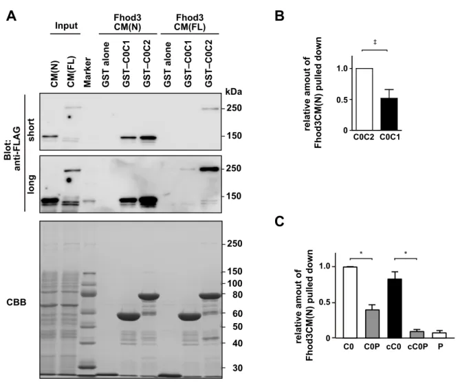
cMyBP-C is a multidomain protein with 11 Ig or fibronectin-like domains, designated C0 through C10 from the N terminus (SI Appendix, Fig. S6A). The C-terminal region of cMyBP-C con- stitutively binds to and stabilizes the thick myosin filament through interactions with myosin rod or titin, whereas the N-terminal so-called regulatory domains (C0C2) extend from the thick filament to regulate the actin-myosin association (6). We therefore prepared the N-terminal regions of cMyBP-C as GST- fused proteins and performed an in vitro pull-down binding assay using lysates of HEK 293 cells expressing various Fhod3 proteins (Fig. 2D). As shown in Fig. 2EandSI Appendix, Fig. S7A, the full length of Fhod3 [Fhod3CM(FL)] in the lysate of HEK293 cells was effectively pulled down with GST–C0C2 and weakly pulled down with GST–C0C1, indicating that the N-terminal region of cMyBP-C is sufficient for Fhod3 association. In contrast, the full length of Fhod3CM-S [Fhod3CM-S(FL)], a short-splice variant that lacks 151 amino acids in the N-terminal region (23) (Figs. 1Eand 2D) was not pulled down with either GST–C0C2 or GST–C0C1, suggesting that the spliced-out N-terminal region of Fhod3 is re- sponsible for the binding. Consistently, the N-terminal fragment of Fhod3 [Fhod3CM(N)] was sufficiently pulled down with GST–
C0C1 or GST–C0C2 (Fig. 2F), but not with GST–C3C6 or GST–
C7C10 (SI Appendix, Fig. S6B). To further investigate whether or not Fhod3 directly interact with cMyBP-C, we purified the N-terminal region of Fhod3 proteins and performed an in vitro
Matsuyama et al. PNAS | vol. 115 | no. 19 | E4387
CELLBIOLOGYPNASPLUS
pull-down binding assay using the purified N-terminal regions of cMyBP-C. As shown in Fig. 2G, the purified Fhod3CM(N) was effectively pulled down with GST–C0C1 and GST–C0C2, but that of Fhod3CM-S [Fhod3CM-S(N)] was not. Thus, Fhod3 directly interacts with cMyBP-C, and this interaction is mediated by the N-terminal regions of both Fhod3 and cMyBP-C.
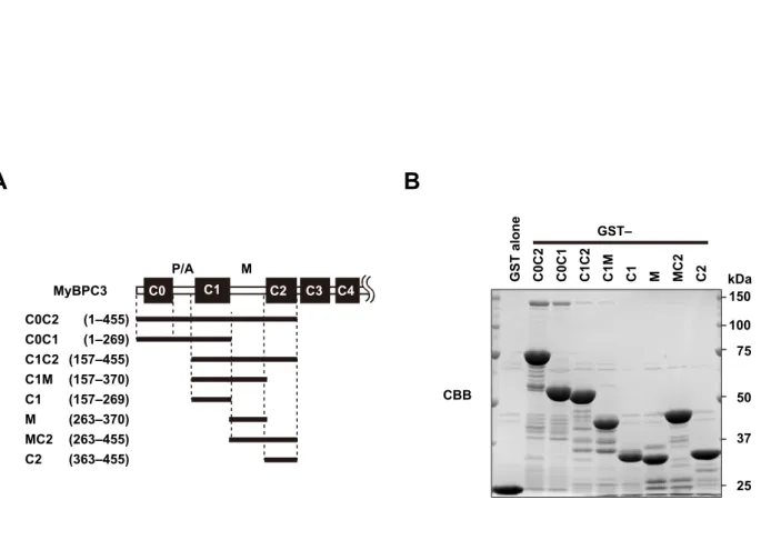
To identify the domains in the N-terminal cMyBP-C respon- sible for the interaction, we further prepared a series of smaller fragments of cMyBP-C (Fig. 2H). C0C2 bound to Fhod3CM(N) more strongly than C0C1 did (Fig. 2IandSI Appendix, Fig. S7B).
Deletion of the C0 domain resulted in a markedly reduced in- teraction (Fig. 2I and SI Appendix, Fig. S8), whereas the C0 domain alone was sufficient for strong binding to Fhod3 (Fig.
2I). Compared with the human cMyBP-C, the mouse homolog has a unique N-terminal extension of eight amino acids (Gen- Bank accession no. NM_008653). To exclude the possibility that the interaction is mediated via the mouse-specific N-terminal sequence, we further prepared the N-terminally truncated core C0 domain (cC0), which corresponds to the human C0 domain (Fig.
2J). As shown in Fig. 2K, cC0 sufficiently bound to Fhod3CM(N), indicating that the N-terminal extension specific to mice is dis- pensable for the interaction. In contrast, the addition of the P/A region to C0 or to cC0 attenuates the interaction, indicating that the P/A region seems to negatively regulate the binding. The extent of the inhibitory effect of the P/A region seems higher in the case of cC0P than that of C0P (SI Appendix, Fig. S7C), possibly suggesting that the inhibitory effect of the P/A region is attenuated by the presence of the N-terminal extension. The M and C2 domains might also attenuate the inhibitory effect. These findings indicate that the core C0 domain of cMyBP-C is sufficient for the interaction with Fhod3, whereas the binding may be regulated by other domains in a complex manner and differently in mice and human.
Mode of Interaction of the C0 Domain of cMyBP-C with the N- Terminal Region of Fhod3. Phosphorylation of the M domain of cMyBP-C is known to modulate the interaction of cMyBP-C with the thin and thick filaments, thereby affecting the cross-bridge cycle and cardiac hemodynamics (5). In addition, familial HCM
C A
B
phalloidin merge MF20 mergeMF20 merge
Fhod3 Fhod3
cMyBP-C
merge cMyBP-C
D
primary culture
Fhod3 cMyBP-C
F
FH1
FH2
3 5 13
11
8 2 5 2 1 2 5
1
0 2
1 2 4 6789 10 12 1416 17 18 19 2223242627
E
Fhod3CM
Fhod3CM-S
T(D/E)5XE Ile
*
*
Fhod3CM-IA
I1127A Exon
structure
Domain structure
Tg (Fhod3CM-IA) Tg (–) Tg (Fhod3CM-WT)
Fhod3-actininmerge
1127
Fig. 1. Colocalization of Fhod3 with cMyBP-C in the cardiac sarcomere. (A) Primary culture of embryonic C57BL/6 mouse cardiomyocytes was subjected to immunofluorescent double staining for endogenous Fhod3 [anti–Fhod3-(650–802); red] and cMyBP-C (green). (Scale bar, 5μm.) (B) Sections of adult hearts from C57BL/6 mice were subjected to immunofluorescent staining for Fhod3 [anti–Fhod3-(650–802); red] and cMyBP-C (green) followed by phalloidin staining (not depicted in merge). (Scale bar, 5μm.) (CandD) Sections of adult hearts from C57BL/6 mice were subjected to immunofluorescent staining for myosin heavy chain (green) and Fhod3 [anti–Fhod3-(650–802)] (C) or cMyBP-C (D) (red). (Scale bars, 5μm.) (E) Schematic presentation of exon structure (Top), domain structure (Middle), and various constructs (Bottom) of mouse Fhod3. Alternative splicing exons 11, 12, and 25 are indicated by gray boxes (Top). The al- ternative splicing regions are in gray boxes, and the residue responsible for actin binding (Ile1127) is indicated by asterisk (Middle). Adapted with permission from ref. 25. (F) Sarcomeric localization of endogenous and exogenous Fhod3 in the heart. Sections of adult hearts from nontransgenic mice (Left) and from transgenic mice expressing Fhod3CM-WT (Center) and Fhod3CM-IA (Right) were subjected to immunofluorescent double staining for Fhod3 [anti–Fhod3- (650–802); red] andα-actinin (green). Images were taken under the same conditions except with photomultiplier tubes (PMT) voltage (800 and 600 V for nontransgenic and transgenic mice, respectively). (Scale bar, 5μm.)
E4388 | www.pnas.org/cgi/doi/10.1073/pnas.1716498115 Matsuyama et al.
missense mutations have been identified in the M domain (10).
To determine whether phosphorylation or a mutation in the M domain affects the interaction between Fhod3 and cMyBP-C, we prepared various mutant fragments of cMyBP-C and examined the interaction with Fhod3 (Fig. 3A). Nonphosphorylatable ala- nine substitutions (3SA and 4SA) or phosphorylation mimetic substitutions (3SD and 4SD) for serines in the M domain had no effect on the binding. The HCM missense mutations in the M domain (R279H and R288W: corresponding to R273H and R282W in human cMyBP-C, respectively) also did not cause any
effects. Thus, no substitutions in the M domain examined showed any effects on the interaction with Fhod3.
We further investigated the effect of substitutions in the C0 domain. Similar to the HCM mutations in the M domain, those in the C0 domain, G13R and R43W (corresponding to G5R and R35W in human cMyBP-C, respectively), did not affect the interaction (Fig. 3 B and C). In contrast, substitutions of lysine or arginine residues positioned on the surface of the C0 domain (28) to glutamate effectively abolished the interac- tion. Because the electrophoretic mobility of mutant proteins is
C
A B D
E
IP
Lysate Control IgG Anti-Fhod3
IP
Lysate Control IgG Anti-Fhod3
anti-Fhod3
Blot:
anti-cMyBP-C
Input GST alone
GST–
C0C1 GST–
C0C2
CBB CBB
kDa
250
37 50 75 100 150
25 250 150
250
37 50 75 100 150
25 250 150 kDa anti-cMyBP-C
IP
Lysate Control IgG Anti-cMyBP-C
anti-cMyBP-C anti-Fhod3 Blot:
Input GST alone
GST–
C0C1 GST–
C0C2
F
200
30 45 66 116 kDa Input GST
alone GST–
C0C1 GST–
C0C2
G
Blot:
CBB Fhod3CM
CM(FL) CM-S(FL) CM(N) CM-S(N)
splicing
region FH1 FH2
(1–1586)
J
H I K
MyBPC3 C0C2 C0C1 C1C2 C0P PC1 PC1C2 C0
(1–455) (1–269) (157–455) (1–162) (101–269) (101–455) (1–104)
M
C0 C1 C2 C3
P/A
MyBPC3 C0 C0P cC0 cC0P P
(1–104) (1–162) (9–104) (9–162) (101–162)
C0 P/A C1
GST alone C0C2 C0C1 C1C2 C0P PC1 PC1C2 C0
250
37 50 75 100 150 kDa
C0 C0P cC0 cC0P P
200
45 66 116 GST–
GST–
Fhod3
CM-S(FL) CM(FL) Marker CM-S(FL) CM(FL) CM-S(FL) CM(FL) CM-S(FL) CM(FL) CM(N) CM(FL) Marker CM(N) CM(FL) CM(N) CM(FL) CM(N) CM(FL)
Fhod3
CM(N) CM-S(N) CM(N) CM-S(N) CM(N) CM-S(N) CM(N) CM-S(N)
250
150 250
shortlong 150 Blot: anti-FLAG
longshort
Blot: anti-FLAG
200 Blot: anti-FLAG 116
150
200 116 kDa 150
250 kDa
150 250 kDa
150 kDa
Blot: anti-FLAG Blot: anti-FLAG
CBB CBB
20
20 102 163
102 163 262 369454
983
1586
400
1 552
1500 1039
Fig. 2. Interaction between Fhod3 and cMyBP-C. (AandB) Proteins in lysates of hearts from transgenic mice expressing Fhod3CM-WT were immunopre- cipitated with the anti–Fhod3-C-20 (A) or the anti–cMyBP-C (B) antibodies, and then analyzed by immunoblot with the indicated antibodies. Uncropped images are shown inSI Appendix, Fig. S5. (C) Proteins in lysates of hearts from nontransgenic C57BL/6 mice were immunoprecipitated with the anti–Fhod3-C- 20 or control IgG, and then analyzed by immunoblot with the anti–cMyBP-C antibody. Uncropped images are shown inSI Appendix, Fig. S5. (D) Schematic structures of various Fhod3 constructs used in the present pull-down binding assay (E–K). (E) FLAG-tagged Fhod3CM(FL) or Fhod3CM-S(FL) in the lysate of HEK-293F cells (input) was incubated with GST–cMyBP-C (C0C1 or C0C2) or GST alone and pulled down with glutathione-Sepharose 4B beads. Precipitated proteins were subjected to SDS/PAGE and analyzed by immunoblot with the anti-FLAG antibody (Upper) or stained with Coomassie Brilliant Blue (CBB) (Lower). Positions for marker proteins are indicated in kilodaltons. (F) FLAG-tagged full-length (FL) or N terminus (N) of Fhod3CM in the lysate of HEK-293F cells (input) was incubated with GST–cMyBP-C (C0C1 or C0C2) or GST alone and pulled down with glutathione-Sepharose 4B beads. Precipitated proteins were analyzed as inE. (G) Purified N-terminal region of Fhod3 proteins [Fhod3CM(N) or Fhod3CM-S(N)] tagged with FLAG was incubated with GST–cMyBP-C (C0C1 or C0C2) or GST alone and pulled down with glutathione-Sepharose 4B beads. Precipitated proteins were analyzed as inE. (HandJ) Schematic structures of various cMyBP-C constructs used inIandJ, respectively. (IandK) FLAG-tagged Fhod3CM(N) in the lysate of HEK-293F cells was incubated with indicated GST-fused fragments of cMyBP-C and pulled down with glutathione-Sepharose 4B beads. Precipitated proteins were analyzed as inE. The ar- rowheads indicate the position of Fhod3CM(N).
Matsuyama et al. PNAS | vol. 115 | no. 19 | E4389
CELLBIOLOGYPNASPLUS
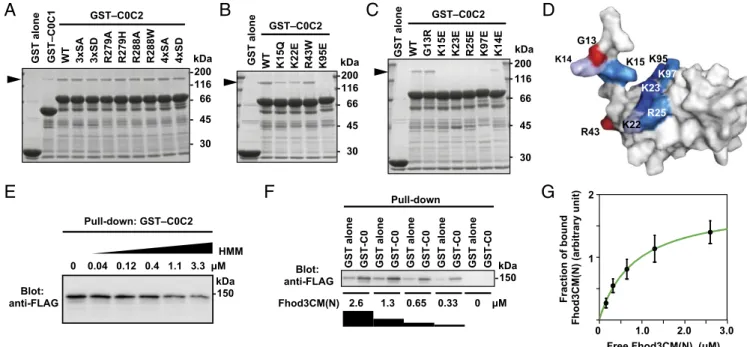
not markedly altered, these substitutions of surface residues are expected not to cause major structural changes. Intriguingly, the residues responsible for Fhod3 binding (Fig. 3D) partially over- lapped with those for binding to the RLC of myosin (28), sug- gesting that the interaction between Fhod3 and cMyBP-C is affected by the presence of myosin. Indeed, as shown in Fig. 3E, heavy meromyosin attenuated the interaction in a dose-dependent manner, suggesting the possibility that the C0C2-mediated bind- ings to Fhod3 and myosin are mutually exclusive. Furthermore, we titrated in different concentrations of Fhod3CM(N) to estimate a dissociation constant (Fig. 3F) and found that Fhod3CM(N) bound to the C0 domain in a concentration-dependent manner with an estimatedKdvalue of about 0.8μM (Fig. 3G). Although the binding affinity of cMyBP-C for Fhod3 is higher than that for the RLC of myosin (28) or for the S2 segment of myosin (29), cumulative contribution of the RLC and S2 segment may affect the overall affinity of myosin head. Thus, the cardiac formin Fhod3 interacts with cMyBP-C via a charge-dependent interaction between the N-terminal region of Fhod3 and the cardiac-specific C0 domain of cMyBP-C.
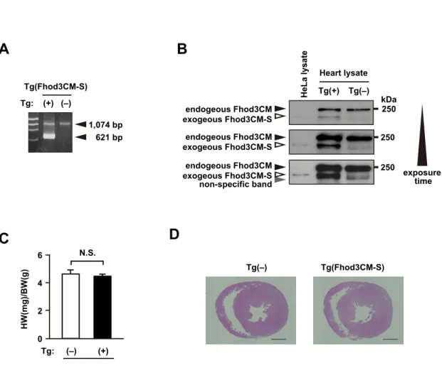
Role of the Interaction with cMyBP-C in Fhod3 Localization.We next investigated whether or not the interaction between Fhod3 and cMyBP-C is required for the in situ localization of Fhod3 in the sarcomeres. To investigate this, we generated transgenic mice expressing the short form (Fhod3CM-S) (Fig. 1E), defective in binding to cMyBP-C (Fig. 2E), under the control of theα-MHC promoter [Fhod3Tg(α-MHC–Fhod3CM-S)]. From over 200 injections, we obtained only one transgenic line expressing Fhod3CM-S protein at a low level (SI Appendix, Fig. S9A and B). In the
Fhod3CM-S–expressing cells shown in Fig. 4AandSI Appendix, Fig. S3, the intense signals for Fhod3, which reflect the locali- zation of exogenously expressed Fhod3CM-S, distributed in a diffuse pattern and failed to show the characteristic localization pattern to the C-zones. Consistent with this observation, the N- terminally tagged short variant of Fhod3 also failed to localize at the C-zone in neonatal rat cardiomyocytes (SI Appendix, Fig. S1) (13). Thus, the binding with cMyBP-C seems to be necessary for the in situ localization of Fhod3 in the sarcomeres. It should be noted that Fhod3CM-S was expressed only in a small sub- population of cardiomyocytes in the cardiac section (SI Appen- dix, Fig. S3), indicating that, similar to Fhod3CM-IA, the Fhod3CM-S transgene is inactivated in some cardiomyocytes.
Although the precise mechanism for this is presently unknown, a toxicity of the aberrant Fhos3CM-S protein dislocated from the C- zone may explain the reason. Full expression of the toxic peptide in the cardiomyocytes is supposed to impair normal cardiac de- velopment and result in lethality. If the exogenous expression of the toxic peptide is suppressed by inactivation of the transgene, the lethality could be overcome. Indeed, the obtained transgenic line expressing Fhod3CM-S protein at a very low level appears phenotypically normal (SI Appendix, Fig. S9CandD).
To confirm the crucial role of the interaction with cMyBP-C on Fhod3 localization in the physiological state, we prepared cMyBP-C–null mice from cMyBP-C mutant mice carrying the targeted trap allele (SI Appendix, Fig. S10). The cMyBP-C–null mice showed cardiomyopathic changes at morphological and histological levels (Fig. 5DandFandSI Appendix, Figs. S10C andDand S14) but did not display any significant alterations in
C
A B D
E
GST–C0C2 GST–C0C2
GST alone GST–C0C1 WT 3xSA 3xSD R279A R279H R288A R288W 4xSA 4xSD WT K15Q K22E R43W K95EGST alone WT G13R K15E K23E R25E K97E K14E
GST–C0C2
GST alone
200
45 66 116 kDa
30
200
45 66 116 kDa
30
200
45 66 116 kDa
30
Pull-down: GST–C0C2
HMM
Blot:
anti-FLAG 150
kDa
G F
GST alone GST-C0 GST alone GST-C0 GST alone GST-C0 GST alone GST-C0 GST alone GST-C0
2.6 1.3 0.65 0.33 Blot:
anti-FLAG 150
kDa Pull-down
Fhod3CM(N)
K15 G13 K14
R43
K23 R25
K95 K97
K22
Fraction of bound Fhod3CM(N) (arbitrary unit)
0 1.0 2.0 3.0
Free Fhod3CM(N) ( 1
2
Fig. 3. Specific interaction of the C0 domain of cMyBP-C with Fhod3. (A–C) Effect of amino acid substitutions on the interaction of C0C2 with the N-terminal region of Fhod3. FLAG-tagged Fhod3CM(N) in the lysate of HEK-293F cells was incubated with GST-fused C0C2 fragment carrying the indicated amino acid substitution: 3xSA, the S281A/S290A/S310A substitution; 4xSA, the S281A/S290A/S310A/S315A substitution. Proteins were pulled down with glutathione- Sepharose 4B beads. Precipitated proteins were subjected to SDS/PAGE and stained with CBB. The arrowheads indicate the position of Fhod3CM(N). (D) Mapping of the residues responsible for the Fhod3 interaction on the surface of the C0 domain. The figure is drawn using PyMOL from the structure at PDB ID code 2K1M (28). Amino acid residues responsible for the interaction with Fhod3 inBandCare shown in blue, and residues associated with cardiomyopathy are shown in red. (E) Competitive effects of heavy meromyosin on Fhod3 binding to cMyBP-C–C0C2. GST–cMyBP-C–C0C2 was incubated with the lysate of HEK-293F cells expressing FLAG-tagged Fhod3CM(N) (final concentration of 2.0μM) in the presence of various concentration of heavy meromyosin. (F) Representative pull-down assay for quantification of binding between cMyBP-C–C0 and Fhod3CM(N). GST–cMyBP-C–C0 or GST alone immobilized to glu- tathione particles was incubated with the indicated concentrations of the lysate of HEK-293F cells expressing FLAG-tagged Fhod3CM(N). To avoid disturbing the binding equilibrium, bound Fhod3CM(N) were analyzed directly without washing by immunoblot with the anti-FLAG antibodies followed by fluorescence measurement. (G) Quantitative analysis for binding of cMyBP-C–C0 to Fhod3CM(N). Fractions of specifically bound Fhod3-N were determined as fraction bound to GST–C0 minus fraction to GST alone inF.
E4390 | www.pnas.org/cgi/doi/10.1073/pnas.1716498115 Matsuyama et al.
the sarcomere structure (SI Appendix, Fig. S13), which is in line with previous reports (30–33). Consistent with our hypothesis, in the cardiac section of the cMyBP-C–null mice shown in Fig. 4C andSI Appendix, Fig. S11, Fhod3 did not show a periodic ac- cumulation pattern, but instead demonstrated a diffuse distri- bution, although the expression level of Fhod3 was not affected in the cMyBP-C–null mice (Fig. 4B). Thus, cMyBP-C is required for the characteristic localization of Fhod3 to the C-zone in the sarcomere. Furthermore, these findings raise the intriguing question of whether or not the mislocalization of Fhod3 is re- sponsible for the pathogenesis of cardiomyopathy in cMyBP-C–
null mice.
Exacerbation of Cardiac Dysfunction by Overexpression of Fhod3 in cMyBP-C–Null Mice.To investigate the physiological significance of Fhod3 anchoring to the C-zone and to explore the consequences of Fhod3 mislocalization, we examined the effect of Fhod3 over- expression in cMyBP-C–null mice. If the cardiomyopathic phenotype of cMyBP-C–null mice worsens by overexpression of Fhod3, the aberrant Fhod3 protein is likely toxic, possibly in a similar manner to that of the Fhos3CM-S protein. By crossing cMyBP-C–null mice with transgenic mice expressing wild-type Fhod3 (Fhod3CM) under the control of the α-MHC promoter [Fhod3Tg(α-MHC–Fhod3CM)], we generated cMyBP-C–null mice overexpressing Fhod3 in the heart [Mybpc3−/−;Fhod3Tg(α-MHC–Fhod3CM)] (Fig. 5A). As shown in Fig.
5B, theseMybpc3−/−;Fhod3Tg(α-MHC–Fhod3CM)pups all died around 2 wk after birth. Although the heart-to-body weight ratio at postnatal day 9 (P9) did not differ significantly betweenMybpc3−/−; Fhod3Tg(α-MHC–Fhod3CM)mice andMybpc3−/−mice (Fig. 5C), dilatation of the right ventricle with severe liver congestion was evident (Fig. 5D, E, andG), suggesting thatMybpc3−/−; Fhod3Tg(α-MHC–Fhod3CM)mice died due to cardiac failure. In con- trast, lung congestion was not apparent (SI Appendix, Fig. S12).
Disarray of cardiomyocytes, which is a characteristic change for cMyBP-C–null heart (31, 32), was also more prominent in Mybpc3−/−;Fhod3Tg(α-MHC–Fhod3CM) mice (Fig. 5F). Consistent with these findings, the expression of the cardiac remodeling- associated fetal genes was significantly elevated in Mybpc3−/−; Fhod3Tg(α-MHC–Fhod3CM)mice compared withMybpc3−/−mice (Fig.
5H and I). Thus, the overexpression of Fhod3 seems to induce exacerbation of cardiac dysfunction in cMyBP-C–null mice, al- though it does not affect the cardiac function of wild-type mice.
Disorganization of the Sarcomeric Structure Due to Overexpression of Fhod3 in the cMyBP-C–Null Heart. To clarify the mechanism by which Fhod3 overexpression induces the exacerbation of cardiac dysfunction only in cMyBP-C–null mice, we examined the sarco-
mere structure in the heart ofMybpc3−/−;Fhod3Tg(α-MHC–Fhod3CM)
mice. There were no significant differences in sarcomeric struc- tures comprising the Z-line marked with the antibody toα-actinin and thin actin filaments stained with phalloidin (SI Appendix, Fig.
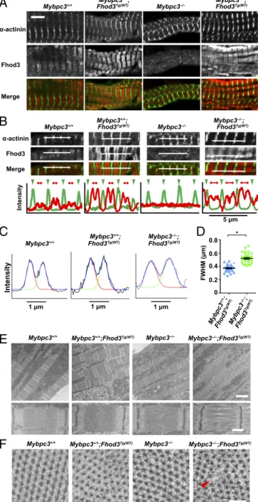
S13). However, somewhat to our surprise, the localization pattern of Fhod3 showed striking differences; the intense signals for overexpressed Fhod3 inMybpc3−/−;Fhod3Tg(α-MHC–Fhod3CM)mice distributed in a periodic pattern despite the absence of cMyBP-C, although weak signals for endogenous Fhod3 inMybpc3−/−mice distributed diffusely in the absence of cMyBP-C (Fig. 6A), as al- ready shown in Fig. 4C. Intriguingly, the periodic pattern of overexpressed Fhod3 in cMyBP-C–null mice was apparently dif- ferent from the characteristic C-zone pattern of endogenous Fhod3 or that of overexpressed Fhod3 in wild-type mice; the ac- cumulation of overexpressed Fhod3 in cMyBP-C–null mice was not restricted to the central C-zone, but it expanded to the outer peripheral region of the A-band. The difference in localization pattern was evident in line scan graphs of fluorescent intensity along the long axis of sarcomeres (Fig. 6B). The width of the Fhod3 distribution pattern (full-width at half-maximum, FWHM) was significantly larger in Mybpc3−/−;Fhod3Tg(α-MHC–Fhod3CM)
mice than inFhod3Tg(α-MHC–Fhod3CM)mice (Fig. 6CandD). Thus, in the absence of cMyBP-C, overexpressed Fhod3 protein, seems to distribute along the entire region of the A-band.
To determine the consequences of the abnormal distribution of Fhod3 throughout the entire A-band, we further examined the ultrastructure of sarcomeres using transmission electron mi- croscopy. Fhod3 overexpression in the presence of cMyBP-C did not affect the ultrastructure of sarcomeres: intact sarcomeres with Z-lines, M-lines, and A-bands were observed (Fig. 6E).
Depletion of cMyBP-C per se also did not alter the sarcomere organization (Fig. 6E) as previously reported (30–32). In con- trast, in Fhod3-overexpressing cMyBP-C–null mice, disorder of the array of myosin filaments was observed: gaps between thick filaments are frequently detected at the center of the sarcomere (Fig. 6E). In some sarcomeres, thick filaments appear to be bundled or clumped at the level of the M-line, suggesting the disordering of hexagonal myosin filament lattice. Consistent with this, cross-sections of sarcomeres showed disorder of the hex- agonal lattice of myosin filaments in Fhod3-overexpressing cMyBP-C–null mice at the level of the M-line or of non- overlapping regions that contained only thick filaments (Fig. 6F).
Thus, an overexpression of Fhod3 in the cMyBP-C–null heart induces the abnormal accumulation of Fhod3 to the entire A- band, thereby leading to a loss of sarcomeric integrity, which may be responsible for the cardiac dysfunction and death of those mice. These findings suggest that an excess amount of aberrant
250
50 150
A
mergeB
mergeFhod3
+/+ +/– –/–
Fhod3 Fhod3
+/+
C
–/–
Fig. 4. Interaction between Fhod3 and cMyBP-C is required for Fhod3 localization to the C-zone. (A) Sarcomeric localization of Fhod3CM-S in the heart.
Sections of adult hearts from transgenic mice expressing Fhod3CM-S were subjected to immunofluorescent double staining for Fhod3 [anti–Fhod3-(650–802);
red] andα-actinin (green). (Scale bar, 5μm.) (B) Immunoblot analysis of total proteins from the heart of wild-type (+/+), cMyBP-C+/−(+/−), and cMyBP-C−/−(−/−) mice using antibodies against Fhod3 (anti–Fhod3-C-20), cMyBP-C, orα-tubulin. (C) Sarcomeric localization of endogenous Fhod3 in the heart from cMyBP-C– null mice. Sections of adult hearts from cMyBP-C–null mice were subjected to immunofluorescent double staining for Fhod3 [anti–Fhod3-(650–802); red] and α-actinin (green) followed by phalloidin staining (not depicted in merge). (Scale bar, 5μm.)
Matsuyama et al. PNAS | vol. 115 | no. 19 | E4391
CELLBIOLOGYPNASPLUS
Fhod3 functions deleteriously as a toxic peptide in cMyBP-C–
null mice.
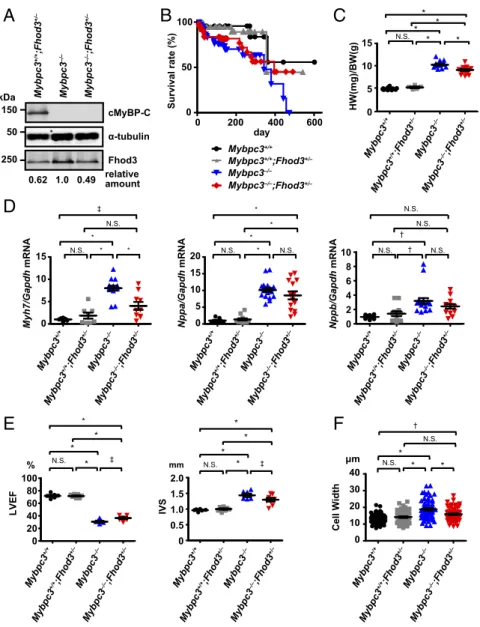
Modulation of the Cardiac Function in cMyBP-C–Null Mice by Reduced Fhod3 Protein Levels.To evaluate the pathogenic role of the ab- errant Fhod3 in cMyBP-C–null mice, we finally investigated the effect of the reduction of Fhod3 protein levels in cMyBP-C–null mice. By crossing cMyBP-C–null mice with heterozygous Fhod3 knockout mice (Fhod3+/−) (15), we generated cMyBP-C–null mice expressing half the normal amount of Fhod3 protein (Mybpc3−/−;Fhod3+/−) (Fig. 7A). Although no significant differ- ence in the long-term survival over a year was observed between Mybpc3−/−;Fhod3+/−mice andMybpc3−/−;Fhod3+/+mice, about 50% of Mybpc3−/−;Fhod3+/−mice were alive at the time point when allMybpc3−/−;Fhod3+/+mice died (Fig. 7B). The heart-to- body weight ratio of Mybpc3−/−;Fhod3+/− mice at 16 wk was significantly lower than that in Mybpc3−/−;Fhod3+/+mice (Fig.
7C). The expression of the cardiac remodeling-associated fetal geneβ-MHC was consistently decreased inMybpc3−/−;Fhod3+/−
mice compared withMybpc3−/−;Fhod3+/+mice, although differ- ences in the expressions of ANF and BNP were not statistically significant (Fig. 7D). An echocardiographic analysis revealed that a reduction of the ventricular ejection fraction observed in cMyBP-C–null mice was slightly but significantly recovered inMybpc3−/−;Fhod3+/−mice (Fig. 7E). Although the changes in ventricular dimensions were not significant, the ventricular wall thickening was significantly recovered in Mybpc3−/−;Fhod3+/−
mice (Fig. 7E). Consistent with this, the width of individual cardiomyocytes was significantly reduced inMybpc3−/−;Fhod3+/−
mice (Fig. 7F). Such improvement was also observed at 36 wk of age (SI Appendix, Fig. S14). Thus, reductions in protein levels of Fhod3 in cMyBP-C–null mice appeared to partially antagonize the hypertrophic changes in cMyBP–C-null mice.
Discussion
In the present study, we show that the cardiac formin Fhod3 directly interacts with the cardiac isoform of MyBP-C; the
A
D
Mybpc3+/+;Fhod3Tg(WT) Mybpc3–/–;Fhod3Tg(WT)E
F
G
C B
I
250 50 150 kDa
Mybpc3–/–
Mybpc3+/+ Mybpc3–/–
Mybpc3+/+ Mybpc3–/–
Mybpc3+/+
Mybpc3+/+;Fhod3Tg(WT) Mybpc3–/–;Fhod3Tg(WT)
Mybpc3+/+;Fhod3Tg(WT) Mybpc3–/–;Fhod3Tg(WT) α-tubulin
Fhod3 cMyBP-C 3cpbyM+/+ 3cpbyM+/+3dohF;)TW(gT 3cpbyM–/–3dohF;)TW(gT
Mybpc3–/–
Mybpc3+/+
Mybpc3+/+;Fhod3Tg(WT) Mybpc3–/–;Fhod3Tg(WT) Mybpc3–/–
0 5 10 15 20
Days 0
50 )%(etarlavivruS100
0 5 10 15
20 N.S. N.S.
HW/BW (mg/g)
Mybpc3
+/+
Mybpc3
+/+;Fhod3
Tg(WT)
Mybpc3
–/–;Fhod3
Tg(WT)
Mybpc3
–/–
* *
hdpaG/appNANRm
* *
*
0 5 10 15 20 25
Mybpc3
+/+
Mybpc3
+/+;Fhod3
Tg(WT)
Mybpc3
–/–;Fhod3
Tg(WT)
Mybpc3
–/–
0 5 10 15 20 hdpaG/bppNANRm
*
*
†
*
*
Mybpc3
+/+
Mybpc3
+/+;Fhod3
Tg(WT)
Mybpc3
–/–;Fhod3
Tg(WT)
Mybpc3
–/–
H
WB/WL)g/gm(
Mybpc3
+/+
Mybpc3
–/–;Fhod3
Tg(WT)
Mybpc3
–/–
0 10 20 30 40
‡ * †
1.0 28 1.0 21 relative amount
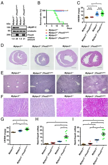
Fig. 5. Physiological effects of transgenic overexpression of Fhod3 in cMyBP-C–null mice. (A) Immunoblot anal- ysis of Fhod3, cMyBP-C, and α-tubulin using whole heart tissue lysates from wild-type (Mybpc3+/+), Fhod3- Tg [Mybpc3+/+;Fhod3Tg(WT)], cMyBP-C–null (Mybpc3−/−), and cMyBP-C–null mice overexpressing Fhod3 [Mybpc3−/−; Fhod3Tg(WT)] at P9. (B) Survival curves of mice of the indicated genotypes [Mybpc3+/+, n = 52; Mybpc3+/+; Fhod3Tg(WT), n = 38; Mybpc3−/−, n = 50; Mybpc3−/−; Fhod3Tg(WT),n=52]. (C) Heart-to-body weight ratio of cMyBP-C–null mice overexpressing Fhod3 [Mybpc3−/−; Fhod3Tg(WT),n=23] and control mice [Mybpc3+/+,n= 30;Mybpc3+/+;Fhod3Tg(WT),n=14;Mybpc3−/−,n=24] at P9. Values are means (long bars)±SEM (short bars). *P<
0.001; N.S., not significant. (D–F) Histological analysis of hearts and livers of cMyBP-C–null mice overexpressing Fhod3 [Mybpc3−/−;Fhod3Tg(WT)] and control mice at P9.
Short-axial sections of hearts (D), liver tissues (E), and short- axial–sectioned lateral wall of the left ventricles (F) were stained with H&E. (Scale bars: 1 mm inDandEand 100μm inF.) (G) Liver-to-body weight ratio of cMyBP-C–null mice overexpressing Fhod3 [Mybpc3−/−;Fhod3Tg(WT),n=7] and control mice (Mybpc3+/+,n=7;Mybpc3−/−,n=7) at P9.
Values are means (long bars)± SEM (short bars). *P <
0.001,†P<0.01,‡P<0.05. (HandI) Quantitative real-time PCR analysis of fetal cardiac gene expression in cMyBP-C– null mice overexpressing Fhod3 [Mybpc3−/−;Fhod3Tg(WT), n=11] and control mice [Mybpc3+/+,n=8;Mybpc3+/+; Fhod3Tg(WT),n =7;Mybpc3−/−,n=9] at P9. Values are means (long bars)±SEM (short bars).Nppa, encoding ANF;
Gapdh, encoding GAPDH;Nppb, encoding BNP. *P<0.001,
†P<0.01.
E4392 | www.pnas.org/cgi/doi/10.1073/pnas.1716498115 Matsuyama et al.
![Fig. 1. Colocalization of Fhod3 with cMyBP-C in the cardiac sarcomere. (A) Primary culture of embryonic C57BL/6 mouse cardiomyocytes was subjected to immunofluorescent double staining for endogenous Fhod3 [anti – Fhod3-(650 – 802); red] and cMyBP-C (green)](https://thumb-ap.123doks.com/thumbv2/123deta/9878436.1905317/4.877.68.820.83.618/colocalization-sarcomere-primary-embryonic-cardiomyocytes-subjected-immunofluorescent-endogenous.webp)