~
Melli. Fac. Agr. Kinki Univ. 31 :35~40
(199) 35Effects of Sesamin and Related Lignan Compounds on Fatty Acid Metabolism in Hep G2 Cell
Norifumi SHIRASAKA, Chizuko SAYOU, Tetsuo MURAKAMI and Hajime YOSHIZUMI
Deperlllleni of Food science and Nuln"tiol1. Fuculty of Agriculture.
Kinki university. Nakalllachi. Nara 631-8505. Japan
Synopsis
The effects of sesamin and episesamin, lignan compounds which are present in sesame seeds, on fatty acid biosynthesis, and on the docosahexaenoic acid (DHA) biosynthetic pathway in particular, were investigated in the human hepatoma cell line, Hep G2.
Eicosapentaenoic acid (EPA), sesamin and episesamin were incorporated into the cell depending upon their concentrations, and EPA was metabolised into docosapentaenoic acid (DPA) and DHA. However, no significant changes in fatty acid composition were observed. This result indicates that sesamin and episesamin do not inhibit the biosynthesis of DHA from EPA.
Introduction tied from cellular lipid of Schizochytrium sp. SR21
61. All other authentic fatty acids were obtained Sesame has long been used extensively as a from Sigma (St. Louis, MO). DME medium was traditional health food in the Orient for various from NISSUI Pharmaceutical Co. (Tokyo, Japan) purpose. The effective components, especially lig- and fetal bovine serum (FBS) from GIBCO BRL nan compounds, in the health food have recently (Rockville, USA). All other reagents were of ana- been investigated. Sesame oil contains relatively lytical grade.
large quantities of sesamin and related lignan com- Cell culture. Hep G2 cells derived from human pounds, and they have an antioxidant action in vitro hepatoma were from American Type Culture Col-
1·3) lection (Bethesda, USA) and subcultured at the ra-
Akimoto et al. reported that sesamin and related tio of 1 :4. Confluent monolayer of Rep G2 cells was lignan compounds present in sesame seeds or their treated with 0.25% trypsin. Isolated cells were oil are specific inhibitors of ~5-desaturase, which seeded into 60 mm dishes with 4 ml of DME con- catalyzes the desaturation of dihomo-
r
-linolenic taining 10% FBS at a concentration of 1.5 x 10 6 acid to arachidonic acid in PUFA biosynthesis in cells/dish. The Hep G2 cells were incubated at both microorganisms and animals 4). 37 in atomosphare of 5% CO2-95% air. After 3 In this report, we investigated the effect of sesa- days of incubation, cells reached subconfluent and min and episesamin on docosahexaenoic acid (DRA) were used in the experiment.biosynthesis from eicosapentaenoic acid (EPA) in Incorporation and metabolism of eicosapentaenoic human hepatoma cell line, Rep G2. acid (EPA), sesamin and episesamin in Hep G2 cell.
The solution of EPA methyl ester, sesamin and Materials and Methods episesamin were added to the culture medium as ethanol solution. The cells were incubated for 24 or Chemicals. 8,11,14, 17-Eicosatetraenoic acid (20:4 48 h at 37 in the 5% CO2-95% air. After incu- n-3) was purified from mycelial lipid of Mortierella bation, the medium was removed and cells were a/pina M226-9 grown with linseed oilS). 7, 10, 13, harvested by a rubber policeman.
16, 19-Docosapentaenoic acid (22: 5 n-3) was puri- Lipid analysis. Cellular lipids were extracted by
36 . IIIR.~ "AKA cl at.
the method of Folch et al. 7, An internal standard of fatty acid, 20 III of margaric acid (17: 0, 400 mM solution), was added to the cellular lipid extracts.
The resultant lipid extract was evaporated to dry- ness under reduced pressure at 35 C and then used for experimint.
For analysis of the fatty acid composition, each extarct was transmethylated with 10% methanolic HCI at 80 C for 2h. The resultant fatty acid methyl esters were extracted with 4 ml of n-hexan which was washed out with 2% KHC03, concentrated in a centrifugal evaporator at 70 C, and then dissolved in acetonitrile for analysis by capillary gas-liquid chromatography (GLC). The analytical conditions were as follow: apparatus, GC-14B ( himadzu, Japan) equipped with a flame ionization detector (FID) with a split injector; column, ULBO HR-
-10 (0.25 mm x 50m; hinwa Chemical Indus- tries, Japan); column temperature, 200 C ; injection port temperature, 250 C; carrier gas, He (inlet pressure, 200 kPa); make up gas, 2 (60 ml/min);
air and H2 , 60 kPa; and split ratio, 25: l.
For analysis of the cellular level of lignan com-
-
~o-
J: 100 75i
o...
tJl 50 Qj
U Ql>
:;:
lU 25
Qj a:
o
pounds in Hep G2 cell, each extarct was dissolved in chloroform for analysis by high performance liquid chromatography (HPLC). The analytical conditions were as follows: HPLC: pump, LC-5A ( himadzu);
column, Inertsil OD -2 (4.6 x 250 mm; GL cience, Japan); detector, PD-2A ( himadzu); wavelength, 290 nm; mobile phase, m thanol/water (80: 20, vol/vol); flow rate, 1 mL/min; and column tem- perature, 30 C.
Other methods. Protein content of cell was d - termined by the method of Lowry et al. 8 and each fatty acid concentration in the cells was express d as nmol acid/mg protein.
Results and Discussion
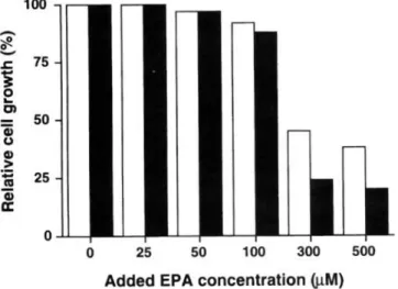
Effect of EPA concentration on growth of Hep G2 cell. Hep G2 cells were incubated with various concentrations of EPA at 37 C for 24 or 48 h. As shown in Fig. 1, the cell growth wa not inhibited at EPA concentration less than 100 tIM, but further incr a s in the EPA concentration caus d drastical decr ases in the cell growth.
o
25 50 100 300 500 Added EPA concentration ij.I.M)Fig. 1. Effects of EPA concentration on the growth of Hep G2 cell. The cells were incubated for 24 h (open bars) or 48 h (meshed bar) in a DME m dium containing the indicated conc ntrations of EPA. All value are means of four d terminations, and the standard deviation is less than 700 .
Incorporation and me/abol; m of EPA in Hep G2 c II.
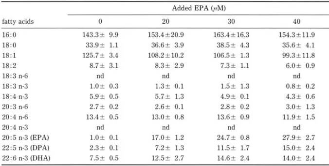
There was no significant difference between fatty acid levels of incorporation at 48 hand 24 h (Table 1).
EPA was incorporated into cells depending upon the concentration of EPA concentration ( 40t1M) and metabolized to OHA. Furth r increases in the
EPA concentration (> 40tIM) resulted in increases in the EPA and DPA levels in the c II, but the cel- lular level of DHA was not increased. This result indicate that EPA was easily metabolized to docosap ntaenoic acid (OPA), but cellular activity of conver ion of OPA to OHA was weak.
37 Effects of e amin and Related Lignan Compounds on Fatty Acid Metabolism in Hep G2 Cell
Table 1. Effects of EPA concentration and incubation period on the incorporation and metabolism of n-3 polyunsaturated fatty acids in Hep G2
1) 24 hour
Added EPA (pM)
fatty acids 0 20 30 40
16:0 143.3± 9.9 153.4±20.9 163.4±16.3 154.3±11.9
18:0 33.9± 1.1 36.6± 3.9 38.5± 4.3 35.6± 4.1
18:1 125.7± 3.4 108.2±10.2 106.5± 1.3 99.3± 11.8
18:2 8.7± 3.1 8.3± 2.9 7.3± 1.1 6.0± 0.9
18:3 n-6 nd nd nd nd
18:3 n-3 1.0± 0.3 1.3± 0.1 1.5± 1.3 0.8± 0.2
18:4 n-3 5.9± 0.5 5.7± 1.3 4.9± 0.1 4.3± 0.6
20:3 n-6 2.7± 0.2 2.6± 0.1 2.8± 0.2 3.0± 1.3
20:4 n-6 13.4± 0.5 13.0± 0.8 13.6± 0.9 11.9± 1.5
20:4 n-3 nd nd nd nd
20:5 n-3 (EPA) 1.0± 0.1 17.0± 1.2 24.7± 0.8 27.9± 2.7
22:5 n-3 (DPA) 2.3± 0.1 7.2± 1.3 11.5± 1.7 15.0± 2.4
22: 6 n-3 (DHA) 7.5± 0.5 12.5± 2.7 14.6± 2.4 14.0± 2.4
(nmol/mg protein) 2) 48 hour
Added EPA (liM)
fatty acids 0 20 30 40
16:0 134.0±22.7 145.9±29.7 145.6±17.9 155.4±15.6
18:0 36.5± 5.9 41.8± 8.8 42.2± 6.4 45.8± 7.5
18:1 130.5±22.0 122.3±28.1 119.6±19.3 114.1 ± 19.7
18:2 7.6± 1.6 7.9± 1.4 9.6± 2.4 9.5± 3.0
18:3 n-6 nd nd nd nd
18:3 n-3 1.2± 0.0 1.5± 0.3 1.5± 0.1 1.5± 0.6
18:4 n-3 5.7± 1.2 5.4± 1.7 4.9± 0.8 5.2± 0.1
20:3 n-6 3.0± 0.4 2.8± 0.8 2.9± 0.3 3.0± 0.1
20:4 n-6 14.7± 2.2 14.9± 2.8 14.2± 1.7 14.8± 0.8
20:4 n-3 nd nd nd nd
20:5 n-3 (EPA) 1.2± 0.0 15.7± 1.6 20.8± 1.3 28.0± 1.0
22: 5 n-3 (DPA) 2.3± 0.2 6.9± 1.2 10.1± 1.1 14.9± 0.9
22: 6 n-3 (DHA) 8.1± 1.1 15.1± 4.3 16.7± 3.7 18.7± 3.1
Values indicate mean± .D. (n=3). (nmol/mg protein)
Abbreviation used: nd, not detected; EPA, eicosapentaenoic acid; DPA, docosapentaenoic acid; DHA, docosahexaenoic acid.
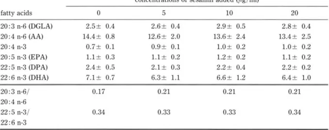
Effect of lignan compounds on PUFA metaboli m concentration added (less than 20 pg/ml), and no in Hep G2 cell. The cellular fatty acid level in the inhibitory effects on the cell growth was observed presence of sesamin and episesamin without ex- under the condition (data not shown). The addition ogenous EPA supplement was showed in Table 2. of sesamin and episesamin caused a slight increase About 40 % of sesamin and episesamin added to in DGLA/ AA ratio and DPA/DHA ratio, but no m dium were found in the cells depending upon the significant changes in the fatty acid levels were ob-
38 N. SHIRASAKA et al.
served. The fatty acid compositions of the cell culture medium, and the fatty acid levels in the cells without exogenous fatty acid supplement were were so low that fatty acid metabolism in the cell similar to those of serum which was added to the was unclear.
Table 2. Effects of sesamin (a) and episesamin (b) on the metabolism of n-6 and n-3 polyunsaturated fatty acids by Rep G2 cell
(a)
concentrations of sesamin added (f.lg/ml)
fatty acids 0 5 10 20
20: 3 n-6 (OGLA) 2.5± 0.4 2.6± 0.4 2.9± 0.5 2.8± 0.4
20: 4 n-6 (AA) 14.4± 0.8 12.6± 2.0 13.6± 2.4 13.4± 2.5
20:4 n-3 0.7± 0.1 0.9± 0.1 1.0± 0.2 1.0± 0.2
20:5 n-3 (EPA) 1.1± 0.3 1.1± 0.2 1.2± 0.2 1.1± 0.2
22:5 n-3 (OPA) 2.4± 0.5 2.1± 0.3 2.2± 0.4 2.2± 0.2
22: 6 n-3 (ORA) 7.1± 0.7 6.3± 1.1 6.6± 1.2 6.4± 1.0
20:3n-6/ 0.17 0.21 0.21 0.21
20:4 n-6
22:5 n-3/ 0.34 0.33 0.33 0.34
22:6 n-3
(nmol/mg protein) (b)
concentrations of episesamin added (/lg/ml)
fatty acids 0 5 10 20
20: 3 n-6 (OGLA) 2.5± 0.4 3.3± 0.8 2.7± 0.3 2.9± 0.4
20 :4 n-6 (AA) 14.4± 0.8 15.5± 1.9 16.6± 4.7 12.8± 2.0
20:4 n-3 0.7± 0.1 0.9± 0.2 0.8± 0.2 0.9± 0.2
20:5 n-3 (EPA) 1.1± 0.3 1.3± 0.2 0.9± 0.3 1.1± 0.4
22:5 n-3 (OPA) 2.4± 0.5 2.5± 0.5 2.5± 0.7 2.5± 0.7
22: 6 n-3 (ORA) 7.1± 0.7 6.3± 1.8 6.3± 1.3 6.2± 1.8
20:3 n-6/ 0.17 0.20 0.21 0.23
20:4 n-6
22:5 n-3/ 0.34 0.40 0.39 0.40
22:6 n-3
Values indicate means±S.O. (n=4) (nmol/mg protein)
Abbreviation used: OGLA, dihomo-r-linolenic acid; AA, arachidonic acid. For other abbreviations, see legend to Table 1.
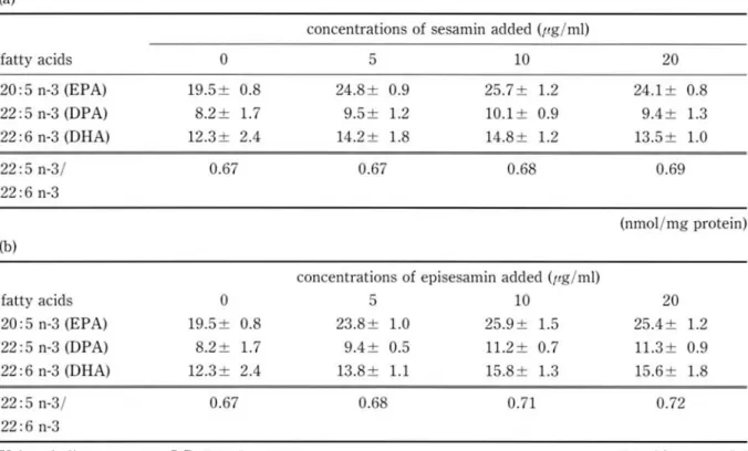
The cellular levels of EPA, OPA and ORA and min added with no signifcant difference.
OPA/ORA ratio in the presence of sesamin and In the previous report, sesamin exhibited no in- episesamin with exogenous EPA (30 11M) were hibitory effects on 116 desaturases, which catalyzes showed in Table 3. The cellular levels of EPA, the desaturation of linoleic acid (18: 2n-6) to r-lino- OPA and ORA were slightly increased by addition lenic acid (18: 3n-6) in microorganism and animal of both sesamin and episesamin, but were indepen- ~I. On the other hand, the recent report demon- dent of the concentrations of sesamin and episesa- strated that the pathway of ORA synthesis in
39 Effects of esamin and R lated Lignan ompounds on Fatty Acid etabolism in Hep G2 Cell
animal cells involves the microsomal chain elonga- study well agreed with these facts. But the effect of tion of DPA (22: 5n-3) to 24: 5n-3, followed by its esamin and episesamin on DBA biosynthesis in desaturation to 24: 6n-3. This microsomal product Bep G2 was not completely clear because of a low is then metabolized to DBA (22: 6n-3) via perox- activity in the fatty acid metabolism in Bep G2 cells.
isomal 3-oxaidation. In this pathway, it i thought It is necessary that additional studies with radio that desaturation of 24: 5n-3 to 24: 6n-3 was cata- isotope labeled compounds are required to clarify lyzed by 6-desaturase 91. The result in the present these problems.
Table 3. Effects of sesamin (a) and episesamin (b) on DBA formation from EPA
(a)
concentrations of sesamin added (ftg/ml)
fatty acids 0 5 10 20
20:5 n-3 (EPA) 19.5± 0.8 24.8± 0.9 25.7± 1.2 24.1± 0.8
22:5 n-3 (DPA) 8.2± 1.7 9.5± 1.2 10.1± 0.9 9.4± 1.3
22: 6 n-3 (DBA) 12.3± 2.4 14.2± 1.8 14.8± 1.2 13.5± 1.0
22:5 n-3/ 0.67 0.67 0.68 0.69
22: 6 n-3
(nmol/mg protein) (b)
concentrations of epis samin added (ftg/ml)
fatty acids
o
10 2020: 5 n-3 (EPA) 19.5± 0.8 23.8± 1.0 25.9± 1.5 25.4± 1.2
22:5 n-3 (DPA) 8.2± 1.7 9.4± 0.5 11.2± 0.7 1l.3± 0.9
22: 6 n-3 (DBA) 12.3± 2.4 13. ± 1.1 15.8± 1.3 15.6± 1.8
22: 5 n-3/ 0.67 0.68 0.71 0.72
22:6 n-3
Values indicate means± .D. (n=4) (nmol/ mg protein)
Abbreviation us d: s e legend to Table 1.
References
1 ) Y. F K OA, T.O A\\'A and M. A~IIKI: Agric. Bioi. R , OALL: j. BioI. Cllem.. 193, 265-275 (1951) 11 111 .. 49, 301-306 (198 ) 9) A. Vo s, M. REI. HART, . A)lK RAPPA and H. PRE HER
2) T.O A\\·A. T. AGATA M. A IIKI, and Y. Fl KUD. : : J BioI. CII 111.. 266, 19995-20000 (1991) Agric. Bioi. lIem .. 49, 3351-3355 (1985)
3) Y. F KlOA. T. A .ATA. T. OSA\\'A and ~IIKI: j. (Receiv d: ept mb r 30 1997) Am. Oil 11 m. oc.. 63, 1027-1032 (1986)
4) . HIMIZlI, K. KI~IOTO, Y. HI. IE., H. KA\\' HIMA, M.
UGA. 0 and H. Y A~1. OA: Lipid. 26, 512- 16 (1991)
~ ) H. KA\\'ASHI~IA . KAMAOA, E. AKlIRADA)lI. K. AKIMo·
TO, K. Ko. ISHI and . HI ilZl : J Am. Oil lIem. oc..
74, 455- 59 (1997)
6) T. AKAHARA, T. YOKOCHI. T. HICASHIH R, . TA. AKA, T. Y AGl' HI and D. Ho:-m : j. Am. Oil lIem. OC.. 73, 1 21-1426 (1996)
7 ) ]. FOL H, M. LEES and G. H. LO )lEo. TA. LY: j. Bioi.
11 m.. 266, 497-509 (1957)
O. H. LO\\,RY, N.]. RO-EBRO CH, . L. F RR and R.].
40
ヒ ト肝癌 由来培養細胞株 He pG2 の高度不飽和脂肪酸生合成系への ゴマ リグナン類 の影響
白坂 憲 章 ,佐 用 知 寿 子 , 村 上 哲 男 , 書 栖 箆
近攻大学農学部食品栄頚学科
要 約
ヒ ト肝癌 由来増発細胞株 HepG2は,培地に添加 したエイコサペンタエン酸 (EPA)を,濃度依存的に細胞 内 に取 り込む ことが確認 された。 また,微弱ではあるが低浪度 (添加丑40〃M以下)においては濃度依存的に ドコ サヘキサエン酸 (DHA)へ と変換する解が確認 され HepG2細胞 には EPAか ら DHAを生合成する経路が存 在することが確認 された。
また,胡麻に含まれる リグナン頬であるセサ ミソおよびその鏡像異性体であるエビセサ ミソほ添加浪度に依存 して細胞 内への取 り込 まれることが確認 されたが,添加に よる脂肪酸組成の顕著な変化は認め られず, HepG2 細胞における EPAか らの DHAの生成を阻雪するという結果は得 られなかった。