Abbreviations: AI, apoptotic index; LMS, leiomyosarcoma; MCM2, minimichrosome 2; PCNA, prolif-erating cell nuclear antigen; PI, p53 index; TUNEL, terminal deoxynuleotidyl transferase-mediated dUTP-biotin nick end labeling
Apoptotic Cell Death and p53 Expression in Leiomyosarcoma of
Soft-Tissue Origin
Haruhiko Yoshida, Mari Watanabe*, Kohei Shomori*, Hisao Ito* and Takeshi Minamizaki†
Department of Pathobiological Science and Technology, *First Department of Pathology and †Department of Orthopaedic Surgery, Tottori University Faculty of Medicine, Yonago 683-0826 Japan
Leiomyosarcoma (LMS) of soft-tissue origin was studied on the expression and the biological significance of apoptosis in relation to p53 oncoprotein. Immunohistochemical analysis was performed initially on the paraffin-embbeded sections taken from 29 surgical tissues (20 cases) including 9 recurrent/metastatic tumors. The results are as follows: A positive correlation was observed between the p53 indices (PIs) and the proliferative markers designated by Ki-67, PCNA and MCM2, both of which increased significantly in the high-grade malignant LMS much more than in the low-grade one (P < 0.001). Apoptotic cells were detected by the TUNEL method and evaluated as the apoptotic index (AI). A high AI-level was shown in the high-grade malignant LMS, especially in cases of the recurrent/metastatic sites in comparison with the tumors of the primary site (P < 0.05). The AI was statistically higher in the p53-positive cases of high-grade malignant LMS than in the p53-negative cases of low-grade malignant LMS. In conclusion, apoptotic activity paralleled the overexpression of p53 protein along with an increasing grade of malignancy and may be related intimately to the increased malignant potential, especially to recurrence/metastasis.
Key words: apoptosis; immunohistochemistry; leiomyosarcoma; p53 protein
Leiomyosarcoma (LMS) is now known as one of the most frequent malignant tumors occur-ring in the soft-tissue of adults. It has been di-vided into 3 categories according to location by Enzinger and Weiss (1988), because of its clini-cal and biologiclini-cal behavior. Hashimoto et al. (1986) emphasized a prognostic difference be-tween superficial and deep forms of LMS, an adverse prognosis of the deep form as compar-ed to the superficial form. Histologically, con-ventional LMS is classified currently into well, moderately and poorly/anaplastic differentiated types depending on the degree of differentia-tion, but it is very difficult to evaluate the ma-lignancy in predicting metastasis despite the multiple factors of size, cellularity, atypia and
necrosis which are known as prognostic factors. Even at the level of mitotic activity, a serious decision can hardly be made on the biological behavior of LMS (Enzinger and Weiss, 1988).
Apoptosis is an active process of cell death occurring in both normal and neoplastic condi-tions and is characterized as the orderly parti-cipation of biological, morphological and mo-lecular genetic events (Kerr et al., 1972, 1994; Wyllie, 1981; Umansky, 1982). It is induced by a variety of agents including oncogenic pro-teins, mainly p53 protein, the bcl-2 gene family and Fas/Fas legand among the process and re-gulation of apoptosis (Kerr et al., 1994). Recent studies have disclosed 2 pathways for the ex-pression of apoptosis (Clarke and Purdie, 1993;
Endo et al., 1999): p53 gene-dependent (Haupt et al., 1966; Chen et al., 1996) and -independent pathways, the latter of which can be induced by growth factors, hormones, chemotherapeutic agents and radiation (Clarke and Purdie, 1993; Endo et al., 1999). In considering malignant neoplasma, apoptosis may play an important role in the regulation of tumor growth in intimate relation to the p53 gene regulating the cell cycle in conjunction with cell proliferation. The relationship between p53 gene status and apoptosis, however, has rarely been reported on soft-tissue sarcoma (Staunton and Gaffney, 1995; Yuki et al., 1995; Nakanishi et al., 1997; Valenti et al., 1998). We examined the relationship between the p53 protein and apoptosis in 29 surgical specimens from 20 cases of LMS of soft-tissue origin which were classified histologically from low-grade to high-grade malignancies according to the mitotic rate and proliferative cell markers. In addition, we discussed the significance and evaluation of apoptotic activity as a biological and prognostic factor of LMS.
Materials and Methods
This study was performed on 29 paraffin-embedded tumor tissues from 20 cases of LMS of soft-tissue origin, all of which were removed surgically. The histological diagnosis of these cases were determined in addition to immuno-histochemical findings of vimentin, alpha-smooth muscle actin and HHF 35, and classifi-ed into 3 groups according to tumor cell differ-entiation from poorly to well differentiated types. Mitotic activity is an another aid in eval-uating tumor malignancy and thus, the number of mitoses was counted in 10 high-power fields selected randomly as a mitotic rate: under 10 as low-grade, 10 to 20 as intermediate and over 20 as high-grade malignancies.
The clinical data for these cases were col-lected from medical records. Each case in this study was analyzed according to the tumor nodes metastases classification developed by the Union Internationale Contra le Cancer to evaluate the relation of apoptotic activity in LMS to their biological behavior.
Immunohistochemistry
An immunohistochemical study was performed by the streptavidin-biotin-complex method us-ing the followus-ing monoclonal antibodies: anti-p53 nuclear protein (DO-7, 1:50, Dako, Glostrup, Denmark), anti-Ki-67 (MIB-1, 1;50, Immunotech, Marceille, France), anti-proliferating cell nu-clear antigen (PCNA) (PC10, Nichirei, Tokyo, Japan) and anti-minimichrosome maintenance 2 protein (MCM2). MCM proteins are com-posed of 6 isoforms ranging from MCM2 to MCM7. They are expressed in the G1/S phase of the cell cycle and bind to DNA strands for a replication of DNA under the presence of DNA polymerase. Monoclonal antibody (2H10 clone) against MCM2 protein was produced by the im-munization of oligopeptide of N-terminal from MCM2 protein with 892 amino acids and was reported as a proliferative marker available to examine the grading of malignancy*. The de-paraffinized sections were pretreated with cit-rate buffer (0.01 mol/L citric acid: pH 6.0) at 92˚C for 30 min in a microwave oven (Azumaya MI-77, Osaka, Japan) for all antibodies except anti-PCNA antibody and then incubated at 4˚C overnight with the primary antibodies. The positive cells were counted and evaluated as the ratio of immunoreactive cells to the total num-ber of 1,000 tumor cells.
TUNEL
Terminal deoxynucleotidyl transferase-medi-ated dUTP-biotin nick end labeling (TUNEL) was employed for the detection of apoptotic cells using an ApopTag Plus in situ apoptosis detection kit (Oncor, Gaithersburg, MD) which was followed by the method originally devel-oped by Kerr et al. (1972). Briefly, the sections were incubated with 20 µg/mL proteinase K (Behringer Mannheim/Yamanouchi, Tokyo) for 15 min at 37˚C after blocking endogenous peroxidase in methanol for 30 min at room tem-perature. Thereafter, the sections were layered and incubated on terminal deoxynucleotidyl transferase with digoxigenin-11-dUTP in a *Shomori K. Personal communication.
moist chamber for 90 min at 37˚C. After stopping the reaction, anti-digoxigenin-peroxidase was applied to the reaction mixture for 30 min at room temperature. The positive reaction was estimated with 3,3'-diamino-benzidine reaction at room temperature. The positive cells were counted and evaluated as the ratio of TUNEL-positive cells to a total number of more than 1,000 tumor cells.
Results
The clinicopathologic data are shown in Table 1. The patients were comprised of 7 males and 13 females. The age distribution was from 35 to 76 years old (mean age, 65.6 years). The pri-mary sites of the tumors involved 12 from the retroperitoneum and abdomen, 4 from the lower extremities, 1 from the upper extremity and 1 from the trunk in addition to 1 from the uterus. Recurrent/metastatic cases were included in 4 cases (10 specimens). Twenty nine specimens from 20 cases of LMS of soft-tissue origin were histologically evaluated into 3 categories of malignancy based on tumor cell differentiation and the mitotic index: 9 low-grade, 8 inter-mediate and 12 highly malignant LMSs (Table 2). The cases of low-grade malignancy appear-ed very clear in cell differentiation showing less than 10 mitotic figures per 10 high-power fields selected randomly. The cases of intermediate malignancy were well to moderate in cell dif-ferentiation associated with 10 to 20 mitotic figures, and in high-grade malignancy, the tu-mor cells were poorly differentiated assuming
Table 1. Clinical data of the cases used in this study
Age range (mean)* 35–76 (65.6)
Sex ratio (male:female) 7:13 Primary site
Retroperitoneum and abdomen 11 Upper extremity 1 Lower extremity 4 Body 1 Uterus 1 Recurrence/metastasis 5 *year.
Table 2. Comparative studies on LMS categorized in 3 groups according to the mitotic rate
Malignancy Number Cell AI PI Cell proliferation
of
differ-specimens entiation Ki-67 PCNA MCM2
Low-grade (< 10) 9 Well 0.4 ± 0.2 0.4 ± 1.8 2.0 ± 0.6 6.8 ± 1.2 3.2 ± 0.8 Intermediate (10–20) 8 Moderately 1.2 ± 0.5 4.6 ± 2.7 5.0 ± 3.2 16.9 ± 2.6 9.7 ± 2.1 High-grade (20 <) 12 Poorly 1.3 ± 0.9 23.7 ±15.0 16.7 ± 5.0 33.4 ± 6.8 22.1 ±10.7 Mean ± SD.
( ), mitotic rate/10 high-power field.
AI, apoptotic index; LMS, leiomyosarcoma; MCM2, minimichrosome 2; PCNA, proliferating cell nuclear antigen; PI, p53 index; TUNEL, terminal deoxynuleotidyl transferase-mediated dUTP-biotin nick end labeling.
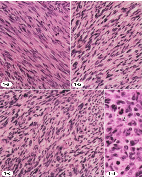
pleomorphic giant cells abundantly. The mitot-ic figures were easily encountered at a ratio of over 20 per 10 high-power fields (Figs. 1a–d).
TUNEL labeling for apoptotic cells
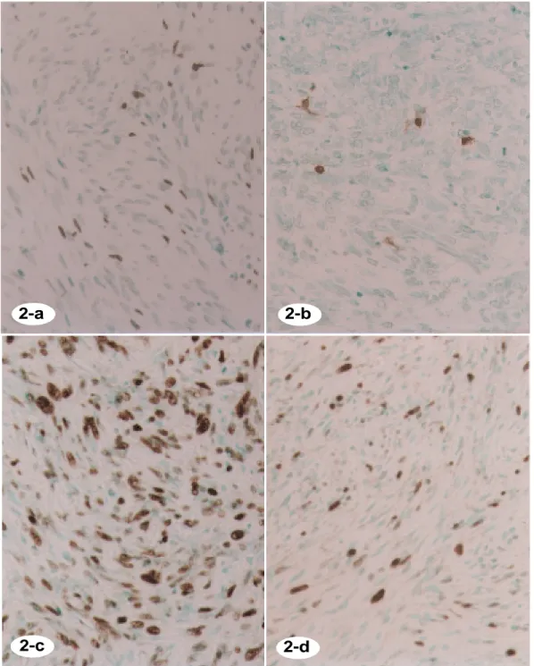
Apoptotic cells were determined by the TUNEL method and evaluated as the apoptotic index (AI). Apoptotic cells were distinguished as cells with condensed and fragmented nuclei separated by a clear halo in hematoxylin and eosin stain (Fig. 1d). TUNEL-positivity was observed at a varying degree to the cell nuclei with condensed and fragmented nuclei and oc-casionally to tumor cells which looked normal (Figs. 2a and b). Table 2 shows the AI, posi-tivities of p53 and the proliferative markers (Ki-67, PCNA and MCM2) of LMS categorized into 3 groups according to the mitotic rate. The immunohistochemical findings of p53 as well as Ki-67, PCNA and MCM2 are shown in Figs. 2c and d. Nine low-grade malignant LMSs showed 0.4 ± 0.2 of AI (mean ± SD) and 0.4 ±
Fig. 1. Histological findings of LMS. Hematoxylin and eosin stain, × 200.
a: Well-differentiated LMS; mitotic cells, 9/10 HPF, low-grade malignancy, 71/F.
b: Moderately-differentiated LMS; mitotic cells, 10/10 HPF, intermediate malignancy, 76/F. c: Poorly-differentiated LMS; mitotic cells, 34/10 HPF, high-grade malignancy, 63/F. d: Arrow indicates an apoptotic cell.
1-a
1-b
1-d
1-c
Figs. 2a and b. TUNEL for apoptotic cells. Positive for the tumor cell nuclei at a varying degree. a: AI = 5.1; third-recurrent tumor, 63/F. b: AI = 0.8; poorly-differentiated LMS, primary, 61/F.
Fig. 2c. Immunohistochemical stain of p53 protein. Strongly positive for the tumor cell nuclei. PI = 57, poorly-differentiated LMS, 74/F.
Fg. 2d. Immunohistochemical stain of Ki-67. Positive for the tumor cell nuclei at a various degree; 21.5%, poorly-differentiated LMS, 74/F.
2-a
2-b
2-d
2-c
0.18 of PI, whereas 12 high-malignant LMSs were statistically higher at a rate of 1.1 ± 0.9 and 23.9 ± 15.0, respectively (P < 0.05). Three kinds of proliferative markers of Ki-67, PCNA and MCM2 proteins were examined to assess the proliferating activity of the tumor cells di-vided into 3 categories of malignancy. The nu-clear positivities of Ki-67, PCNA and MCM2 increased statistically in the high-grade malig-nant LMSs at a rate of 16.7 ± 6.8 in Ki-67, 33.4
± 6.8 in PCNA and 22.1 ± 10.7 in MCM2, more than those of the low-grade ones, respectively (2.0 ± 0.6 in Ki-67, 6.8 ± 1.2 in PCNA and 3.2 ± 0.8 in MCM2).
Relationship between TUNEL indices and p53 expression
TUNEL and p53 positivities were examined comparatively in primary LMSs (3 cases) and their recurrent/metastatic tumors (9 tumors) as shown in Table 3. TUNEL indices (AIs) of the recurrent/metastatic tumors were statistically
higher (mean AI: 2.0 ± 0.7) in comparison with the primary site (0.4 ± 0.1). p53-positive cells increased significantly in cases of the recurrent/ metastatic tumors (mean PI: 12.3 ± 3.1) than in those of the primary tumors (0.6 ± 0.5) (P < 0.05) in the same way.
Expression of apoptotic cells between p53-positive and -negative LMSs
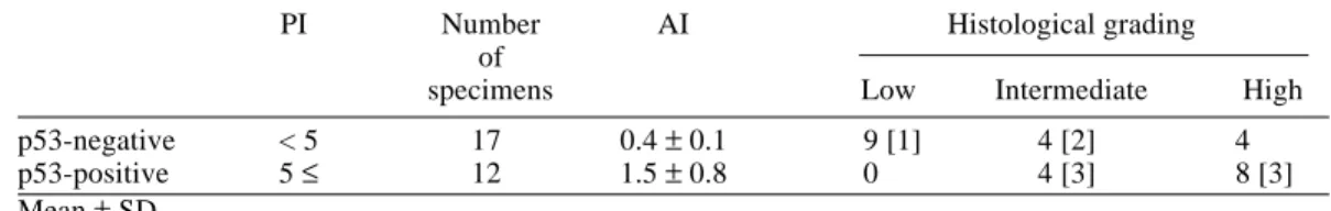
Low-grade malignant LMS showed a rate of PI under 5 as shown in Table 2, and thus we figur-ed that p53-positive tumors were ratfigur-ed over 5 PI. The p53-positive LMSs were found in 12 of 29 tumors as shown in Table 4 and revealed the AI at a mean of 2.2 ± 1.6, statistically higher than that (0.4 ± 0.1) of the p53-negative LMSs (P < 0.05). Histologically, the p53-positive group included 8 (66.7%) of 12 high-grade and 4 (50%) of 8 intermediate malignancies, where-as 9 (100%) low-grade and 4 (50%) of 8 interme-diate malignant LMSs were included in the p53-negative group. The recurrence/metastasis-free
Table 4. AI between p53-negative and positive cases in 3 histological groups
PI Number AI Histological grading
of
specimens Low Intermediate High
p53-negative < 5 17 0.4 ± 0.1 9 [1] 4 [2] 4
p53-positive 5 ≤ 12 1.5 ± 0.8 0 4 [3] 8 [3]
Mean ± SD.
[ ], number of recurrent/metastatic cases. AI, apoptotic index; PI, p53 index.
Table 3. Comparative study on immunohistochemistry by the TUNEL method between 3 primary tumors and their recurrent/metastatic tumors
Histological Number AI PI Cell proliferation
grading of
mitoses Ki-67 PCNA MCM2
Primary tumor [3] Low 2– 9 0.4 ± 0.1 0.6 ± 0.5 0.5 ± 0.2 8.4 ± 2.4 3.8 ± 2.7 Recurrent/meta-
static tumor* [9] high 11–34 2.0 ± 0.7 12.3 ± 3.1 12.8 ± 3.5 22.1 ± 9.6 18.1 ± 7.9 Mean ± SD.
[ ], number of specimens. * Recurrent period: 2–7 years.
AI, apoptotic index; MCM2, minimichrosome 2; PCNA, proliferating cell nuclear antigen; PI, p53 index; TUNEL, terminal deoxynuleotidyl transferase-mediated dUTP-biotin nick end labeling.
survival rate was compared between the p53-positive and -negative groups (Fig. 3). The pa-tients with p53-positive tumors was significant-ly worse than those with p53-negative tumors (log-rank test: P = 0.047).
Discussion
In this study, we evaluated the frequency of apoptotic cell death in an increasing malignant potential of LMS based on mitotic rate as well as expression of the proliferative markers of Ki-67/PCNA/MCM2 responsible of tumor growth. Secondly, the relationship between p53 over-expression and AIs was investigated to clarify the significance of apoptosis on tumor malig-nancy and prognostic factor. Our results show-ed a positive correlation between mitotic rate and increasing grade of histological malignancy on the one hand and the proliferative markers of Ki-67/PCNA/MCM2 on the other. These find-ings were exactly similar to those reported be-fore by others (Drobijak et al., 1994; Yoo et al., 1997; Konomoto et al., 1998). MCM2 protein is part of the MCM family which is composed of 6 isoforms of MCM2 to MCM7. They are expressed in the G1/S phase of the cell cycle
and bind to DNA strands for replication of DNA under the existence of DNA polymerase. The positivity for anti-MCM2 antibody ranged between those of Ki-67 and PCNA.
We observed p53 positivity in 12 (41.4%) of 29 LMSs, of which histological grading was high-malignant in 8 and intermediate malignant in 4. In the current study, p53-immunostaining was positive at a rate of 20 to 64% in various types of soft-tissue sarcomas (Yoo et al, 1997; Stefanou et al, 1998) and 21.6 to 43% of LMS of soft-tissue origin (O’Reilly et al., 1997; Konomoto et al., 1998). The mutated p53 gene loses its inherent transcriptional activity, result-ing in enhancement of tumor cell proliferation (Endo et al., 1999). Yoo et al. (1997) suggested the role of the p53 mutation as the pathogenesis of soft-tissue sarcomas. Regarding the biolog-ical behavior or prognostic factor, however, the overexpression of p53 protein was estimated as being diverse from the one suggesting a posi-tive correlation between p53 protein and prog-nosis (Drobijak et al., 1994; Toffoli et al., 1994; Yoo et al., 1997) to the others that indicated no relation between p53 expression and biological behavior (O’Reilly et al., 1997; Stefanou et al., 1998). We would like to emphasize here that the overexpression of p53 protein could be highly valuable as a prognostic factor taken from the findings of an increasing accumulation of p53 protein with an intimate relation to the grading of tumor malignancy.
Detection and identification of apoptotic cells was successfully car-ried out on the paraffin-embedded sections by using the TUNEL meth-od which was intrmeth-oduced and devel-oped by Gavrieli et al. (1992) and Leoncini et al. (1993) and is based on detecting naturally occurring DNA strand breaks. TUNEL-positive, apoptotic cells were easily recog-nized as the cells with nuclear con-densation, fragmentation separated by a clear halo around the cells in hematoxylin and eosin staining, in addition to the number of normal-looking cells as mentioned previ-Fig. 3. Recurrence/metastasis-free survival curves of patients
with p53-positive and -negative leiomyosarcoma (LMS). Patients with p53-positive tumors were significantly worse than those with p53-negative tumors (log-rank test: P = 0.047). [ ], number of specimens. 48 96 100 50 (%) (month) p53 (–) [17] p53 (+) [12] Degree of freedom = 1 P = 0.047
Time after diagnosis
Recurrence/metastasis-free sur viv al cur v e
ously by others (Lowe et al., 1994; Aihara et al., 1994; Arai and Kino, 1995).
We compared AI and PI with histological grading resulting in a positive correlation of AI to PI which increased statistically from low-grade to high-low-grade malignancy. The AI value, furthermore, was statistically much higher in p53-positive cases of high-grade malignant LMS than in p53-negative cases. Thus, in cases of high-grade malignant LMS, especially recur-rent/metastatic tumors, a more rapid growth of tumors caused by increased proliferative activ-ity was probably due to a loss of regulation by p53. Apoptosis is regulated and controlled in its expression by various oncogens and sup-presser genes (Kerr and Hormon, 1994). p53 gene is one of the major regulators of apoptosis which can easily allow apoptosis in association with MDM2 by regulating the cell cycle in the manner of growth arrest at G1/S or G2/S phase or apoptotic cell death, but the mutative p53 gene suppresses apoptosis by a loss of tran-scriptional activity to cyclin-dependent kinase (Aihara et al., 1994). Ikeda et al. (1998) de-monstrated that naturally occurring apoptosis appeared to be induced predominantly via a p53-independent pathway from the observa-tion of early and advanced gastric carcinoma. Kawauchi et al. (2000) suggested that apoptosis in cases of synovial sarcoma may be thought to be caused by a multiple apoptosis-regulating mechanism other than p53 protein. Our find-ings of a positive correlation between an in-creased amount of apoptotic cells and p53 posi-tivity, however, is most likely to indicate that apoptotic status was induced via a p53-depen-dent pathway.
There has been a positive correlation re-ported between apoptotic cells and higher malignancies of non-Hodgkin’s lymphomas (Leoncini et al., 1993), prostatic carcinomas (Aihara et al, 1994) and colorectal adenomas (Arai and Kino, 1995). Tatebe et al. (1996) also reported that apoptosis and cell proliferation were more frequent in metastatic foci than in primary lesions of colorectal carcinoma and suggested that apoptosis might reflect not only cell loss, but proliferative activity. A recent study on synovial sarcoma indicated that
apo-ptosis may be an indicator of poor prognosis (Kawauchi et al., 2000). Our results support this hypothesis from the comparative exami-nation of primary tumors and their recurrent/ metastatic tumors. We used the materials of LMS originating in deep soft-tissue except for one from organ-specific LMS of the uterus and intestine which represented rather low mitotic activity. In fact, it is suggested that the suscep-tibility of undergoing apoptosis may reflect the histological differences between tumor types (Staunton et al., 1995). Therefore, the mecha-nism and role of apoptosis in LMS still remain to be elucidated.
Acknowledgments: This study was presented at the 33rd Annual Musculoskeletal Tumor Meeting of the Japanese Orthopaedic Association held on 14 July, 2000 in Kumamoto. The authors thank Mr. Hisayuki Matsumoto and Miss Yuki Torigai, both of whom are students at the College of Medical Care Technol-ogy, Tottori University.
References
1 Aihara M, Truong LD, Dunn JK, Wheeler TM, Scardino PT, Thompson TC. Frequency of apo-ptotic bodies positively correlates with Gleason grade in prostatic Cancer. Hum Pathol 1994; 25:797–801.
2 Arai T, Kino I. Role of apoptosis in modulation of the growth of human colorectal tubular and vil-lous adenomas. J Pathol 1995;176:37–44. 3 Enzinger FM, Weiss SW. Soft tissue tumors. 2nd
ed. St Lous: CV Mosby, 1988. Chapter 15. Leio-myosarcoma; p. 402–421.
4 Chen J, Wu X, Lin J, Levine AJ. mdm-2 inhibits the G1 arrest and apoptosis functions of the p53 tumor suppressor protein. Mol Cell Biol 1996; 16:2445–2452.
5 Clarke AR, Purdie CA, Harrison DJ, Morris RG, Bird CC, Hooper ML, et al. Thymocyte apopto-sis induced by p53-dependent and independent pathways. Nature 1993;362:849–852.
6 Endo K, Sakatani T, Watanabe M, Yoshida H, Nannba E, Ito H. Wild-type p53 transfer resulted in cell cycle arrest, but not apoptosis of newly established human malignant fibrous histiocyto-ma cell line. Internat J Oncol 1999;15:935–942. 7 Drobijak M, Latres E, Pollack D, Karpeh M,
Dudas M, Woodruff JM, et al. Prognositic impli-cations of p53 nuclear overexpression and high
proliferation index of Ki-67 in adult soft-tissue sarcomas. J Natl Cancer Inst 1994;86:549–554. 8 Hashimoto H, Daimaru Y, Tsuneyoshi M, Enjoji
M. Leiomyosarcoma of the external soft tissues. Cancer 1986;57:2077–2088.
9 Haupt Y, Barak Y, Oren M. Cell type-specific inhibition of p53-mediated apoptosis by mdm2. EMBO J 1996;15:1596–1606.
10 Gagvrielli Y, Sherman Y, Ben-Sasson SA. Iden-tification of programmed cell death in situ via specific labeling of nuclear DNA fragmentation. J Cell Biol 1992;119:493–501.
11 Ikeda M, Shomori K, Endo K, Makino T, Matsuura T, Ito H. Frequent occurrence of apoptosis is an early event in the oncogenesis of human gastric carcinoma. Virchows Arch 1998;432:43–47. 12 Kawauchi S, Fukuda T, Oda Y, Saito T, Takeshita
M, Yokoyama K, et al. Prognostic significance of apoptosis in synovial sarcoma: correlation with clinicopathologic parameters, cell proliferative activity, and expression of apoptosis-related pro-teins. Mod pathol 2000;13: 755–765.
13 Kasagi N, Gomyo Y, Shirai H, Tsujitani S, Ito H. Apoptotic cell death in human gastric carcinoma: analysis by terminal deoxynucleotidyl transferase-mediated dUTP-biotin nick end labeling. Jpn J Cancer Res 1994;85:939–945.
14 Konomoto T, Fukuda T, Hayashi K, Kumazawa J, Tsuneyoshi M. Leiomyosarcoma in soft tissue: examination of p53 status and cell proliferating factors in different locations. Hum Pathol 1998; 29:74–81.
15 Kerr JFK, Wyllie AH, Currie AR. Apoptosis: a basic biological phenomenon with wide-ranging implications in tissue kinetics. Br J Cancer 1972; 26:239–257.
16 Kerr JFR, Winterford CM, Hormon BV. Apopto-sis: its significance in cancer and cancer therapy. Cancer 1994;73:2013–2026.
17 Leoncini L, Del Vacchio MT, Megha T, Barbini P, Galieni P, Pileri S, et al. Correlations between apoptotic and proliferative indices in malignant non-Hodgkin’s lymphomas. Am J Pathol 1993; 142:755–763.
18 Lowe SW, Jacks T, Housman DE, Ruley HE. Abrogation of oncogene-associated apoptosis allows transformation of p53-deficient cells. Proc Natl Acad Sci USA 1994;91:2026–2030. 19 Nakanishi H, Ohsawa M, Naka N, Uchida, Ochi
T, Aozasa K. Immunohistochemical detection of bcl-2 and p53 proteins and apoptosis in soft tissue sarcoma: their correlations with prognosis. On-cology 1997;54:238–244.
20 S t e f a n o u D G , N o n n i A V , A g n a n t i s N J , Athnassiadou SE, Briassoulis E, Pavlidis N. p53/ MDM-2 immunohistochemical expression cor-related with proliferative activity in different types of human sarcomas: a ten-year follow-up study. Anticancer Res 1998;18:4673–4681. 21 O’Reilly PE, Raab SS, Niemann TH, Rodgers JR,
Robinson RA. p53, proliferating cell nuclear antigen, and Ki-67 expression in extrauterine leiomyosarcoma. Mod Pathol 1997;10:91–97. 22 Staunton MJ, Gaffney EF. Tumor type is a
deter-minant of susceptability to apoptosis. Am J Clin Pathol 1995;103:300–307.
23 Tatebe S, Ishida M, Kasagi N, Tsujitani S, Kaibara N, Ito H. Apoptosis occurs more frequently in metastatic foci than in primary lesions of human colorectal carcinomas: analysis by terminal-deoxynucleotidyl-transferase-mediated dUTP-biotin nick end labeling. Int J Cancer 1996;65: 173–177.
24 Toffoli G, Doglioni C, Cernigoi C, Frustaci S, Perin T, Canal B, et al: p53 over-expression in human soft tissue sarcomas: relation to biological aggressiveness. Ann Oncol 1994;5:167–172. 25 Umansky SR. The genetic program of cell death.
Hypothesis and some applications: transforma-tion, carcinogenesis, aging. J Theor Biol 1982; 97:591–602.
26 Valenti MT, Azzarello G, Vinate O, Manconi R, Balducci E, Guidolin D, et al. Differentiation, proliferation and apoptosis levels in human leio-myoma and leiomyosarcoma. J Cancer Res Clin Oncol 1998;124:93–10
27 Wyllie AH. Cell death: a new classification separating apoptosis from necrosis. In: Bowen ID, Lockshin RA, eds. Cell death in biology and pathology. London: Chapman and Hall; 1981. p. 9–34.
28 Yoo J, Lee HK, Kang CS, Park WS, Lee JY, Shim SI. p53 gene mutation and p53 protein ex-pression in human soft tissue sarcomas. Arch Pathol Lab Med 1997;121:395–399.
29 Yuki M, Yokota K, Okuyama S. Cell prolifera-tion and cell death (apoptosis) as indices differ-entiating malignant from benign gastrointestinal myogenic tumors. Nippon Shokakibyo Gakkai Zasshi 1995;92:206–216 (in Japanese with Eng-lish abstract).
Received April 12, 2001; accepted May 17, 2001 Corresponding author: Prof. Haruhiko Yoshida