Yonago Acta Medica 2017;60:59–63 Patient Report
Corresponding author: Hiroaki Saito, MD, PhD sai10@med.tottori-u.ac.jp
Received 2016 November 14 Accepted 2016 November 28
Abbreviations: CT, computed tomography; EUS, endoscopic ultra-sound; FDG, fluorodeoxyglucose; GI, gastrointestinal; GIST, gas-trointestinal stromal tumors; HPF, high-power field; PET, positron emission tomography; SUV, standardized uptake value
Gastric Schwannoma with Enlargement of the Regional Lymph Nodes Resected
Using Laparoscopic Distal Gastrectomy: Report of a Patient
Shota Shimizu, Hiroaki Saito, Yusuke Kono, Yuki Murakami, Hirohiko Kuroda, Tomoyuki Matsunaga, Yoji Fukumoto, Tomohiro Osaki and Yoshiyuki Fujiwara
Division of Surgical Oncology, Department of Surgery, School of Medicine, Tottori University Faculty of Medicine, Yonago 683-8504, Japan
ABSTRACT
Preoperative differential diagnosis of gastric submuco-sal tumors has generally been difficult because they are covered with normal mucosa. However, recent advances in endoscopic ultrasound (EUS)-guided sampling of submucosal gastrointestinal lesions have made it pos-sible to achieve preoperative differential diagnosis of gastric submucosal tumors. A 76-year-old woman was referred to our hospital with a gastric submucosal tumor. The tumor was observed in the antrum of the stomach. It was preoperatively diagnosed as a schwannoma after immunohistochemical evaluation of a biopsy speci-men, obtained using endoscopic ultrasound-guided fine needle aspiration. A computed tomography scan of the abdomen revealed lymphadenopathies near the tumor indicating the possibility of lymph node metastasis from the gastric tumor. The patient underwent laparoscopic distal gastrectomy with D1 + lymph node dissection. The resected tumor was a submucosal tumor measuring 65 × 45 × 35 mm; it was histopathologically diagnosed as a schwannoma. Resected lymph nodes were enlarged in the absence of lymph node metastasis as a result of reactive lymphadenopathy. A definitive preoperative diagnosis of gastric schwannoma is possible using im-munohistochemical staining techniques and EUS-guid-ed sampling techniques. After definitive preoperative diagnosis of gastric schwannoma, minimal surgery is recommended to achieve R0 resection.
Key words gastric schwannoma; laparoscopic surgery; submucosal tumor
Mesenchymal tumors of the gastrointestinal (GI) tract mainly comprise a spectrum of spindle cell tumors that include gastrointestinal stromal tumors (GISTs), leiomy-omas, leiomyosarcleiomy-omas, and schwannomas.1
Immuno-histochemical examination of the specimens obtained from a tumor is essential to make a definitive diagnosis. Preoperative differential diagnosis of gastric submucosal tumors has generally been difficult because they are cov-ered with normal mucosa; this makes it difficult to ob-tain specimens. However, recent advances in endoscopic
ultrasound (EUS)-guided sampling of submucosal GI lesions have made it possible to achieve preoperative differential diagnosis of gastric submucosal tumors;2 this
enables physicians to easily decide on the optimal treat-ment strategy. In this report, we present a 76-year-old woman with preoperative diagnosis of gastric schwan-noma with enlargement of the regional lymph nodes of the stomach.
PATIENT REPORT
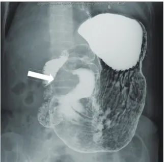
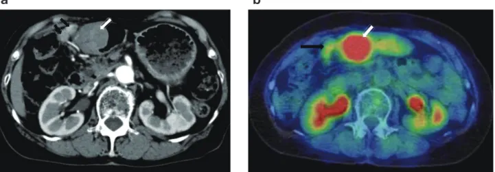
An asymptomatic 76-year-old woman was referred to our hospital because of the presence of a gastric submu-cosal tumor. The patient underwent appendectomy as a child and cholecystectomy in her 40s. There was no significant past medical, family, or psychosocial histo-ry. The upper GI series showed a smooth-marginated protruded lesion at the greater curvature of the antrum of stomach (Fig. 1); upper GI endoscopy revealed that this lesion measured 38 mm (Fig. 2a). Endoscopic ul-trasonography (EUS) showed a well-defined hypoecho-ic mass arising from the proper muscle layer of the stomach; an endoscopic ultrasound-guided fine needle aspiration biopsy was performed (Fig. 2b). Histological evaluation revealed a spindle cell tumor with a fascicular pattern. Immunohistochemical analysis indicated that the tumor was positive for S-100 and negative for c-kit, CD34, α-SMA, and desmin. The preoperative histolog-ical diagnosis was gastric schwannoma. A computed tomography (CT) scan of the abdomen demonstrated a protruded lesion measuring 45 × 37 mm at the antrum of the stomach and lymphadenopathies near the tumor (Fig. 3a). The 18-fluorodeoxyglucose (FDG) positron emission tomography (PET) and CT scans indicated high uptake of 18-FDG in the tumor at the antrum of the stomach [standardized uptake value (SUV) max, 5.89–6.81] and slight uptake in the enlarged lymph node
S. Shimizu et al.
Shimizu et al. Figure 1
(a)
(b)
Shimizu et al. Figure 2
Fig. 1. Upper gastrointestinal X-ray showing the
smooth-margin-ated protruded lesion at the greater curvature of the antrum of the stomach (arrow).
Fig. 2. Endoscope image. a: Upper gastrointestinal endoscopy showing the smooth protruded lesion measuring 38 mm at the greater
curvature of the antrum of the stomach. b: Endoscopic ultrasonography image showing a well-defi ned hypoechoic mass arising from the proper muscle layer of the stomach (arrow).
(SUVmax, 2.07–2.02)(Fig. 3b). Because of the possibili-ty of lymph node metastasis, we performed laparoscopic distal gastrectomy with D1 + lymph node dissection.
Macroscopically, the gastric mass covered by mu-cosa measured 65 × 45 × 35 mm, with the cut surface having the appearance of a firm lobulated yellowish white tumor. The tumor was located mainly in the prop-er muscle layprop-er (Fig. 4). Microscopic examination of the resected and H and E-stained specimens revealed a spindle cell neoplasm arranged in a palisade manner
with abundant collagen fi bers. There was lymphoid cuff-ing (Fig. 5a). Immunohistochemical staincuff-ing revealed that the tumor cells were positive for S-100 and negative for c-kit and neurofi laments (Figs. 5b–d). There were no tumor cells in the resected enlarged lymph nodes, indi-cating reactive lymphadenopathies. These histopatholog-ical and immunohistochemhistopatholog-ical findings are consistent with the presence of a gastric schwannoma with reactive lymphadenopathies.
DISCUSSION
Schwannomas, in other words neurinomas or neurilem-momas, are benign neurogenic tumors originating from Schwann cells, which normally wrap around the axons of the peripheral nerves. Theoretically, schwannomas can develop anywhere along the peripheral course of a nerve. However, they most commonly occur in the head and neck, but rarely in the GI tract. In the GI tract, the stomach is the most common gastrointestinal site of schwannoma, which constitute 0.2% of gastric neo-plasms.3, 4 The typical presentation of gastric
schwanno-ma is as a submucosal tumor with spindle cell histology. GIST, which are the most common mesenchymal tumor of the stomach, have the same presentation as gastric schwannoma. Despite strong morphological similarities, these tumors are heterogeneous regarding their im-munophenotypes. In 1988, Daimaru et al. successfully identifi ed schwannoma as a primary GI tumor based on positive S-100 staining.5 GIST also became a distinct GI
tumor diagnostic category when it was discovered that nearly all GIST cells express c-kit protein.1, 6 Before the
Gastric schwannoma resected using laparoscopic surgery
(a)
(b)
Shimizu et al. Figure 3
(a)
(b)
Shimizu et al. Figure 4
Fig. 3. CT image. a: CT image showing a protruded lesion measuring 45 × 37 mm at the antrum of the stomach (white arrow), and
en-largement of the regional lymph nodes of the stomach (black arrow). b: 18-FDG positron emission tomography CT image showing high uptake of 18-FDG in the tumor at the antrum of the stomach (SUVmax, 5.89–6.81) (white arrow) and slight uptake in the lymph nodes (SUVmax, 2.07–2.02) (black arrow). CT, computed tomography; FDG, fl uorodeoxyglucose; SUV, standardized uptake value.
Fig. 4. The tumor was located mainly in the proper muscle layer. a: Gastric mass covered by mucosa measuring 65 × 45 × 35 mm, b: with
the cut surface showing a fi rm lobulated yellowish white tumor.
recognition of S-100 antigen and c-kit antigen in gastric schwannomas and in GISTs, these neoplasms were most often classified as leiomyomas, leiomyosarcomas, or gastrointestinal autonomic nerve tumors.1, 4–6 With the
advent of immunohistochemical staining techniques and ultrastructural evaluation, it is now possible to identify these neoplasms based on their distinct immunophe-notypes. Furthermore, recent advances in EUS-guided sampling of gastrointestinal submucosal lesions make it possible to make a preoperative differential diagnosis
of gastric submucosal tumors. In fact, in our case pre-operative diagnosis of gastric schwannoma was made by means of an immunohistochemical examination of specimens obtained from the tumor by EUS-guided sampling.
Surgical resection is the only possible treatment for gastric schwannoma.3, 7 The size and location of the
tumor, as well as its relationship with the surrounding organs, are important factors in determining the type of operation. Local extirpation, wedge resection, partial,
a
a
b
S. Shimizu et al.
subtotal or even total gastrectomy, are all acceptable operations for the complete removal of the tumor.8
Lapa-roscopic techniques can also be used.9 Lymph node
dis-section is not required because it has been reported that gastric schwannoma is a benign tumor and is unlikely to metastasize to the lymph nodes. In our case, however, there were enlarged lymph nodes with moderate uptake of 18-FDG; this indicated the possibility of lymph node metastasis and the presence of another tumor such as malignant lymphoma that had developed in the lymph nodes. Shayanfar et al. reported a schwannoma that originated in the regional lymph nodes of the stom-ach.10 Furthermore, Murase et al. reported a malignant
schwannoma of the esophagus with lymph node me-tastasis.11 Considering the possibility of the presence of
tumor cells in the enlarged lymph node, we performed laparoscopic distal gastrectomy with D1+ lymph node dissection in the present case. Postoperative histopatho-logical examination of the enlarged lymph nodes after resection revealed that there were no tumor cells present, indicating the presence of reactive lymphadenopathies. The correlation between gastric schwannoma and en-larged lymph nodes remains unclear. Because enen-larged lymph nodes are regional lymph nodes and are located near the tumor, it is likely that gastric schwannoma can induce reactive lymphadenopathies. In this regard, the 18-FDG positron emission tomography and CT scans
showed high uptake in gastric schwannoma in our case. Komatsu et al. reported the fi rst case of gastric schwan-noma that exhibited increased accumulation of 18-FDG on PET imaging.12 It has not been clarifi ed why benign
schwannomas show increased 18-FDG uptake on PET; Kawabe et al. reported that 18-FDG accumulated in infl ammatory tissue involving lymphocyte proliferation in chronic tonsillitis.13 It has been reported that cuff-like
lymphoid aggregates are recognized around the tumor in 97% of gastric schwannomas;14 this also occurred in
the present case. Therefore, it is likely that lymphocyte proliferation in gastric schwannoma is related to enlarge-ment of the regional lymph node observed in our case. Thus far, the majority of previous series in the literature have demonstrated no lymph node metastasis regarding gastric schwannoma.15, 16 Therefore, local resection of the
stomach and extirpation of enlarged lymph nodes might be suffi cient in the present case.
Gastric schwannoma is considered a benign tumor. It is unclear whether pathological parameters such as tumor size and mitotic rate have prognostic signifi cance in this tumor, as in the cases of GISTs reported thus far; this is because gastric schwannoma is rare compared with GIST and experience concerning their long-term follow-up is limited. In this regard, Voltaggio et al. re-ported that some gastric schwannomas exceed 10 cm in size, and a minority have a mitotic rate > 5/50
high-pow-Shimizu et al. Figure 5
(a)
(b)
(c)
(d)
Fig. 5. Photomicrographs. a: There was lymphoid cuffi ng. Bar = 200 µm. b: Tumor cells were positive for S-100 and c: negative for c-kit. d:
Immunostained neurofi laments. Bar = 50 μm.
a
c
b
Gastric schwannoma resected using laparoscopic surgery
er fi elds (HPFs); none of these 51 gastric schwannoma cases showed evidence of aggressive behavior.17 Despite
this, these authors advised caution regarding gastric schwannomas with higher mitotic rates (> 10/50 HPFs), because experience from their long-term follow-up was limited.17 In fact, one case of malignant gastric
schwan-noma has been recently reported;18 in this case,
immu-nohistochemistry revealed that the tumor cells were pos-itive for S-100 protein and negative for c-kit and smooth muscle actin. Mitosis was scattered with 10 mitoses per 50 HPFs, indicating the aggressiveness of this tumor. This patient died at 5 months after surgery as a result of multiple liver metastases. Therefore, careful follow-up is required in gastric schwannoma patients with higher mitotic rates (> 10/50 HPFs) after surgery.
A definitive preoperative diagnosis of gastric schwannoma is possible using immunohistochemical staining techniques and EUS-guided sampling tech-niques. After defi nitive preoperative diagnosis of gastric schwannoma, minimal surgery is recommended to achieve R0 resection.
The authors declare no confl ict of interest. REFERENCES
1 Nishida T, Hirota S. Biological and clinical review of stro-mal tumors in the gastrointestinal tract. Histol Histopathol. 2000;15:1293-301. PMID: 11005253.
2 Mekky MA, Yamao K, Sawaki A, Mizuno N, Hara K, Nafeh MA, et al. Diagnostic utility of EUS-guided FNA in pa-tients with gastric submucosal tumors. Gastrointest Endosc. 2010;71:913-9. PMID: 20226456.
3 Melvin WS, Wilkinson MG. Gastric schwannoma. Clinical and pathologic considerations. Am Surg. 1993;59:293-6. PMID: 8489097.
4 Sarlomo-Rikala M, Miettinen M. Gastric schwannoma-a clinicopathological analysis of six cases. Histopathology. 1995;27:355-60. PMID: 8847066.
5 Daimaru Y, Kido H, Hashimoto H, Enjoji M. Benign schwan-noma of the gastrointestinal tract: a clinicopathologic and immunohistochemical study. Hum Pathol. 1988;19:257-64. PMID: 3126126.
6 Miettinen M, Majidi M, Lasota J. Pathology and diagnostic criteria of gastrointestinal stromal tumors (GISTs): a review. Eur J Cancer. 2002;38 Suppl 5:S39-51. PMID: 12528772. 7 Prevot S, Bienvenu L, Vaillant JC, de Saint-Maur PP. Benign
schwannoma of the digestive tract: a clinicopathologic and immunohistochemical study of fi ve cases, including a case of esophageal tumor. Am J Surg Pathol. 1999;23:431-6. PMID: 10199472.
8 Bandoh T, Isoyama T, Toyoshima H. Submucosal tumors of the stomach: a study of 100 operative cases. Surgery. 1993;113:498-506. PMID: 8488466.
9 Basso N, Rosato P, De Leo A, Picconi T, Trentino P, Fantini A, et al. Laparoscopic treatment of gastric stromal tumors. Surg Endosc. 2000;14:524-6. PMID: 10890957.
10 Shayanfar N, Shahzadi S. Schwannoma In A Perigastric Lymph Node: A Rare Case Report. Iran J Pathol. 2008;3:43-6. 11 Murase K, Hino A, Ozeki Y, Karagiri Y, Onitsuka A, Sugie
S. Malignant schwannoma of the esophagus with lymph node metastasis: literature review of schwannoma of the esophagus. J Gastroenterol. 2001;36:772-7. PMID: 11757750.
12 Komatsu D, Koide N, Hiraga R, Furuya N, Akamatsu T, Uehara T, et al. Gastric schwannoma exhibiting increased fluorodeoxyglucose uptake. Gastric Cancer. 2009;12:225-8. PMID: 20047128.
13 Kawabe J, Okamura T, Shakudo M, Koyama K, Wanibuchi H, Sakamoto H, et al. Two cases of chronic tonsillitis studied by FDG-PET. Ann Nucl Med. 1999;13:277-9. PMID: 10510887. 14 Hou YY, Tan YS, Xu JF, Wang XN, Lu SH, Ji Y, et al.
Schwannoma of the gastrointestinal tract: a clinicopatholog-ical, immunohistochemical and ultrastructural study of 33 cases. Histopathology. 2006;48:536-45. PMID: 16623779. 15 Fujiwara S, Nakajima K, Nishida T, Takahashi T, Kurokawa Y,
Yamasaki M, et al. Gastric schwannomas revisited: has pre-cise preoperative diagnosis become feasible? Gastric cancer. 2013;16:318-23. PMID: 22907486.
16 Zheng L, Wu X, Kreis ME, Yu Z, Feng L, Chen C, et al. Clin-icopathological and immunohistochemical characterisation of gastric schwannomas in 29 cases. Gastroenterol Res Pract. 2014;2014:202960. PMID: 24688535.
17 Voltaggio L, Murray R, Lasota J, Miettinen M. Gastric schwannoma: a clinicopathologic study of 51 cases and critical review of the literature. Hum Pathol. 2012;43:650-9. PMID: 22137423.
18 Takemura M, Yoshida K, Takii M, Sakurai K, Kanazawa A. Gastric malignant schwannoma presenting with upper gastro-intestinal bleeding: a case report. J Med Case Rep. 2012;6:37. PMID: 22277785.