Abbreviations: FCR, flexor carpi radialis muscle; FCU, flexor carpi ulnaris muscle; FDS, flexor digitorum superficialis mus-cle; PL, palmaris longus musmus-cle; PP, palmaris profundus musmus-cle; PT, pronator teres muscle
A Case of the Bilateral Duplicate Palmaris Longus Muscles
Coupled with the Palmaris Profundus Muscle
Yuichi Takanashi, Masaki Eda, Toshiyuki Kaidoh* and Takao Inoué
Division of Morphological Analysis, Department of Anatomy, School of Medicine, Tottori University Faculty of Medicine, Yonago 683-8503, Japan and *Division of Medical Morphology, Department of Anatomy, School of Medicine, Tottori University Faculty of Medicine, Yonago 683-8503, Japan
The palmaris longus muscle is one of the most variable muscles in human anatomy. During a routine anatomical dissection for medical students at Tottori University, we found duplicate palmaris longus muscles in the bilateral forearms together with the palmaris profundus mus-cle in the right forearm. The bilateral aberrant palmaris longus musmus-cles were observed at the ulnar side of the palmaris longus muscle and their distal tendons were attached to the flexor retinaculum. The palmaris profundus muscle found in the right forearm was located at the radial side of the flexor digitorum superficialis muscle. The proximal tendon was originated from the anterior surface in the middle of the radius, while the distal tendon coursed radial to the median nerve through the carpal tunnel, finally inserting into the distal part of the flexor retinaculum. Both the palmaris longus and aberrant palmaris longus muscles were in-nervated by the median nerve. The palmaris profundus muscle was presumably supplied by the median nerve.
Key words: cadaver; forearm; regional anatomy; skeletal muscle
The palmaris longus muscle (PL) is located in the superficial layer of the anterior surface of the fore-arm. It has a small belly arising from the medial epicondyle of the humerus, and its long thin tendon inserts into the palmar aponeurosis in the hand. The muscle is one of the most variable muscles in the human body (Takafuji et al., 1985). During an ana-tomical dissection for medical students at Tottori University in 2011, we found a pair of the duplicate PL in the bilateral forearms. Furthermore, the pal-maris profundus muscle (PP), which is not usually exist, was found in the right forearm. Although the duplicate PL is frequently reported (Reimann et al., 1944; Hojo, 1976; Kimura et al., 1979; Xue et al., 1998; Zeybek et al., 1998; Kawashima et al., 2002), it is rarely seen in bilateral forearms. In addition, there are no reports on the bilateral duplicate PL
coupled with the PP. In this paper, we describe myo-logical findings and innervation of this rare case.
Materials and Methods
Anomaly of the PL was noticed on both sides of a 57-year-old Japanese male cadaver. We observed closely the origin and insertion of the muscles, finely dissected the region of interest, and examined muscle shape and nerve supplies macroscopically, and photographed. We also measured the size of the muscles and tendons using a vernier caliper. The length of a muscle belly was measured between the origin (medial epicondyle) and the most distal part of the muscle-tendon border. The length of PL tendon was measured from the most distal part of
the muscle-tendon border to the base of palmar aponeurosis, whereas that of aberrant PL was done from the identi-cal point to the attachment to the flexor retinaculum. The width of muscle belly was measured at the maximum width. The width and thickness of the both PL and aberrant PL were measured at the midpoint of the entire length of the tendon.
Results
The duplicate PL and their nerve sup-ply found in the bilateral forearms are shown in Figs. 1 and 2, respectively. Their measurements, myological find-ings and nerve innervations are sum-marized in Table 1. The PP in the right forearm is shown in Fig. 3. No other muscular anomaly was recognized in the body.
Duplicate PL in the right forearm
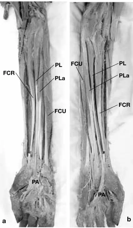
The aberrant PL was found ulnar to the PL (Fig. 1a). The bellies and ten-dons of both the aberrant PL and PL were closely apposed with each other. The duplicate PL arose from a com-mon tendinous origin of flexor muscles [PL, flexor carpi radialis muscle (FCR), flexor carpi ulnaris muscle (FCU), pro-nator teres muscle (PT) and a part of the flexor digitorum superficialis mus-cle (FDS)] at the medial epicondyle of the humerus.
In the PL, the muscle belly mea-suring 110 mm in length and 12.6 mm
in width, was followed by a long tendon distally at the middle of the forearm. The tendon coursed over the flexor retinaculum, finally inserting into the pal-mar aponeurosis (Table 1).
The morphology of the aberrant PL was quite similar to that of the PL. The muscle belly mea-suring 135 mm in length and 10 mm in width was
Fig. 1. Photographs of superficial flexor muscles of the forearm. Note
PLa between PL and FCU. a: right side, b: left side. FCR, flexor carpi radialis muscle; FCU, flexor carpi ulnar muscle; PA, palmar aponeurosis; PL, palmaris longus muscle; PLa, aberrant palmaris longus muscle.
followed by the tendon distally at the middle of forearm, and finally inserting into the flexor reti-naculum (Table 1).
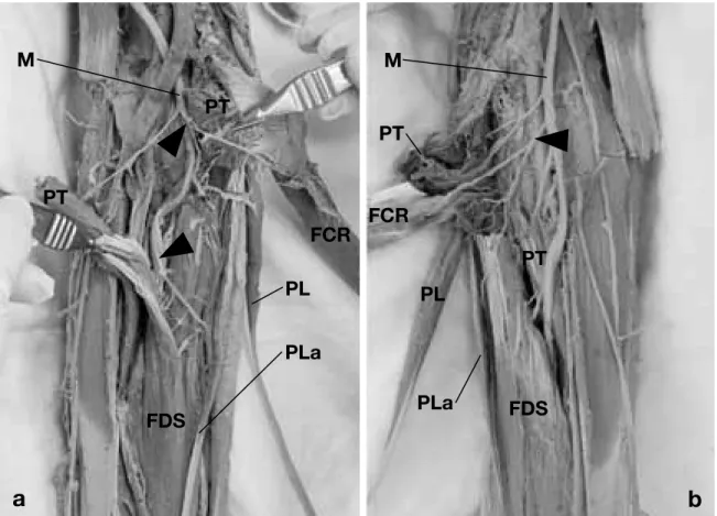
Both the PL and aberrant PL were innervated by branches of the median nerve in the proximal third of the forearm (Fig. 2a). The branches to the FCR, PL and FDS arose as a common trunk from
Fig. 2. Photographs showing the median nerve and its branches to flexor muscles. The belly of the pronator teres is cut
and opened for the demonstration of the innervation. Arrowheads show the common trunk of the median nerve to the PL and PLa. a: right side, b: left side. FCR, flexor carpi radialis muscle; FDS, flexor digitorum superficialis muscle; M, me-dian nerve; PL, palmaris longus muscle; PLa, aberrant palmaris longus muscle; PT, pronator teres muscle.
the median nerve, whereas those to the aberrant PL and FDS arose as another common trunk which branched distally.
Duplicate PL in the left forearm
The morphological findings of the duplicate PL in the left forearm are fundamentally identical to those in the right forearm.
The aberrant PL was also found ulnar to the PL (Fig. 1b). The bellies and tendons of both the aberrant PL and PL were closely apposed with each other. The duplicate PL arose from a common ten-dinous origin of flexor muscles (PL, FCR, FCU, PT and a part of the FDS) at the medial epicondyle of the humerus.
In the PL, the muscle belly measuring 130 mm in length and 10 mm in width was followed
by a long tendon distally at the middle of the fore-arm. The tendon coursed over the flexor retinacu-lum, finally inserting into the palmar aponeurosis (Table 1).
The morphology of the aberrant PL was quite similar to that of the PL. The muscle belly measur-ing 145 mm in length and 8.3 mm in width was followed by the tendon distally at the middle of forearm, and finally inserting into the flexor reti-naculum (Table 1).
Similarly to the right forearm, both the PL and aberrant PL were innervated by branches of the me-dian nerve in the proximal third of the forearm (Fig. 2b). However, the ramification of the branch was different. The branches to the FCR, PL, aberrant PL and FDS arose as a proximal common trunk from the median nerve, and another single branch to FDS arose distally from the median nerve.
PP in the right forearm
The PP in the right forearm was located at the ra-dial side of the middle part of FDS. It was spindle in shape with slender tendons on both ends. The proximal tendon was apparently independent to the radial head of the FDS and originated from the anterior surface in the middle of the radius. The muscle belly measured 77 mm in length. The distal tendon measuring 45 mm in length coursed radial to the median nerve under the flexor retinaculum. It passed through the carpal tunnel together with the median nerve, and finally inserted into the dis-tal part of flexor retinaculum (Fig. 3). Since the PP coursed in the vicinity of the median nerve, the muscle was presumably supplied by the median nerve. The branch to the muscle could not be mac-roscopically demonstrated.
Discussion
The variation of the PL was classified by Reimann et al. (1944) as follows: complete agenesis, variation in the location and form of the fleshy portion, aber-rancy in the attachment at either extremity, duplica-tion or triplicaduplica-tion, accessory slips or substituduplica-tions of similar form or position. In particular, duplicate PL have been frequently reported (Reimann et al., 1944; Hojo, 1976; Kimura et al., 1979; Xue et al., 1998; Zeybek et al., 1998; Kawashima et al., 2002). The occurrence of duplicate PL in Japanese is 1.9%
(Koganei et al., 1903), 2.1% (Adachi, 1910), 2.5% (Sato et al., 1974), whereas 3.1% in Russians (Gruber, 1868) and 0.8% in Americans (Reimann et al., 1944).
Most of the duplicate PL thus far reported were unilateral. As far as we know, there was only one report of the duplicate PL in both forearms (Kimura et al., 1979). As for the occurrence of the aberrant PL in the duplicated PL, it is mostly locat-ed at the ulnar side of PL as observlocat-ed in the present study (Xue et al., 1998), but occasionally at the ra-dial side (Kawashima et al., 2002). The aberrant PL is innervated by the median nerve as was observed in the present study (Kimura et al., 1979; Takafuji et al., 1985; Xue et al., 1998; Kawashima et al., 2002), but occasionally doubly innervated both by the me-dian and ulnar nerve (Xue et al., 1998).
The PP is another case of the anomaly of the PL. It originates from the anterior surface of the radius terminating to the flexor retinaculum of the deeper layer of the palmar aponeurosis (Reimann et al., 1944). Yoshida et al. (1983) observed variations of the deep flexor group of the antebrachial muscles and classified them into four types. Our case cor-responds to the type 2 (palmaris profundus longus muscle) of their classification. The present case is also analogous to that reported by Stark et al. (2010). Humphry (1872) proposed the idea that the superfi-cial layers of the forearm flexor muscles are derived from radial, intermediate, and ulnar sectors in the ventral matrix of the elbow (Howell, 1936a). Ac-cording to this hypothesis, the PT and FCR are differentiated from the radial sector, the FCU from
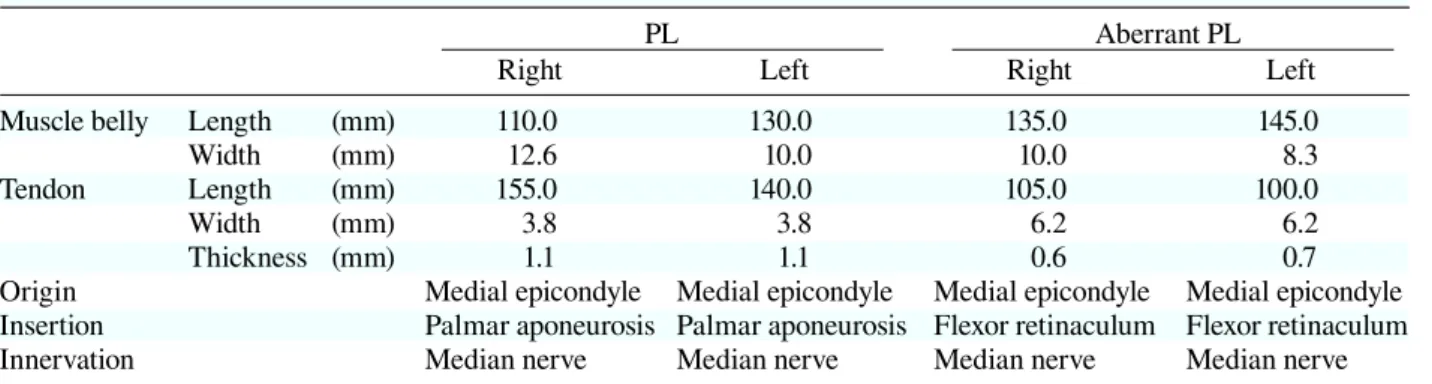
Table 1. Summary of PL and aberrant PL
PL Aberrant PL
Right Left Right Left
Muscle belly Length (mm) 110.0 130.0 135.0 145.0
Width (mm) 12.6 10.0 10.0 8.3
Tendon Length (mm) 155.0 140.0 105.0 100.0
Width (mm) 3.8 3.8 6.2 6.2
Thickness (mm) 1.1 1.1 0.6 0.7
Origin Medial epicondyle Medial epicondyle Medial epicondyle Medial epicondyle Insertion Palmar aponeurosis Palmar aponeurosis Flexor retinaculum Flexor retinaculum
Innervation Median nerve Median nerve Median nerve Median nerve
Fig. 3. Photograph of the anterior side of the right
fore-arm, showing the palmaris profundus muscle (PP with arrow). The flexor retinaculum is transected and turned over. The tendon of the flexor carpi radialis is cut for the demonstration of the distal tendon of the palmaris profun-dus muscle. FCR, flexor carpi radialis muscle; FCRt, ten-don of flexor carpi radialis muscle; FDS, flexor digitorum superficialis muscle; FR, flexor retinaculum; PP, palmaris profundus muscle.
the ulnar sector, and the PL from the intermediate sector. Straus (1942) pointed out that the aberrant PL is differentiated from the radial or ulnar sectors.
As for the PP, Takisawa et al. (1979) considered that the PP is derived from the radial sector.
From a viewpoint of phylogeny, the PL is absent in the lowest class of mammals such as Monotremata (Platypus) (Howell, 1936b). With the progress of the locomotion in hands, the palmar aponeurosis has appeared. Although the PL inserts to the aponeurosis in the human, the final insertion is different in animals: the proximal phalanx in the unau (Humphry, 1869), kinkajou and genet (Parsons, 1898); the flexor retinaculum in the tamandua (Windle and Parsons, 1899); both the palmar apo-neurosis and flexor retinaculum in the chimpanzee and cynocephalus (Champneys, 1871). Since the aberrant PL and PP insert into the flexor retinacu-lum, these muscles are considered to be a kind of atavism.
The PL and PP are of clinical importance from viewpoints of tendon grafts and carpal tunnel syndrome, respectively. The PL is frequently used as a donor tendon for ligament reconstruction in-cluding elbow, wrist and so on (Nestor et al., 1992; Ogunro, 2007; Whitaker et al., 2008. Although the duplicate PL is not so frequently occurred as mentioned above, it will provide a useful grafts in tendon surgery when found in patients. Since the distal tendon of the PP courses in the vicinity of the median nerve as seen in this study, it sometimes compresses the median nerve, causing a carpal tun-nel syndrome (Floyd et al., 1990; Server et al., 1995; Miyoshi et al., 2000). It is interesting that the PP reflecting the old phylogenetic pattern occasionally cause a carpal tunnel syndrome. It is also important to pay a special attention of the presence or absence of the PP in this syndrome.
References
1 Adachi B. Beiträge zur anatomie der Japaner. XII Die Statistik der Muskelvarietäten. Z Morphol Anthropol 1910;12:261–312.
2 Champneys F. The muscles and nerves of a chimpanzee (troglodytes niger) and a cynocephalus anubis. J Anat Physiol 1871;6:176–211.
3 Floyd T, Burger RS, Sciaroni CA. Bilateral palmaris profundus causing bilateral carpal tunnel syndrome. J Hand Surg Am 1990;15:364–366.
4 Gruber W. Über die varietäten des musculus an palma-ris longus. Mem I’Acad Imp Sci St Petersbourg 1868; S711:1–26.
5 Hojo T. A case of the duplicate palmaris longus muscle. Acta Anat Nippon 1976;51:422–425.
6 Howell AB. Phylogeny of the distal musculature of the pectoral appendage. J Morphol 1936a;60:287–315. 7 Howell AB. The musculature of antebrachium and
ma-nus in the platypus. Am J Anat 1936b;59:425–432. 8 Humphry. The myology of the limbs of the unau, the aï,
the two-toed anteater, and the pangolin. J Anat Physiol 1869;4:17–78.
9 Humphry. On the disposition of muscles in vertebrate animals. J Anat Physiol 1872;6:293–376.
10 Kawashima T, Kikushima S, Yokota E, Ohkubo F, Yamada Y, Sato F, et al. A case of an accessory pal-maris longus muscle and a duplicate palpal-maris longus muscle with special reference to their nerve supply— morphologic significance of a common innervation trunk. Okajimas Folia Anat Jpn 2002;79:75–81.
11 Kimura K, Konishi M, Takahashi Y. A case of the duplicate palmaris longus muscle. Boei Ikadaigakko Zasshi 1979;4:57–59 (in Japanese with English abstract). 12 Koganei Y, Arai H, Shikinami J. Varietätenstatistik der
musculn. Tokyo Igakkai Zasshi 1903;17:127–131 (in Japanese with German abstract).
13 Miyoshi S, Kanaya F, Futenma C, Ibaraki K. Carpal tunnel syndrome caused by palmaris profunda. Seikei-geka To SaigaiSeikei-geka 2000;49:877–879 (in Japanese with English abstract).
14 Nestor BJ, O’Driscoll SW, Morrey BF. Ligamentous re-construction for posterolateral rotatory instability of the elbow. J Bone Joint Surg Am 1992;74:1235–1241. 15 Ogunro O. Dynamic stabilization of chronic
scapholu-nate dissociation with palmaris longus transfer: a new technique. Tech Hand Up Extrem Surg 2007;11:241– 245.
16 Parsons FG. The muscles of mammals, with special relation to human myology. J Anat Physiol 1898;32:721– 752.
17 Reimann AF, Dasaler EH, Anson BJ, Beaton LE. The
palmaris longus muscle and tendon. A study of 1600 extremities. Anat Rec 1944;89:495–505.
18 Sato Y, Takeuchi R, Umeno K, Chen S, Takafuji T. A contribution to the study of the M palmaris longus. Acta Anat Nippon 1974;19:318–319.
19 Server F, Miralles RC, Galcerá DC. Carpal tunnel syn-drome caused by an anomalous palmaris profundus tendon. J Anat 1995;187:247–248.
20 Stark ME, Dell MM, Wisco JJ. A variation of the pal-maris profundus muscle. IJAV 2010;3:36–38.
21 Straus WL. The homologies of the forearm flexors: uro-deles, lizards, mammals. Am J Anat 1942;70:281–316. 22 Takafuji T, Azuma S, Tozawa T, Kawashima T,
Takeuchi R, Sato Y. A morphological study on the hu-man palmaris longus muscle. Kyorin Igakkai Zasshi 1985;16:341–353 (in Japanese with English abstract). 23 Takisawa A, Fujimaki N, Jinguji Y. [A rare case of m.
palmaris profundus (author’s transl)]. Kaibogaku Zasshi 1979;54:136–139 (in Japanese).
24 Whitaker I, Cairns S, Josty I. The palmaris longus tendon weave: a novel method of reconstructing the transverse carpal ligament. Plast Reconstr Surg 2008;122:227–228.
25 Windle BCA, Parsons FG. On the myology of the eden-tata. Proc Zool Soc Lond 1899;69:314–339.
26 Xue H, Yang C, Kawate T, Atsumi S. A case of the du-plicate palmaris longus muscle. Yamanashi Ikadaigaku Zasshi 1998;13:127–131 (in Japanese with English ab-stract).
27 Yoshida Y, Yasutaka S, Seki Y. Studies on the flexor carpi radialis profundus and palmaris profundus muscle in man. Acta Anat Nippon 1983;58:59–67 (in Japanese with English abstract).
28 Zeybek A, Gurunluoglu R, Cavdar S, Bayramicli M. A clinical reminder: a palmaris longus muscle variation. Ann Plast Surg 1998;41:224–225.
Received September 27, 2012; accepted October 26, 2012 Corresponding author: Yuichi Takanashi