Articles
A standardized protocol for
the mulberry cells and mulberry bodies in the urinary sediment of Fabry disease
Y
okoYamaTakashi
1, 2, H
oritaShigeru
1, C
Hu-S
uYu
3, m
anabeShun
4, k
ataokaHiroshi
4, 5, t
okuokaYoshikazu
*,
m
oCHizukiToshio
4, 5and n
ittaKosaku
4(Received Date: July 11, 2020)
I. Introduction
Fabry disease is one of the lysosomal storage diseases that was first reported by the German der- matologist Fabry1) and the British dermatologist Anderson2) in 1898. The incidence is about three thousandths. It is an X-linked inherited disease associated with genetic deficiency of α-galactosi- dase A (GLA), one of the lysosomal enzymes. Be- cause of the deficiency of this enzyme, hydrolysis of sphingolipids is impaired in the lysosomes, and glycolipids such as GL-3 and Gb-3 accumulate in various cells of the body, such as the vascular en- dothelial cells and cardiomyocytes. According to a study of the renal histopathological findings in childhood-pubertal Fabry disease, the swelling of the glomerular epithelial cells, the microvesicular degeneration of the endoplasmic reticulum, and a zebra pattern within the glomerular epithelial cells are already evident before the appearance of overt proteinuria. A deposit called a skin-like inclusion body, the zebra body, is also recognized3). There-
fore, the detection of mulberry cells and mulber- ry bodies in the urinary sediment is expected to play a role in the diagnosis of Fabry disease at the early stage. It has been reported that the detection of mulberry cells and mulberry bodies is effective for the early detection of Fabry disease because it is non-invasive and simple4–6). Currently, the mul- berry cells and mulberry bodies are not an essen- tially parthological evidence in urine sediment for Fabry disease. Therefore, there is no standardized procedures for the detection of mulberry cells and mulberry bodies in the urine yet.
We encountered two cases of Fabry disease, a mother and her second son, in whom mulber- ry cells and mulberry bodies were detected in the urine. In order to enhance the detection efficiency and the clinical significance of mulberry cells and mulberry bodies in urine, the urine collection, the magnification of microscope, the differentiation for the mulberry cells and mulberry bodies from other fats and fat-containing cells and fatty parti- cles, composition and the origin, and the morpho- logical changes by enzyme replacement therapy
* tokuoka Yoshikazu: Professor, Graduate School of Engineering; Faculty of Biomedical Engineering, Toin University of Yokohama, 1614, Kurogane-cho, Aoba-ku, Yokohama 225–8503, Japan
1 Department of Clinical Laboratory Medicine, Tokyo Women’s Medical University Hospital
2 Graduate School of Engineering; Faculty of Biomedical Engineering, Toin University of Yokohama
3 Department of Laboratory Medicine, National Taiwan University Hospital
4 Department of Nephrology, Tokyo Women’s Medical University
5 Clinical Research Division for Polycystic Kidney Disease, Department of Nephrology
(ERT) were examined.
II. Case
At the age of 37 and 9 years old, the second son had terminal pain in the extremities at the time of fever, and at the age of 12 years, a mother who was a carrier was admitted to the Department of Pediatrics with suspected Fabry disease. A de- crease in the GLA activity was confirmed in the patients, confirming the diagnosis of Fabry dis- ease. At the age of 23, the patient became aware of dizziness and developed seizures, and at the age of 25, although his other renal function parameters were maintained, a urinary test showed proteinuria (2+). Renal biopsy was performed to confirm the degree of renal dysfunction associated with Fabry disease. Since the proteinuria persisted, the pa- tient was started on enzyme replacement therapy with agalsidase beta (Fabrazyme®) at the age of 27 years.
The mother of the male patient had been di- agnosed as having hypertension since she was 40 years old. She had been treated by the department of cardiovascular medicine for a long period, because she had also been diagnosed as having hypertrophy. The urinary sediment showed mul- berry bodies. Electrocardiography showed the ST depression in leads V5 and V6, Echocardiography showed left ventricular hypertrophy (left ven- tricular mass index 186 g/m2), and cardiac MRI showed fat deposition in the myocardium.
Fabry disease was suspected, and a renal bi- opsy was performed to confirm the diagnosis.
Twenty-four glomeruli were collected and 5 were completely indurated. Podocyte swelling was ob- served and the cytoplasm was scattered. Based on the findings she was diagnosed with of Fabry disease and started to be treated with ERT (Fab- razyme®).
III. Materials and Methods
1. Urine collection, specimen preparation and microscopy
For the observation of the urinary sediment, the early morning urine or random urine is accept- able. However, it is recommended to collect the mid-stream of random urine for the convenience to patients. Therefore, the mid-steam random urine was applied. The urine specimens were cen- trifuged at 500×g for 5 minutes. The supernatant was removed, and the pellet was resuspended in a total volume of 200 μL. The suspension was well mixed and 15 μL was loaded on to a slide glass covered by a piece of 18×18 coverslip for the bright field microscopic observation. After wash- ing with normal saline and discarding the superna- tant, the pellet was similarly resuspended stirred and 15 μL was loaded on to a slide glass covered by a piece of 18×18 coverslip for the bright field microscopic observation.
2. Confirmation of the typical morphology of mulberry cells and mulberry bodies
The characteristic of mulberry body excret- ed in the urine was a spiral body. Epithelial cells field with mulberry bodies are called mulberry cells.
3. Confirmation of the origin of mulberry cells and mulberry bodies
After preparation of the urinary sediment, a non-fixed sediment suspension was prepared and processed by Sudan III staining and observed under a polarizing microscope to confirm the na- ture of the spiral structures. In order to confirm the nature of the components exhibiting a spiral structure and the cells filled with them, 100 μL of non-fixed sediment suspension were smeared on to a slide glass coated with silane after cytospin.
We conducted immunohistochemistry (IHC) anal-
ysis using anti podocalyxin goat IgG monoclonal antibody and anti Gb-3 mouse IgG monoclonal antibody (COSMO Bio Co.) as the primary anti- bodies. Alexa Fluor 594TM Donkey anti goat IgG and Alexa Fluor 488TM Donkey anti mouse IgG (Invitrogen TM) were diluted to 500-fold with buffer (1% bovine albumin 0.1% sodium azide phosphate buffer) to be used as the secondary an- tibodies. In order to recognize nuclei, DAPI (In- vitrogen TM) was used after 1000-fold dilution with buffer (1% bovine albumin and 0.1% sodium azide phosphate buffer).
4. The evaluation method for mulberry cells and mulberry bodies after ERT
We observed the urinary excretion of mulberry cells and mulberry bodies after ERT. The evalua- tion method used so far was a qualitative evalua- tion to determine the structures were excreted in the urinary sediment or not (positive or negative).
The existing evaluation method was sufficient as a screening test for Fabry disease. But in this study, we carried out monitoring, focusing on the effect of ERT, and determined the number of cells/bod- ies per low power field (LPF). Daily excretion by calculating the non-centrifugal urinary equivalent (μL) of the whole field (WF) from the equivalent
(μL) other than the centrifuge and dividing by the urinary creatinine value (mg/dL). The counting method for mulberry cells and mulberry bodies is shown in Figure 1.
IV. Results
1. Urine collection, specimen preparation, and microscopy
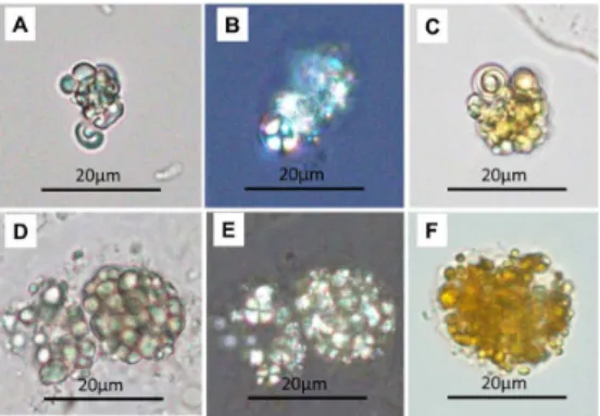
We compared the appearance of mulberry cells and mulberry bodies in non-centrifugated urine specimens and the urine specimens processed by washing with normal saline, as needed. After washing with normal saline, no difference was ob- served in the appearance and the detection rate in the urine. Regarding the scales of magnification used, it was difficult to confirm the structures at a magnification of 100X. The magnification of 200X was sufficient to confirm the magnification of 400X allowed the structures to be visualized in detail (Figure 2).
Figure 1, a typically aggregated mulberry bod- ies and a typical mulberry cell with several mulberry bodies (unstained, 400X). A, there are six mulberry bodies which aggregate. The margin of mulberry body is obviously distinct.
B, there is a mulberry cell with several mul- berry bodies. When an epithelial cell contains mulberry bodies within the cytoplasm, the epithelial cell will be defined as a mulberry cell. The cytoplasm of epithelial cell is blur but the mulberry bodies are dis-tinct, which is a mulberry cell.
Figure 2, two clusters of unstained aggregated mul- berry bodies. Figures A, B, and C, the same and unstained mulberry bodies in different magnification (100X, 200X, and 400X), which aggregate in a cluster. Figures D, E, and F, the same and unstained mulberry bodies in differ- ent magnification, which aggregate in a cluster.
The unstained mulberry bodies are washed by the normal saline. The characteristics of mulberry body are not significantly changed and the margin of mulberry body still distinct with high refraction.
2. Confirmation of the typical morphology of mulberry cells and mulberry bodies
The mulberry cell meant a cell filled with mul- berry bodies with were with typical spiral struc- tures. The mulberry body also had a clear spiral structure (Figure 3).
3. Confirmation of the origin of the mulberry cells and mulberry bodies
The component exhibiting a spiral structure showed weak pale yellow-orange staining by Su- dan III and the Maltese cross birefringence was confirmed under a polarizing microscope. The oval fat body showed a strongly positive pale yellow-orange staining by Sudan III staining and Maltese cross birefringence under a polariz- ing microscope (Figure 4). Glomerular epithelial cells exhibiting vacuolar degeneration in the renal biopsy tissue showed a positive fluorescent anti- body staining for Gb-3. In the smear of the urine sediment, components exhibiting a spiral struc- ture. The spherical cells which were larger than a leukocyte were with a positive staining for anti
podocalyxin goat IgG monoclonal antibody, anti Gb-3 mouse IgG monoclonal antibody, and DAPI.
These cells that were presumed to be derived Figure 3, the unstained mulberry cells and mulberry
bodies from the second son and the mother. Fig- ure A, a typical mulberry cell from the second son. Figure B, a typical mulberry body from the second son. Figure C, a typical mulberry cell from the mother. Figure D, a typical mulberry body from the mother.
Figure 4, the differences between a mulberry cell and an oval fat body. Figures A, B, and C are mulberry cells. Figures D, E, and F are oval fat body. Figure B is the polarized image of figure A, the polarized mulberry bodies with refractive Maltese crosses. Figure E is the polarized image of figure D, the polarized fat droplets of the oval fat body with refractive Maltese crosses.
Figure C (Sudan III stain), a stained mulberry cell. The spiral structure within a mulberry body is obvious. Figure F (Sudan III stain), a stained oval fat body. The stained fat droplets like a swollen orange ball.
Figure 5, the IHC images of the mulberry cells from the urinary sediment smear. The red color is podocalyxin, the green the Gb3, and the blue the nuclei. All of the images are with posi- tive reactions for DAPI (blue). Figure A, the strong positive reaction of podocalyxin and the weak positive reaction of Gb3. Figure B, the strong positive reaction of podocalyxin and the positive reaction of Gb3. Figure C, the weak positive reaction of podocalyxin and the strong positive re-action of Gb3. Figure D, E and F the negative reaction of podocalyxin and the positive reaction of Gb3.
from the glomerular epithelial cells that were positive for anti podocalyxin goat IgG monoclo- nal antibody-negative, anti Gb-3 mouse IgG anti- body-positive, and DAPI-positive (Figure 5).
4. The evaluation method for the excreted mul- berry cells and mulberry bodies after ERT
In the son with classic Fabry disease, 5.1×10⁴ mulberry cells/gCr and 1.31×10⁵ mulberry bod- ies/gCr were excreted in the urine per day during ERT. In the mother, with hemizygous Fabry dis- ease, 3.44×10⁴ mulberry cells/gCr and 3.53×10⁵ mulberry bodies/gCr were excreted in the urine per day before ERT. The excreted mulberry cells and mulberry bodies were 11.73×10⁴ cells/gCr and 15.71×10⁵ bodies/gCr per day during the ERT. Af- ter ERT for 7 months, the excreted mulberry cells and mulberry bodies were 7.69×10⁴ cells/gCr and 6.87×10⁵ bodies/gCr per day.
V. Discussion
In order to expect the effect of suppressing the progression of various organ disorders in Fab- ry disease, sufficient initiation of ERT should be considered at an early stage, and a clinical study that can explain the appropriate initiation timing of ERT and the criteria by which the effect of initi- ation. As one of the indicators, mulberry cells and mulberry bodies in urine sediment are considered to play an important role.
In this study, we demonstrated a noninvasive- ly standardized protocol to identify and quantita- tively evaluate the presence of urinary mulberry cells or mulberry bodies of Fabry disease. There- fore, we proposed the standardized protocol for the mulberry cells and mulberry bodies in urinary sediments as an index for the early diagnosis. This standardized protocol could also be applied for the evaluation of ERT.
Most of the urine specimens for the routine
tests are the random urine from the mid-stream.
The appearance of mulberry cells and mulberry bodies in random urine specimens was compared between unprocessed urine samples and urine samples washed with physiological saline, and the results revealed no differences in the appearance or detection rate. Since washing with physiological saline reduces mulberry cells and mulberry bod- ies and may cause underestimation, urine samples used for testing may be urine at any time, including urine samples used for other tests. it was thought.
In regard to the degree of magnification needed for microscopic examination, it is described in Aims of the Guidelines on Urinary Sediment Examina- tion Procedures Proposed by the Japanese Com- mittee for Clinical Laboratory Standards (JCCLS)
7) (Special Issue) that urinary sediments should be examined at 400 magnification. However, in order to detect Fabry disease efficiently and early, it is necessary to screen mulberry cells and mulberry bodies at 200 magnifications and count mulberry cells and mulberry bodies in all fields at 400 mag- nifications and report the emission amount. We thought that it was desirable.
Regarding the morphology of mulberry cells and mulberry bodies, a typical spiral structure was observed before ERT.
To confirm the origin of the spiral structures and identify mulberry cells and mulberry bodies, we applied Sudan III staining and polarization microscopy. The mulberry cells and mulberry bodies showed pale yellow-orange staining with Sudan III and Maltese cross birefringence by a polarizing microscope. The origin of the mulberry body found in urine was thought to be fat glob- ules containing cholesterol esters and phospho- lipids. Furthermore, since they showed positive immunohistochemical staining with anti-Gb-3 antibody, they were considered as being derived from fat globules in which Gb-3 containing cho- lesterol ester and phospholipid were accumulated.
The oval fat body, which were morphologically
similar components, showed strongly positive pale yellow-orange staining with Sudan III, and although there was a difference in the intensity of staining as compared to that of mulberry bodies, differentiation between the two may not be reli- able by this method. This component also showed Maltese cross birefringence under polarized light microscopy. Becker et al.8) reported that screen- ing for Maltese cross birefringence in lipid par- ticles under a polarizing microscope is effective for the detection of Fabry disease. However, the oval fat bodies excreted in nephrotic syndrome and fat-containing cells excreted in other diseases may also show the Maltese cross birefringence un- der a polarizing microscope, so that it may not be as a usefully useful a screening method for Fabry disease.
There are few reports on the origin of mulber- ry cells excreted in the urine. Using anti-CD77 antibody, it was reported that epithelial cells with accumulated GL3 are exfoliated into the urine9). However, the origin of the GL3-accumulated ep- ithelial cells has not been proven. Therefore, IHC was performed to determine whether glomerular epithelial cells showing accumulation of Gb-3 were excreted in the urine or not. The cells that have a spiral structure and the cells that are larger than the leukocytes that contain them and are larg- er than the leukocytes were anti-podocalyxin an- tibody-positive, anti-Gb-3 antibody-positive, and DAPI-positive are presumed to be derived from the glomerular epithelial cells. Cells that were anti-podocalyxin antibody-negative, anti-Gb-3 antibody-positive, and DAPI-positive were also observed, and were considered to be derived from renal tubular epithelial cells, because the cells were spherical and the marginal structure of the cells was serrated. Until now, the number of uri- nary podocytes in healthy subjects is reported to be
<0.5/mgCr, whereas that in patients with glomer- ular disease is higher than 10/mgCr10). The reason why glomerular epithelial cells can be easily be
detected in the urine of patients with Fabry disease is that the glomerular epithelial cells are impaired by the accumulation of Gb-3 and are easily exfo- liated into the urine. Trmarchi et al.11) reported the presence of glomerular epithelial cells among ex- foliated cells in the urine using podocalyxin and synaptopodin in patients with Fabry disease. Fur- thermore, it is reported that podocalyxin may be lost in patients with Fabry disease. Therefore, it is highly possible that the origin of mulberry cells is glomerular epithelial cells, and it is important to confirm this origin using podocalyxin and synap- topodin. For monitoring Gb-3 accumulation after ERT, LysoGb3 is used as an indicator12). However, since it is expensive and results take a long time, a test that is rapid and cheap and can be performed anywhere is desired. We believe that a test that can meet this expectation is the detection of mulberry cells and mulberry bodies in the urinary sediment.
Urinary sediment examinations for mulberry cells and bodies have so far been qualitative, that is, positive or negative, and detailed evaluation of the effects of ERT was not possible with such evalua- tion. However, our method is useful, in that it en- ables quantitative evaluation of the daily excretion of mulberry cells and mulberry bodies to grasp the accumulation state of Gb-3 in the renal tissue and also evaluate the effect of ERT. Comparison of the daily excretion of mulberry cells and mul- berry bodies between urinary protein-negative and urinary protein-positive patients after ERT in patients with Fabry disease showed that the uri- nary protein-negative patients showed disappear- ance of mulberry cell excretion and decrease of mulberry body excretion, with maintenance of the renal function. On the other hand, in the urinary protein-positive patients, while the amount of mulberry bodies excreted decreased, the excretion of mulberry cells persisted unabated, and the renal function declined, often necessitating dialysis13). These results suggest that quantitative evaluation of the daily excretion of mulberry cells and mul-
berry bodies in the urine may allow evaluation of the effect of ERT and of the renal prognosis in pa- tients with Fabry disease.
It has been reported that before the start of ERT, the higher the urinary protein excretion, the faster the deterioration of renal function14), and the higher the urinary protein excretion, the lower the inhibitory effect of ERT on the renal function de- terioration15). Therefore, it is considered important to monitor the morphology of the mulberry cells and mulberry bodies based on standard testing methods and to quantitatively monitor their daily excretion before proteinuria becomes apparent.
VI. Conclusions
The well distinguishing for the morpholog- ical characteristics about the mulberry cells and mulberry bodies from the cells with fat droplets are important with a bright field microscope. The magnification of 200X is useful for the screening of the mulberry cells and mulberry bodies. The magnification of 400X for the whole field identifi- cation (differential counting for the mulberry cells and mulberry bodies) is necessary. A quantitative count of the mulberry cells and mulberry bodies is a valuable test for the evaluation of ERT.
[References]
1) Fabry J. Ein Beitrag Zur Kenntnis der Purpura haem- orrhagica nodulatis (Purpura papulosa hemorrhagica Hebrae). Arch Dermatol Syphilis 1898; 43: 187–200.
2) Anderson W. A case of “angiokeratoma”. Br J Der- matol 1898; 10: 113–7.
3) Tondel C, Bostad L, Hirth A, et al.: Renal biopsy findings in children and adolescents with Fabry dis- ease and minimal albuminuria. Am J Kidney Dis 51:
767–776, 2008.
4) Nakamichi T, Miyazaki M, Nakayama K, et al.:
Fabry’s disease discovered with chance urinary mul-
berry cells: a case report. CEN Case Rep. 2013; 2:
49–52.
5) Honda T, Komathu E, Furuse S, Mise N. Fabry dis- ease diagnosed based on the detection of urinary mulberry bodies. Int Med. 2016; 55: 2903.
6) Shimohata H, Ogawa Y, Maruyama H, Hirayama K, Kobayashi M. A case of renal variant of Fabry dis- ease diagnosed by the presence of urinary mulberry cells. Int Med. 2016; 55: 3475–8.
7) Japanese Association of Medical Technologists.
Aims of the Guidelines on Urinary Sediment Exam- ination Procedures Proposed by the Japanese Com- mittee for Clinical Laboratory Standards (JCCLS).
Japanese Journal of Medical Technology. 66: No.
J-STAGE-1 Special Issue: Urinary Sediment 2017:
13, 2017.
8) Gavin J. Becker, Kathleen Nicholls: Lipiduria with special relevance to Fabry disease. Clin Chem Lab Med 2015; 53 (Suppl): S1465–S1470.
9) Mathu Selvarajah, Kathy Nicholls, Tim D. Hewit son, et al. Targeted urine microscopy in Ander- son-Fabry Disease: a cheap, sensitive and specific di- agnostic technique. Nephrol Dial Transplant (2011) 26: 3195–3202.
10) Vogelmann SU et al.: Am J Physiol Renal Physiol 2003; 285: F40–8.
11) Hernan Trimarchi, Romina Canzonieri, Cristian Costales-Collaguazo, et al.: Early decrease in the podocalyxin to synaptopodin ratio in urinary podo- cytes. Clinical Kidney Journal, 2019, vol. 12, no. 1, 53–60.
12) Bouwien E, Saskia M Rombach, Johannes MFG Aerts, et al.: Consequences of a global enzyme short age of agalsidase beta in adult Dutch Fabry patients.
Orphanet Journal of Rare Diseases 2011,6: 69.
13) Yumi Aoyama, Yusuke Ushio, Takashi Yokoyama, et al. Urinary Mulberry Cells as a Biomarker of the Efficacy of Enzyme Replacement Therapy for Fabry Disease: Internal Medicine.
14) Warnock DG, Daina E, Remuzzi G, et al.: Enzyme replacement therapy Fabry nephropathy. Clin J Am Soc Nephrol 5: 371–378, 2010.
15) Germain DP, Waldek S, Banikazemi M, et al.: Sus- tained, long-term renal stabilization after 54 months of agalsidase β therapy in patients with Fabry dis- ease. J Am Soc Nephrol 18: 1547–1557, 2007.