九州大学学術情報リポジトリ
Kyushu University Institutional Repository
多毛類イシイソゴカイの雄性生殖細胞の発達過程及 び雌雄における配偶子放出機構の研究
マリア, ジャヌアリ, ピーター
http://hdl.handle.net/2324/4110548
出版情報:Kyushu University, 2020, 博士(農学), 課程博士 バージョン:
権利関係:
Germ cell development in male Perinereis wilsoni and gamete spawning mechanisms in males and females
Dissertation submitted in accordance with the requirement of Kyushu University for the Degree of Doctor of Philosophy
Maria January Peter
Fisheries Research Laboratory
Graduate School of Bioresource and Bioenvironmental Sciences Faculty of Agriculture
Kyushu University
2020
TABLE OF CONTENTS
LIST OF ABBREVIATIONS LIST OF FIGURES
LIST OF PUBLICATIONS
Page No.
CHAPTER 1
General introduction on polychaetes 1
1.1 Taxonomy and distribution 2
1.2 Morphology, habitat and feeding 2
1.3 Reproduction 3
1.3.1 Gametogenesis 4
1.3.2 Endocrine Control 5 1.3.3 Environmental control 5
1.4 Embryonic development and segmentation 6 1.5 Importance of polychaetes 7 1.6 Experimental animal (Perinereis wilsoni) classification 8
1.7 Figures 9
CHAPTER 2 Germ cells development in male Perinereis wilsoni 10
2.1 Abstract 11
2.2 Introduction 12
2.3 Materials and Methods 14
2.3.1 Animals 14
2.3.2 cDNA cloning of Perinereis wilsoni-piwi gene 14
2.3.3 In situ hybridization 14
2.3.4 Histology 15
2.4 Results 16
2.4.1 Germ cell localization in male P. wilsoni 16
2.4.2 Sexual development of gametes in P. wilsoni 17
2.4.3 Figures and Table 19
2.5 Discussions 33
2.5.1 Germ cells localization and gametes production in P. wilsoni 33
2.6 Conclusion 36
CHAPTER 3 Mechanisms of gamete releasing in males and females P. wilsoni 37
3.1 Abstract 38
3.2 Introduction 39
3.3 Materials and Methods 41
3.3.1 Observation of spawning sites 41
3.3.2 Histology 41
3.4 Results 42
3.4.1 Spawning sites in P. wilsoni 42
3.4.2 Gamete spawning sites in male P. wilsoni 42
3.4.3 Gamete spawning sites in female P. wilsoni 42
3.4.4 Figures 44
3.5 Discussions 51
3.5.1 Gamete spawning sites in P. wilsoni 51
3.5.2 The eosinophilic structure associated with gamete during spawning 52
3.6 Conclusion 53
CHAPTER 4 General discussion 55
4.1 Germ cells localization in male and gamete production P. wilsoni 55
4.2 Gametes releasing sites in males and females P. wilsoni 58
ACKNOWLEDGEMENTS 62
REFERENCES 64
LIST OF ABBRIVIATIONS NBT Nitro Blue Tetrazodium
BCIP 5-Bromo-4-Chloro-3-Indoly Phosphate
PBS Phosphate Buffered Saline
Pl. dumerilii Platynereis dumerilii P. wilsoni Perinereis wilsoni Pw-vasa Perinereis wilsoni -vasa Pw-piwi Perinereis wilsoni - piwi
SAZ Segment Addition Zone
LIST OF FIGURES CHAPTER 1
Figure. 1. Experimental animal, Perinereis wilsoni
Figure. 2. Polychaete (Perinereis wilsoni) aquaculture in Oita prefecture, Japan
CHAPTER 2
Figure. 1. Whole mount in situ hybridization with the Pw-piwi probe in an adult male worm in late August.
Figure. 2. Horizontal sections of the hybridized worm showing the localization of the Pw- piwi expression in an adult male in late August.
Figure. 3. Specificity of the Piwi probe in in situ hybridization.
Figure. 4. Germ cell clusters in a juvenile worm.
Figure. 5. Germ cells before and after the start of vitellogenesis.
Figure. 6. Germ cells fill body cavities of each segment in March (mid-body region).
Figure. 7. Monthly development of male germ cells.
Figure. 8. Whole mount in situ hybridization with Pw-piwi in early March.
Figure. 9. Whole mount in situ hybridization of Pw-piwi in end of March.
Figure. 10. Whole mount in situ hybridization of Pw-piwi in April; swimming epitoke.
Figure. 11. Pn-piwi expression in mothnly development of male germ cells.
Figure. 12. Schematic diagram on the gametogenic cycle of P. wilsoni.
Figure. 13. Schematic diagram on the timing of the germ cells migration from the parapodia to the deeper body cavity during segments addition and the start of the vitellogenesis.
CHAPTER 3
Figure. 1. Observation of spawning sites in P. wilsoni. (b) Male (c) female
Figure. 2. Horizontal sections of swimming male worm just prior to spawning in April.
Figure. 3. Sperm spawning area in a tail region of a male.
Figure. 4. Histological observation of sperm releasing sites in P. wilsoni.
Figure. 5. Horizontal sections of swimming male worm just prior to spawning in April.
Figure. 6. Egg spawning area in female.
Figure. 7. The eosinophilic structure associated with gamete release indicated by arrows.
(A)
Spawning male. (B) Spawning female. H&E stain.
LIST OF PUBLICATIONS Peer-reviewed scientific paper:
Maria January Peter, Mercedes Maceren-Pates, Gaudioso Pates Jr, Michiyasu Yoshikuni Yoshihisa Kurita (2020). Germ cell development in male Perinereis wilsoni and gamete spawning mechanisms in males and females. Zoological Science
Scientific conference abstract presentation
Maria January Peter and Maceren-Pates Mercedes: A study of germ cells development in Perinereis wilsoni 90th International conference. Biodiversity, Ecology and Evolution (2019), Amakusa Marine Biology Laboratory, Kyushu University, Japan.
1
CHAPTER 1
GENERAL INTRODUCTION
2 1.1 Taxonomy and distribution of Polychaetes
Annelida is the phylum of the segmented worms. It is one of the most diverse phyla in the animal kingdom. The phylum comprises more than 20000 species which widely distributed in ocean water, fresh water and in moist terrestrial environment (Fauchald and Rouse, 1997;
Kriska, 2013). Over centuries, annelids had been classified into three classes; Polychaeta (marine worms), Oligochaeta (Earthworms) and Hirudinea (Leeches). Among these, polychaeta is the largest group in the phylum, mostly comprised by marine worms and few (about 2%) are fresh water species (Glasby and Timm, 2008; Shoobs et al., 2016). There are about 11,456 species of polychaetes which are distributed in a wide range of habitats;
wetlands, sandflats, intertidal zones and deep seas (Kriska, 2013; Pamungkas et al., 2019).
Based on the development of the anterior appendages and life habits, polychaetes had been classified into two major groups, Errantia and Sedentaria. Errantia includes the active scavengers, the free living and crawler worms of the family Nereididae and Phyllodocida.
Sedentaria are permanent burrowers or tube dwellers, for example, the Capitella. (Hartman, 1968, 1969; Kriska, 2013).
1.2 Polychaetes morphology, habitat and feeding
A body of polychaete is divided into three parts of head, trunk and tail region. The head part includes a prostomium which bears palps, eyes, tentacles and antenna and a peristomium where the mouth is located. The body length ranges from 1mm to 3m with the average size of 5cm to 10cm (Kriska, 2013). Polychaetes occupy a wide range of habitats; burrowing in sediments or sands, living under rocks or corals and some are tube-dwellers which includes some members of the family Oweniidae (Dauvin and Thiẻbaut, 1994). The diversity in life style of polychaetes is greatly influenced by morphological variations and feeding habits as well. There are active hunters such as Phyllodocidae, Syllidae and Nereidae, some are
3
deposit feeders such as Capitellids and Arenicolids while others are filter feeders like Lumbrids, Serpulids and Sabellids (Fauchald, 1977).
1.3 Polychaetes reproduction
Majority of the polychaetes are gonochoric and only few are hermaphrodites. The reproduction mode varies among species depending on their life cycle. In dioecious reproduction, gametes mature in separate sexes. Spawning can be broadcasting with external fertilization as in nereidids (Fauchald, 1977) or viviparity development shown by Syllidae (Franke, 1999). Asexual reproduction involves splitting of the adult worm into two or more individuals or by budding off the new individual from the parent. In most case, budding occurs at the posterior region (reproductive region) and the parent organism will regenerate and survive to reproduce another time. Examples are Pristina leidyi, syllids and oweniids (Oliver, 1984; Franke, 1999; Özpolat and Bely, 2015).
Nereidids are errant polychaetes with unique reproductive strategy called epitoky. Epitoky is a form of reproduction in which sexually immature worm (atoke) transform into sexually mature worm (epitoke), and epitokes emerge from benthic to short pelagic life in spawning season (Fong, 1993; Bartels et al., 1990; Franke, 1999; Hébert-Chatelain et al., 2008). There are two forms of epitoky, Epigamy and Schizogamy. Epigamy is a sexual reproduction when the whole body transforms into the epitoke. Schizogamy is another form of reproduction in which only part of the body will transform into sexual form and detach from asexual part by fission (Pamungkas and Glasby, 2015). In breeding season, ripe animals release gametes after a swimming dance (nuptial) and die shortly within hours (Fong, 1993; Bartels et al., 1990; Hébert-Chatelain et al., 2008). However, some species had evolved into iteroparous and can survive more than one spawning season in a life time such as Eunice fucata (Fischer, 1999). Sexes of mature individuals can easy be differentiated due to differences in gamete
4
colours; for examples in P. wilsoni male has whitish or cream colour and female look greenish, in P. dumerilii , male has cream/whitish colour and female has yellow (Rebscher et al., 2007) and in Nereis fucata lilac or blue (Brown- Gilpin, 1959).
Transformation into pelagic life is energy consuming process which involves tremendous changes in body system so as to adapt the swimming life. Those are addition sets of setae (brisltes), enlarged parapodia, histolysis of the body wall, reorganisation of the body musculature, growth of vascular system and respiratory surface, which are the morphological and physiological changes reported in the previous studies (Fischer, 1999; Hébert-Chatelain et al., 2008).
1.3.1 Gametogenesis
In many polychaetes, the developing germ cells are usually observed floating in the coelomic cavity. The location of the gonads varies among species, and in some species it is not yet identified (Rice and Eckelbarger, 1989; Gambi, 2000). In P. dumerilii, the gonad settles at the neck region where germ cells proliferate and move to fill other body segments (Rebscher, 2007). In Nicole zostericola, the gonad is located beneath the peritoneum in the thoracic region of the body (Eckelbarger, 1975). In P. wilsoni, primordial germ cells settles at the distal end of each parapodium to proliferate before migration to the deeper body cavity (Maceren-Pates et al., 2015).
Vitellogenesis in polychaetes develops intraovarian or extraovarian. In intraovarian development, female germ cells develop and mature inside the ovary and are released after maturation. In exraovarian vitellogenesis, female germ cells are released singly from the ovary to develop in the coelomic cavity, or released as the cluster to develop and then separated as an individual oocyte after development (Eckelbarger, 1983). The start of vitellogenesis can be used as one of the criteria for sex determination due to enlargement of germ cells as the result of vitellogenin accumulation (Fischer, 1974; Gambi, 2000).
5 1.3.2 Endocrine control
The timing of gametogenic cycle of numerous polychaetes is controlled by and endocrine system in coordination with the environmental cues. The process of the germ cells development itself is said to be solely regulated by the endocrine and neural systems responding to external stimulus (Golding, 1967; Bentley, 1992; Lawrence, 1996) reported that a juvenile hormone secreted by a supraphrageal ganglion induced accumulation of oogonia in the coelomic cavity but inhibit the sexual maturation. In Nereis divoscolor, cerebral endocrine activities were observed to be high in most time of the gametogenic cycle and declined as the life cycle terminates (Dilding, 1983). A similar endocrine trend was observed during oocytes development in Nephtys hombergii (Olive and Bentley, 1980).
Fischer and Hausenschild (1974) suggested that a brain hormone might be responsible in controlling the extent of proliferation of oocytes and the inhibin regulated the homogenous development of the oocytes in late stages of maturation in in P. dumerilii. Lawrence (1996) found that gonadotropin promoted egg synthesis in Harmothoe imbricate.
1.3.3 Environmental factors
Gametogenic cycle of numerous polychaetes is highly synchronized with the environmental cues such as the start of their breeding season corresponds with the rise in sea temperature. Studies on temperate species show that the start of gamete development has been synchronised with the low temperature and short photoperiod in the winter season and the gamete reaches the maturity size in the next spring (Olive, 1978; Garwood, 1980;
Bartels-Hardege and Zeeck, 1990; William, 1997; Clark 1998). Rise in sea temperature and increase in day length have been reported to correspond with nutrients productivity and increase in metabolic activities which in turn favour the growth and development of the newly hatched larvae and growing juveniles (Newell et al., 1982; Bartels-Hardege and Zeeck, 1990; William, 1997; Lawrence and Saome, 2009). Spawning events have been also found
6
to relate with certain phases of the lunar cycle. For instance, P. dumerilii and Nereis succinea spawning events happen 18 days after new moon (Rasmussen, 1973).
1.4 Embryonic development and segmentation
In Nereidids, gametes fertilize in the external environment after broadcast spawning.
Within 1 hour post fertilization (hpf), the fertilized eggs covered by the jelly-coat for protection settle to the substratum prior to start the cleavage. The embryos undergo spiral cleavage forming a ball of mass cells. The embryos hatch within 7-8 days post fertilization (Hardege and Bartels-Hardege,1996).
The larvae undergo three stages; trochophore, metatrochophore and nectochate.
Trochophore is the free swimming and non-feeding larvae formed 2-3 days post fertilization.
Metatrochophore at 4-6 dpf starts the parapodium protrusion accompanied with the formation of chaetae, but does not actively move. In this stage, the larvae accomplished to form the adult eyes. Nectochaete (7 dpf) has a three fully-segmented body with well- developed parapodia, ciliary bands and chaetae. Nectochaete larva do not feed and have a mixed pelagic and benthic life style (Fischer et al., 2010).
Segments are added from the pygidium, accordingly, anterior segments are older than posterior segments. In errant polychaetes, the segment addition is active in a late larvae and juvenile stage, and slows down as the animal reaches the sexual maturity stage. The rate of segment addition varies among species. However, the growth and enlargement continues as the germ cells develop (Clack and Clack, 1962; Reish, 1993). The process of segment addition was reported to be controlled by growth-promoting hormones secreted by a supraesophageal ganglion (Clack, 1963). Each segment bears one pair of parapodia on each side of the body which can be used as means for locomotion, sensation, respiration and excretion (Giangrande and Gambi, 1998). In some species, parapodium can also act as a
7
temporal gonad as recent described in P. wilsoni (Maceren et al., 2015). Segments are separated each other by the complete intersegmental septum (Maceren et al., 2015) or the incomplete septum (Fischer, 2004).
1.5 Importance of Polychaetes
Polychaetes play the important roles in the ecosystem as food source for shrimps, fishes and shore birds. Some species are commonly used as live baits by anglers in fishing (Lawrence at al., 1998). Currently, polychaetes play great contributions in the aquaculture industry of crustaceans and fishes because polychaetes are rich in nutrients, especially the polysaturated fatty acids. Several reports have identified that the feeding polychaetes enhances the gonad maturation in shrimps (Olive, 1999; Techaprempreecha et al., 2011;
Kumar et al., 2017; Bischoff, 2014;). Aside nutrients provision, polychaetes have been reported to be useful in the treatment of the wastewater in the mariculture system and bioremediation Giangrande et al., 2005; Palmer, 2010; Stabili et al., 2010; Ito et al., 2011).
A case study conducted on Nereis diversicolor reported that polychaetes enhanced the growth of microorganisms (bacteria) which in return increased decomposition of organic sediments in fish farms (Jadao et al., 2008). Moreover, Lucciano et al., (2007, 2010) described that the ability of filter feeder polychaetes (Branchiomma luctuosum and Sabella spallanzanii) to accumulate bacteria is useful as bioremediator as well as the bioindicator of the microbe pollution in water. In regard with this importance, several studies have suggested that polyachetes can be useful in the intergrated aquaculture, nutrient recycling and also in the removal of wastes (Jadao et al., 2008). Nevertheless, Hutchings (1998) reported that the burrowing and feeding activities of polychaetes enhance sedimentation and nutrient recycling in the ecosystem. Additionally, benthic dweller and scavenger polychaetes such as
8
Capitella sp. provide suitable specimens for the biomonitoring and marine toxicological studies (Bryan and Gibbs, 1987; Nugteren et al., 2009; Aller R, 2001).
1.6 Experimental animal (Perinereis wilsoni) classification and distribution Kingdom : Animalia
Phylum : Annelida Class : Polychaeta Order : Phyllodocida Family : Nereididae Genus : Perinereis Species : P. wilsoni
Perinereis wilsoni (P. wilsoni) is the nereidid polychaete which is commonly found in the intertidal reef flats or rock shores. It is widely distributed Taiwan, Japan, South China Sea and South Korea (Glasby and Hwey- Lian, 2006).
9 Fig. 1. Experimental animal, Perinereis wilsoni
Fig. 2. Polychaete (Perinereis wilsoni) aquaculture in Oita prefecture, Japan
10
CHAPTER 2
Germ cell development in male Perinereis wilsoni
11 2.1 Abstract
Perinereis wilsoni is a fully segmented worm with complete intersegmental septa. A previous study of females revealed that germ cells of this animal originated in the tail end segment, called pygidium. In females, germ cells were duplicated in the pygidium, transferred to a newly generating segment, and then settled in the parapodia. Within each segment, the settled germ cells proliferated in the parapodia and then migrated into a body cavity area to begin meiotic development. However, there is no information about germ cell development in males. In this study, I analyzed the male germ cell development by in situ hybridization with use of germ cell markers, Pw-piwi and Pw-vasa. The marker signals were first detected in the distal areas of the parapodia on both sides of each segment and formed a large germ cell cluster in each parapodium. The large germ cell cluster separated into smaller clusters and the small clusters migrated to the deeper body cavity area during growth of animals. Until the female germ cells began vitellogenesis, it was difficult to identify the sex of germ cells by morphological observations. Thus, male and female P. wilsoni have the same mechanism of germ cell provision to all segments.
12 2.2 Introduction
Annelida is a diverse and widely distributed animal phylum. Polychaeta, the largest class of Annelida, comprises two subclasses: Errantia (active scavengers or free-moving worms) and Sedentaria (permanent burrowers). Most polychaete species are marine, although a few are freshwater worms (Hartman, 1969; Fauchald and Rouse, 1997; Bakken and Wilson, 2005; Kriska,, 2013). Polychaetes play an important role in the marine ecosystem as a food resource for crustaceans, fishes, and shore birds (Pamungkas and Glasby, 2015). Polychaetes are also the essential food in shrimp aquaculture for induction of shrimp gonadal maturation (Olive, 1999; Binh et al., 2008; Techaprempreecha et al., 2011). Most polychaete species live as scavengers, decomposing and recycling nutrients in the marine ecosystem (Fauchald and Rouse, 1997; Nugteren et al., 2009; Ito et al., 2011).
Due to divergence in ecological features such as their habitats, morphologies, and behaviors, polychaetes have evolved various reproductive strategies including hermaphroditism, viviparity, and external fertilization (Pocklington and Hutcheson, 1983; Bartels-Hardege and Zeeck, 1990; Fong, 1993; Simon et al., 2014). Nereididae, one of several families of polychaetes, has long been studied for its unique reproductive strategy, called epitoky. As the breeding season approaches, a sexually immature worm (atoke) transforms into a sexually mature worm (epitoke) with a characteristic pelagic morph. In time with the spawning, the entire body of the epitoke is filled with mature gametes. Epitokes release eggs or sperm with a typical swimming behavior and die shortly after the spawning (Bartels- Hardege and Zeeck, 1990; Fong, 1993; Fischer, 1999; Hébert et al., 2008).
Despite the intensive studies of the reproductive biology of the polychaete, knowledge about gametogenesis and release is still limited, especially in fully segmented species. The previous study described the oogenesis of Perinereis wilsoni (Errantia: Nereididae), which has body segments that are completely compartmentalized by intersegmental septa
13
(Maceren-Pates et al., 2015). During growth, each new segment is added at the anterior border of the pygidium, the tail end segment. Thus, anterior segments are older than posterior segments (Fischer, 1999; Fischer and Dorresteijn, 2004; Bakken and Wilson, 2005). It was found that the pygidium was the only source of germ cells in P. wilsoni, providing germ cells to each new segment at the time of segment generation (Maceren-Pates et al., 2015). This may be a common mechanism of germ cell distribution to each body segment in fully segmented species of polychaetes. Within each segment, the distributed germ cells migrate and settle in distal areas of parapodia at both sides of the segment, then proliferate to form one large germ cell cluster in each parapodium. The cluster separates into smaller clusters, which migrate to a body cavity area to begin meiotic development (Maceren-Pates et al., 2015). However, compared to female germ cell development, male germ cell development has never been well described. Furthermore, the mechanisms of gamete release after epitoky are still unclear. To address these matters, I examined 1) germ cell development in males in comparison to oogenesis using histological analyses and gene expression observations and 2) the characteristics of gamete release in males and females.
14 2.3 Materials and methods
2.3.1 Animals
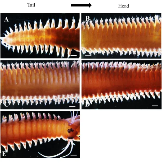
P. wilsoni (Polychaeta – Errantia – Phyllodocida – Nereididae) was purchased from a local hatchery in Oita Prefecture and reared in the Fisheries Research Laboratory (FRL) of Kyushu University. Animals were maintained in filtered running seawater with daily feeding. The natural breeding season in Oita Prefecture is in early April, and animals die shortly after spawning in the next Apr. with one-year life cycle (Fig. 12). The embryos and young juveniles were reared at the FRL. Animals grew by the segment addition at the anterior border of the tail-end body segment, pygidium. This extension growth began just after the spawning in April and declined at the end of July. The segment addition was estimated to occur once per day and reached up to 101 ± 7.5 (standard deviation; n = 30) segments in an adult. About 15 animals were sacrificed at least once per month for the analysis of germ cell development. Prior to fixation, animals were anesthetized with 0.3% ethylene glycol monohexyl ether in seawater and then fixed in 4% paraformaldehyde in phosphate-buffered saline. The photos used in this manuscript show the representative figures of similar animals.
2.3.2 In situ hybridization
Vasa, nanos and piwi genes have been long used as the germline marker in various annelids species (Cox et al., 1998; Kuramochi-Miyagawa et al., 2004; Rebscher et al., 2007; Giani et al., 2011; Beyret and Lin, 2011; Rebscher, 2014; Özpolat and Bely, 2015; Ponz-Segrelles et al., 2018). In our previous experiment, I used the vasa gene to trace germ cells in female P.
wilsoni (Maceren-Pates et al., 2015). In this experiment, I cloned the piwi gene and used to trace germ cells for an improved reliability of germ cell detection. For Pw-piwi cloning in this study, primers were designed from the mRNA sequence of a closely related species, Platynereis dumerilii (Rebscher et al., 2007) (forward: 5'-accacgactccaagcagaag-3'; reverse:
15
5'-atcttgtgggcgtattggca-3'). Total RNA was extracted from unfertilized eggs of mature P.
wilsoni using TRIzol Reagent (Invitrogen) and purified using a RNeasy Kit (Qiagen). cDNA was synthesized using a Prime Script RT-PCR Kit (Takara). Using a pGEM-T Easy Vector System I kit, the 613-bp PCR product was ligated into a plasmid and transformed into Escherichia coli DH5. Probes were labelled with digoxigenin and used for whole-mount in situ hybridization, as described previously (Maceren-Pates et al., 2015). A cDNA sequence of Pw-piwi is registered in DDBJ (Accession No. LC564863).
2.3.3 Histological analysis
After whole-mount in situ hybridization, animals were sectioned for histological observations to detect germ cells inside the body segment, as described previously (Maceren- Pates et al., 2015). The same batch of animals were also prepared for staining with hematoxylin and eosin (H&E). Sections were cut at 7 m.
16 2.4 Results
2.4.1 Germ cell localization in male P. wilsoni
To examine the localization of germ cells in the body segments of males for comparison with the previous observations of female P. wilsoni, I used in situ hybridization with Pw- piwi and Pw-vasa anti-sense probes on adult males at first. Although both probes showed similar expression patterns in whole-mount in situ hybridization, the Pw-piwi probe tended to give a stronger expression compared with the vasa-probe. Therefore, I determined to use the Pw-piwi probe in this study. The Pw-piwi probe was strongly expressed at the distal areas of parapodia on both sides of the segments (Fig. 1). The signal strength gradually decreased from the posterior to the mid-body, then disappeared in the anterior region of the body (Fig.
1). Horizontal sections of worms stained by in situ hybridization showed cell clusters at the parapodia in the posterior region, but no signal was detected in the trunk area (Fig. 2A). In the medial body segments, I observed large cell clusters with the Pw-piwi signal at the distal areas of the parapodia, and several smaller clusters were separated from the large cluster and several Pw-piwi signals in trunk and basal areas of parapodia (Fig. 2B). In the anterior segments, the large cell clusters were not detected in the parapodia, but several signals were observed in the trunk areas of the parapodia and in the deeper body cavity area (Fig. 2C).
The Pw-piwi signals were decreased at the trunk and deeper body cavity areas compared to the clusters at the distal areas of the parapodia. Figure 3 shows no signal detected with use of a sense probe. Piwi signals were still observed at the distal area of the parapodia of the
17
posterior region even in early March (Fig. 8). At the end of March, only a few segments in the posterior were stained weakly with the piwi probe (Fig. 9). No piwi signal was detected in the parapodia in the transformed animal (epitoke) in early April (Fig. 10). Moreover, piwi was expressed in early stages of germ cell maturation (spermatogonia and spermatocytes) and was undetectable in spermatid and sperm stages (Fig. 11).
2.4.2 Sexual development of gametes in P. wilsoni
To study the development and translocation of germ cells, histological observations of P.
wilsoni were conducted from May to the following April. The germ cell clusters formed in the parapodia divided into smaller clusters and migrated toward deeper areas of the body (Fig. 2B). The migration was observed in the early specimens of whole mount in situ hybridization in May, which indicated the migration already began in young juveniles by 40 dpf (Fig. 4), as those were formed earlier than posterior segments. During this migration period, it was not possible to distinguish the sex of each animal because there was no morphological difference in germ cells (Fig. 5A, A’).
In September, the female germ cell became discriminable from the male germ cell by its enlargement with an accumulation of vitellogenin (Fig. 5B, B’). After September, 30 more animals were analyzed and were equally divided into males and females; thus, the sex ratio of P. wilsoni was assumed to be 1:1. The vitellogenesis began simultaneously in all body segments, but the size of vitellogenic germ cells varied, even within the same segment during this season. However, nearly all eggs were observed to attain equal size just before the spawning season (Fig. 6A). By contrast, male germ cells were observed to have a longer proliferation period (Fig. 5C), which lasted until March, and they entered meiosis at the same time, in late March, in all body segments (Fig. 6B, B’). Figure 13 is the schematic diagram which explains the timing of the germ cells migration and the start of the vitellognesis.
18
The developing male germ cells were categorized according to morphological changes in the clusters at each stage (Fig. 7). In the early stage of proliferation, nuclei and nucleoli were clearly visible, and the cytoplasm was filled with granular particles (Fig. 7A). These cells were observed in September and categorized as spermatogonia I. In this stage, the average cell size varied among clusters, even within the same segment. In November, each cluster was filled with proliferating germ cells (Fig. 7B), and by January, the clusters enlarged in size due to the highly proliferating germ cells; the cells decrease in size and the nuclei and nucleoli could not be distinguished due to chromosome condensation. This cluster was categorized as spermatogonia II (Fig. 7C). The enlarged clusters loosened throughout February, divided into smaller clusters (Fig. 7D), and then transitioned into spermatocytes in March (Fig. 7E). In April, a few spermatids were observed as tetrads, although the majority were single sperm cells (Fig. 7F).
19
Fig. 1. Whole-mount in situ hybridization with the Pw-piwi probe in an adult male worm in late August. Strong signals were detected at distal ends of the parapodia in a posterior region and decreased toward an anterior region (A–G). White arrows indicate the Pw-piwi signals at parapodia (H). Body length = 10 cm. The total number of body segments in this individual is 98. Scale bars for A–G and H are 200 and 100 µm, respectively.
Posterior Anterior
D E F
B C
A
G H
20
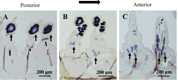
Fig. 2. Horizontal sections of the hybridized worm showing the localization of the Pw- piwi expression in an adult male in late August. (A) Posterior region; strong Pw-piwi signals were observed at large cell clusters in distal areas of parapodia. No clusters were detected in trunk areas. (B) Mid region; the Pw-piwi signals in distal areas were decreased, and several smaller clusters were detected in trunk areas. (C) Anterior region; the Pw-piwi signal was undetectable in distal areas. Smaller clusters were observed in trunk and basal regions of parapodia. Arrows indicate smaller clusters of germ cells. Scale bar, 200 µm.
Posterior Anterior
C
200 µm
B
200 µm
A
200 µm
21
A A’
B B’
100 µm 100 µm
200 µm 200 µm
Fig. 3. Specificity of the Piwi probe in in situ hybridization. (A) Germ cell clusters were shown by in situ hybridization with use of the anti-sense probe of Pw-piwi. (B) No signal shown with use of the sense probe. Arrows indicate germ cell clusters.
22
b’
50 µm
a’
50 µm a
b
A
200 µm
Fig. 4. Germ cell clusters in a juvenile worm. (A) Germ cell clusters shown by in situ hybridization at parapodia of a juvenile worm at 40 dpf. (B) Enlarged
parapodia of the posterior region indicated by a black square (b) in Fig. 4A. Germ cell clusters are shown at distal areas of each parapodia. (C) Enlarged parapodia of the anterior region indicated by a black square (c) in Fig. 4A. Germ cell clusters are not observed at parapodia.
23
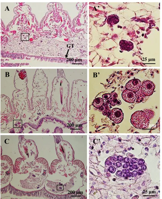
Fig. 5. Germ cells before and after the start of vitellogenesis. (A) It is difficult to distinguish sex with morphological characteristics in germ cells of 4-month-old juveniles in early August. (B) Growing female germ cells with vitellogenesis in September. (C) Proliferating male germ cells in clusters in September. A’, B’, and C’
are enlarged images of the frames in A, B and C, respectively. GT: Gut tract. Stained with H&E.
A’
25 µm
A
GT 200 µm
200 µm
C
25 µm
B’
25 µm
B
200 µm
C’
24
Fig. 6. Germ cells fill body cavities of each segment in March (mid-body region). (A) Female body segments are filled with fully-grown oocytes. (B) Male segments are filled with many germ cell clusters. B’ is the enlarged image of the frame in B. Stained with H
& E.
B
200 µm
B'
10 µm
A
200µm
25
Fig. 7. Monthly developments of male germ cells. (A) Early spermatogonia in Sept. (B) Proliferating spermatogonia in Nov. (C) Proliferating spermatogonia in Jan. (D) A large cluster in early Mar. (E) Spermatocytes in middle March. (F) Spermatids and sperms in Apr. (breeding season). N: Nucleus, n: nucleolus, Cy: Cytoplasm. Black arrows in F show a tetrad spermatid and a single sperm. Stained with H & E. Scale bar; 25 µm.
Cy N
n
A B C
D E F
26
Tail Head
E
D C
B A
Fig. 8. Whole mount in situ hybridization with Pw-piwi in early March. Strong signals were still detected at the distal ends of the parapodia in the posterior region (A), even though germ cell inside the body cavity were in
spermatocytes stage. Scale bar; 200 µm.
27
A B
C D
E
Tail Head
Fig. 9. Whole mount in situ hybridization of Pw-piwi in end of March when the animals were transforming into epitoke. Weak signals were still detected at the distal ends of the parapodia in few segments of the posterior region (A). Scale bar; 200 µm.
28
Fig. 10. Whole mount in situ hybridization of Pw-piwi in April; swimming epitoke. No piwi signals detected at the distal ends of the parapodia region (A). Scale bar; 200 µm.
A B
C D
29
Fig. 11. Pw-piwi expression in monthly development of male germ cells. (A, B) Early spermatogonia in Sept. (C, D) Proliferating spermatogonia in Nov. (E, F) Proliferating spermatogonia in Jan. (G, H) Spermatocytes in middle March. Both genes were undetectable in spermatid and sperm stage. Scale bar A-H, 25µm.
H& E stain Piwi signals
30
Fig. 12. Schematic diagram on the gametogenic cycle of P. wilsoni. (A) Hypothetical germ cells migration from the parapodia to the deeper body cavity in P. wilsoni.
Meiosis
Meiosis
Proliferation
Jan. Sep.
Feb. Oct.
End of Mar. - Early Apr.
Transformation into epitoke
April
Tail A
SAZ Pygidium
31
Fig. 13. Schematic diagram on the timing of the germ cells migration from the parapodia to the deeper body cavity during segments addition and the start of the vitellogenesis. In April, germ cells from the pygidium are supplied to every newly formed segment and settles at the distal end of the parapodia. May, germ cells in the anterior segments started migration to the deeper body cavity. In August, germ cells in the anterior segments have translocated into the deeper body cavity. However, sexes could not be distinguished due to immature state of the germ cells. In Sept., germ cells have reached the deeper body cavity in almost all body segments and start the vitellogenesis simultaneously. During this during period sexes could be differentiated due to enlargement of the oocytes. Black dots represents the hypothetical migrating germ cell clusters. (AS); Anterior segments.
SAZ Pygidium April
Pygidium SAZ
May
Pygidium Tail
SAZ
August September
Tail
Pygidium AS
AS
32
Table 1. Number of segments in adult worm P. wilsoni
Samples No. of segments
in adult worm Samples No. of segments in adult worm
1 94 16 98
2 112 17 107
3 104 18 103
4 94 19 97
4 107 20 108
6 99 21 111
7 104 22 114
8 107 23 98
9 107 24 103
10 107 25 100
11 92 26 101
12 84 27 84
13 101 28 98
14 103 29 92
15 91 30 104
Sum = 3024
Count = 30
Average = 101
STDEV = 7. 53
33 2.5 Discussion
2.5.1 Germ cells localization and gametes production in P. wilsoni
In this study, we compared germ cell development between males and females of P.
wilsoni. P. wilsoni grows by adding new body segments at the anterior boarder of the tail- end segment, the pygidium, and all segments are completely separated by intersegmental septa. In the previous report, an interesting mechanism by which germ cells are allocated to all body segments was identified, that is, a newly generating segment receives copies of germ cells from the pygidium, the only source of germ cells in P. wilsoni (Maceren-Pates et al., 2015). In each segment, germ cells settled first at the parapodia on each side of the segment and proliferated to form a large cell cluster. Then, the large cluster separated into several smaller clusters, which migrated to the inner body cavity area (Maceren-Pates et al., 2015).
During this period, germ cells proliferated and developed in each segment. In the previous paper, it was found that the large germ cell clusters were already formed in the anterior segments of young juveniles at 10 dpf (Fig. 9 of Maceren-Pates et al., 2015). However, at 40 dpf, the Pw-piwi signal disappeared from the parapodia of the anterior segments (Fig. 4).
The germ cell clusters translocated into the deeper body cavity area might be difficult to be observed because the Pw-piwi signal decreased during migration (Fig. 2). These observations indicate that germ cells proliferate to form a large cluster in the parapodia and then migrate to the inner body cavity area within 30 days after the cluster formation.
However, at this stage, it was impossible to distinguish the sex of animals because there were no morphological differences between males and females (Fig. 5A, A’).
In September, all germ cells began vitellogenesis in females, which was recognizable by the increase in size (Fig. 5B, B’). By contrast, male germ cells continued proliferation (Fig.
5C, C’). With these differences, animals could be identified as male or female. At this time,
34
the sex ratio of P. wilsoni was 1:1, and it was the same in the breading season, similar to other species (Reish, 1954, 1957; Barels-Hardege and Zeeck, 1990; Fischer and Dorresteijn, 2004). Before the start of vitellogenesis, although I did not identify the sex of the animals used for the histological analysis, I assumed that the animals included males and females at a ratio of 1:1. Therefore, I may conclude here that there is no clear morphological difference between male and female germ cells in the early developmental phase.
In late September, germ cells had reached the inner body cavity area in all body segments and simultaneously began vitellogenesis. Naturally, the germ cells in the anterior segments reached the body cavity area earlier than those in the posterior segments. However, the anterior germ cells delayed vitellogenesis while waiting for the posterior germ cells to reach the body cavity area. This may suggest that there is some unidentified mechanism to control the initiations of migration and vitellogenesis in female germ cells. As the oocytes reached a maximum size before the spawning season, vitellogenesis seemed to continue until just before spawning. The oocytes varied in size in the early phase of vitellogenesis, and vitellogenesis was not well synchronized among germ cells, even in the same segment (Fig.
5B, B’). This variation in oocyte sizes in the early stages has been observed in several polychaete species (Olive and Garwood, 1981; Fischer, 1974; Golding and Yuwono, 1994).
However, as the breeding season approached, all oocytes attained equal size (Fig. 6A).
By contrast, males had a longer germ cell proliferation phase to maximize the number of sperm produced, and they entered meiosis in late March, as shown in Figure 6B & B’. The developing male germ cells in P. wilsoni were clearly distinct in morphology between stages (Fig. 7). In many polychaetes, morphological characteristics have been used as the criteria for germ cell development. In Nereis virens, spermatogenesis was categorized into five stages based on the morphology of germ cells; spermatogonia I, spermatogonia II, spermatocytes, tetrads, and spermatozoa (Schenk and Hoeger, 2010). In this study, the
35
observed P. wilsoni spermatogenesis stages closely resembled those of N. virens (Schenk and Hoeger, 2010). In P. wilsoni, male germ cells in September might be categorized as spermatogonia I by clear visibility of big nuclei and nucleoli (Fig. 7A). However, as proliferation continued through January, it resulted in the formation of thick and tightly packed clusters of germ cells, which were categorized as late spermatogonia II (Fig. 7B, C).
In February, this tightly packed cluster (spermatogonia II) loosened and began to form several smaller clusters; these were clearly separated by March and were categorized as spermatocytes (Fig. 7D, E). In April, the breading season, some germ cells were observed in groups of four spermatids, whereas the majority were individual sperm cells (Fig. 7F).
However, although the Pw-piwi signal was clearly detected in the stages of spermatogonium and spermatocyte, it was significantly decreased in the stages of spermatid and sperm (Fig.
11). The decrease of the Pw-piwi signal in the late stages of spermatogenesis especially in sperms, had been also reported in other animals (Lin and Spradling, 1997; Houwing and Berezikov,2008; Xiao et al., 2013; Hempfling et al., 2017)
In P. wilsoni segments addition ceases towards the end of July and by September, most of the germ cells has finished migrating to the deeper body cavity and ready to enter vitellogenesis. However, not all germ cells in the posterior segments translocated to the deeper body cavity, as revealed by the detection of piwi signals at the distal area of the parapodia throughout March (Fig. 8). Probably there was not enough time for all the posterior germ cells to migrate since the younger segments were formed lately before the start for the vitellogenesis. Piwi signals weakens as the worms transforms into epitoke (Fig.
9). Piwi signals were not detectable in epitokes (Fig. 10), the reason for disappearance of signals is still unknown, and however it might be related to the transformation process.
36 2.6 Conclusion
This study explored the process of the gamete development in details in a fully segmented worm, Perinereis wilsoni. The findings provide new insights on understanding the timing of the germ cells translocation from the parapodia to deeper body cavity and the start of gamete maturation. The male has shown similar features of germ cell distribution, proliferation and translocation in each segment to those of the female. Germ cells translocate to the deeper body cavity during growth (segment addition phase). The translocation correlates with age of the segments, however, the further development in the body cavity area starts simultaneously in all body segments and all mature gametes are spawn at one time in the breeding season. This may suggest that there should be under the some regulating mechanism to synchronize gamete development among all segments of the whole body.
37
CHARTER 3
Mechanisms of gamete spawning in males and females
Perinereis wilsoni
38 3.1 Abstract
In the previous studies, I described the germ cell development and gamete maturation in a fully segmented worm. In P. wilsoni, germ cells proliferate, differentiate and mature separately in each segment. Like other nereidids, P. wilsoni also undergoes transformation into epikoke for the breeding season. The present research aimed to understand the characteristics of gamete releasing sites in males and females by histological techniques. I found that the complete septa were broken, gametes were mixed between segments prior to spawning and released from the posterior region of the body. However, the releasing sites were differently allocated between males and females. Sperms were released from nephridipores at 2nd through 15th segments from the pygidium, while eggs were released through raptures of skin of 2 – 3 segments between 10th and 30th segments from the tail. The rapturing point varied among females.
39 3.2 Introduction
Nereidids are errant polychaetes whose spawning behaviours had been long studied due to their unique reproductive strategy called epitoky. Epitoky is a form of reproduction in which sexually immature worm (atoke) transform into sexually matured worm (epitoke) and emerge from benthic to short pelagic life in spawning season (Bartels-Hardege and Zeeck, 1990; Fong, 1993; Franke, 1999; Hébert-Chatelain et al., 2008; Fischer and Dorresteijn, 2004).
Nereidids are broadcasting spawners as many of other marine invertebrates. To achieve successful fertilization, the effective communication among partners is of great importance.
Majority of species use the chemical pheromone to insure the synchronous spawning. In polychaetes, the pheromone is found in the coelomic cavity of the ripe epitoke. The pheromone is released and spreads in water, and detected by the opposite partners which will release more pheromones (Jorg et al., 1998). The pheromone is species specific to have the interspecies effects. For instance, Zeeck et al., (1988) identified a ketone compound (5- methly-3-heptanone) in three species, P. dumerilii, Nereis succinea and Pereinereis var.
brevicirrus. The compound induces the swarming behaviour (nuptial dance) and also enables to recognize other individuals of the same species. In Nereis japonica, a different ketone compound (3,5-octadiene-2-one) was found which had the same function to that found in P.
dumerilii (Hardege et al., 1995). Along with the pheromones communication, many species release gametes in strings, streams or plumes with mucus to reduce gametes dilution by rapid and active water mixing (Denny and Shibata, 1989; Olive and Pillai, 1983b; Thomas, 1994;
Eckelbarger et al., 2001; Lotterhos and Levitan, 2010). In breeding season, some of the polychaetes such as syllids modify few nephridia of the posterior into gonoduct (metanephromixia) for gametes release (Franke, 1999; Goodrich, 2009). On the other hand,
40
majority of the nereidids have been reported to release gametes through ruptures of the body wall (Fischer and Dorresteijn, 2004).
Despite the fact that, spawning mechanisms on several nereidids have been explore, the knowledge on gamete releasing sites is still limited especially to a fully segmented worm.
The present research was undertaken to characterize the gamete spawning sites in P. wilsoni.
41 3.3 Materials and Methods
3.3.1 Spawning sites observation
Swimming epitokes (2 – 4 males per female) were placed in a transparent container filled with filtered seawater and observed until spawn occurred naturally. This observation was repeated several times to locate the spawning site in the body. Some epitokes were quickly fixed at the beginning of spawning to analyse the spawning site. After collecting fertilized eggs, we reared embryos for further use in the experiments.
3.3.2 Histology
Two sets of samples were collected for histological analysis of internal morphology of the spawning; swimming worms (ready to spawn) and spawning worms. In both cases, males and females were sampled separately. Animals were anesthetized with 0.3% ethylene glycol monohexyl ether in sea water prior to fixation and then fixed in 4% paraformaldehyde in PBS fixed in 4% paraformaldehyde for 24 hrs at 4℃. Fixed animals were cut into about 1cm blocks from the anterior to the posterior, then dehydrated in a series of ethanol (70-100) % and embedded in paraffin. Serial sections were cut in 7 m and stained with haematoxylin and eosin (H&E) for the microscopic observations.
42 3.4 Results
3.4.1 Spawning sites in P. wilsoni
P. wilsoni body segments are completely separated each other by intersegmental septa.
Each segment is filled with proliferated germ cells by the breeding season, and all those germ cells are released during the characteristic swimming. However, it has never been clearly described how P. wilsoni releases gametes. To examine the spawning mechanism, we observed the animal behaviour during spawning to identify the gamete releasing site, and also examined the fine structure of the releasing site.
The intersegmental septa were broken during the swimming at spawning and gametes were mixed between segments in male and female (Fig. 2, 6). Moreover, the release of gametes was observed to associate with unidentified structures of varies size which stained dark pink with H &E (Fig. 7).
3.4.2 Gamete spawning sites in male P. wilsoni
In male, sperms were continuously released as streams from the several segments in the tail region (Fig. 3A). The total numbers of segments with sperm release were varied among individuals. Those were 10 – 14 segments from 2nd up to 15th segments from the tail end (Fig. 3B). The gamete releasing site were located near the intersegmental septum at the anterior side of each segment (Figs 4A, A’), and a tube like structure from the body cavity was observed connecting to this opening site which could possibly be a “nephridium” (Fig.
4B). The sites were opened/splitted during sperm release (Fig. 4C).
3.4.2 Gamete spawning sites in female P. wilsoni
In female, eggs were released from only 2 – 3 segments in the tail region (Fig. 4A). Exact segments were different between individuals, but were in the range of 10th to 30th from the pygidium. As the epithelial structure was severely ruptured by egg spawning, it was difficult to identify the exact point and its detailed structure for egg release (Figs 6B, c, d). Ten
43
animals were fixed and sectioned to analyse the internal structures for each sex. Figures 3 to 6 show typical examples.
44
Fig. 1. Observation of spawning sites in P. wilsoni. (b) Male (c) female spawning.
A b c
Sperms Eggs
45
Fig. 2. Horizontal sections of a swimming male worm just prior to spawning in April. Arrows indicate broken septa. Stained with H&E.
200 µm
46
Fig. 3. Sperm spawning area in the tail region of a male. (A) The square indicates the area of sperm spawning. (B) A horizontal section of the tail indicated in (A).
Arrows indicate sperm released from both sides of the limited segments. Stained with H&E.
A
B
200 µm
47
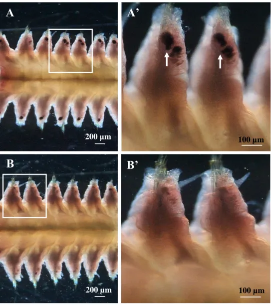
Fig. 4. Histological observation of sperm release sites in P. wilsoni. (A) A horizontal section of sperm-releasing segments. (A’) An enlarged sperm release site indicated by the frame in (A). (B) A sperm release site with a tube-like structure indicated by a black arrow. (C) Sperm release from the opening of a structure similar to that shown in (A’). The square indicates the opening point. Stained with H&E.
25 µm 200 µm
25 µm 25 µm
A ’
A
B C
48
Fig. 5. Horizontal sections of a swimming female worm just prior to spawning in April. Arrows indicate broken septa. Stained with H&E.
200 µm
49
50 µm
b’
A a
b
B
200µm
50 µm
a’
100µm
Fig. 6. Histological observation of egg release sites in P. wilsoni. (A) The white arrow indicates the segments where eggs were released. (B) The egg release sites are indicated by black frames a, b. Eggs are released from breaks at both sides of the segment. a’ and d’ are enlarged images of (a) and (b). Black arrows indicate the break points of the egg- releasing segment. Animals were fixed during spawning. Stained with H&E.
50
A
25 µm
B
100 µm
Fig. 7. Eosinophilic structure associated with gamete release indicated by arrows. (A) Spawning male. (B) Spawning female. H&E stain.
51 3.5 Discussion
3.5.1 Gametes spawning sites in males and females P. wilsoni
P. wilsoni has the complete septum to separate each segment of the body. Therefore, I had thought P. wilsoni would spawn mature gametes form various area of the body. But, in this study, both male and female were found to release gametes from the limited regions in the posterior body (Fig. 3, 6). Males released sperms from 10 – 13 segments and females released eggs from only a few segments. Most germ cells were spawn from those segments during a typical spawning swimming. The spawning swimming was characterized by a strong and flowing serpentine motion of the body. I presume that this strong motion may help to break the intersegmental septa and move germ cells toward the posterior region. This septum breakage looked to precede the germ cell release in both sexes (Fig. 2). If the serpentine motion effectively pushed germ cells into the posterior region, it would increase the internal pressure to break the skin to release germ cells. In female, the germ cell release occurred from only a few neighbouring segments, but those appeared in the back one third region of the body (10 – 30th segments form the tail). This may indicate the egg release site is formed randomly in female. In contrast, in males, sperms were released from about 10 segments in the tail region (Fig. 4). It seemed no shift in location of the release sites among individuals, which suggested there was some common structure related to the sperm release unlike female. There are several precedent papers reporting the site of germ cell release in polychaetes. Most of the nereidids are reported to spawn by rupturing their body wall (Fischer and Dorresteijn, 2004) or by dehiscence (Goodrich, 1895). However, in Syllidae, metanephridia of the fertile segments are modified into metanephromixia during sexual maturity to act as the gonoducts (Franke, 1999; Goodrich, 1895). In this study, I observed that sperms were released by splitting/opening at the “nephridipore” area in P. wilsoni (Fig.
4). It was quite similar to Syllidae. However, I could not identify the detailed structures as
52
metanephromixia. Although these opening sites was basically set in the most segments, the sperm release occurred at the limited “nephridipore” in the tail 10 segments. Therefore, in male, there might be some unknown mechanism involved those limited nephridipore to open for the sperm release.
3.5.2 The eosinophilic structure associated with gamete during spawning
In the present study, I observed irregular structures of different shapes which associated with gametes release. The structures stained dark pink with H&E stain (Fig. 7A, B). In many broadcasting marine invertebrates including polychaetes, the release of gametes had been reported to associate with some adaptive factors which could be biological, chemical or physical factors which offers physical support to minimize gametes dilution in active water movement which will decrease fertilization rates (Denny and Shibata, 1989, Crimaldi and Browning, 2004). In P. n. brevicirrus eggs and sperm bond within 2 threads of mucus (Hardege and Bartels-Hargede, 1996)). In Phragmatopoma californica, sperms are released in strings, streams or plumes (Thomas, 1994). Similarly, in the present study the release of gametes were observed to associate with physical elongated structures. In male, the structures infused inside the sperm cluster while in female the structures surrounded eggs and some in between eggs (Fig. 7). The release of gametes in companion with those structures might suggest that, probably they have a supportive role of keeping the gametes intact along the water column to ensure successful fertilization.
53 3.6 Conclusion
The present study, managed to identify the gamete releasing sites in fully segmented worm.
P. wilsoni gametes were released from the posterior region regardless of the fact that, during gametogenesis, germ cells develop and mature separately in each segment. Males released sperms from nephridipores at 2nd through 15th segments from the pygidium, while female released eggs through raptures of skin of 2 – 3 segments between 10th and 30th segments from the tail. The rupturing point varied in between females.
54
CHAPTER 4
GENERAL DISCUSSION
55 4. General discussion
The present study describes the germ cell development in male Perinereis wilsoni in comparison with characteristics of the female germ cell studied in the previous study by Mercedes et al., (2015). This study also analysed the mechanisms of gamete spawning in males and females.
4.1 Germ cell localization in male P. wilsoni and gamete production
In the previous study in females, it was revealed that the body segments of P. wilsoni were separated by the complete intersegmental septa. Germ cells of this species are originated from the pygidium and distributed to each body segment during the segment addition phase and settled at the distal ends of each parapodium on both sides of the segments. During gamete development in females, germ cells at the parapodia proliferated and formed clusters which then separated into a single female germ cell as they moved towards the coelomic cavity where they started vitellogenesis (Maceren Pates et al., 2015). The present study aimed to focus on understanding the germ cell development in male P. wilsoni in comparison with the female findings so as to expand the knowledge on reproductive biology of P. wilsoni as a representative of fully segmented polychaetes with the complete intersegmental septa.
To find out the localization of germ cells in male, this study analysed a mRNA expression of a germ cell marker gene in adult males of 5 months old, since the sex of this species can be distinguished by the enlargement of germ cells by vitellogenesis. Sampling was carried out through the observation of the coelomic cavity under a microscope and females were distinguished from males by the presence of growing vitellogenic oocytes. Two germline markers were used for whole mount in situ hybridization. The Pw-vasa was cloned in the previous study held by Maceren Pates et al. (2015) and the Pw-piwi was cloned in this study (Chapter 2). In the result, both genes showed the similar patterns of expressions as the strong signals at the distal area of the parapodia and the strength of the signal decreased from the
56
posterior towards the anterior region. These expressions were quite similar to the previous study of females (Maceren Pates et al., 2015). These results indicate that both male and female germ cells in P. wilsoni have the same starting points at the distal area of the parapodia. In the posterior region, strong signals of piwi were detected only at distal area of the parapodia. In the mid body region, the strength of the signals decreased at the parapodia and several small clusters were detected in the trunk and deeper coelomic cavity. Strong signals at the parapodia and small cluster inside the body cavity suggests that the smaller clusters might have divided from the large cluster at the parapodia and migrated into the deeper body cavity and this migration corresponds with the age of the segment (Chapter 2).
To clarify the beginning of germ cell migration and vitellogenesis, the monthly histological observations were conducted through May to September. In May, the piwi signals began to decrease first in the anterior body region about 40 days after fertilization. This decreasing of the piwi signal occurred as a result of germ cell migration into the inner body cavity area from the parapodia, which was shown by the vertical section of hybridized worms. The migrating germ cells were thought to be in a primordial germ cell stage and could not be distinguished the sex. The migration process continues through the months of June to September. In September, most of the germ cells reached the deeper body cavity area in all segments and starts vitellogenesis, simultaneously. Even though the germ cells in the anterior segments migrated early, they looked to arrest the start of vitollegenesis in Sept.
This arrest might be the waiting for the posterior germ cells until their reach in the body cavity area. This arrest of the anterior germ cells may suggest that there is some unknown mechanism to arrest germ cells to enter the vitellogenesis phase. As the beginning of vitellogenesis is related to the cell stage changing from the oogonium to the oocyte, there may be a hormonal control for the arrest of beginning of the vitellogenesis until Sept.
(Chapter 2)