はじめに
ステント留置術は,経皮的バルーン血管形成術の際に 生じる,弾性反跳による血管拡張不良や血管壁解離など の問題を解消するために考案された.Sigwart ら26)は, 1986年に初の冠動脈ステント留置術を実施した.初期の 冠動脈ステント留置術は,決して満足できる治療成績で はなかったが,心筋においてはステント留置後血栓症が 薬物療法で解消され,冠動脈ステント留置術は爆発的に 世界に普及した.一方,頚動脈のアテローム性動脈硬化 連絡先:秋山恭彦,〒 693 8501 出雲市塩冶町 89 1 島根大学医学部脳神経外科Address reprint requests to:Yasuhiko Akiyama, M.D., Department of Neurosurgery, Shimane University Faculty of Medicine, 89 1 Enya cho, Izumo shi, Shimane 693 8501, Japan
頚動脈狭窄症に対する血管内治療
―今日までの治療経験の集積と次世代に向けて―
秋山 恭彦,宮嵜 健史,萩原 伸哉,中右 博也,神原 瑞樹,吉金 努, 将大,
藤原 勇太,内村 昌裕,永井 秀政
島根大学医学部脳神経外科
Carotid Artery Stenting Progress to Date and the Next Steps
Yasuhiko Akiyama, M.D., Takeshi Miyazaki, M.D., Shinya Hagiwara, M.D., Hiroya Nakau, M.D., Mizuki Kambara, M.D., Tsutomu Yoshikane, M.D., Masahiro Tsuji, M.D., Yuta Fujiwara, M.D., Masahiro Uchimura, M.D., and Hidemasa Nagai, M.D.
Department of Neurosurgery, Shimane University Faculty of Medicine
Carotid artery stenting(CAS)is an alternative to carotid endarterectomy(CEA)that promises long term efficacy. Patients indicated for carotid revascularization often have systemic atherosclerotic disease, therefore CAS has advantage in its ability to reduce periprocedural systemic complications including myo-cardial infarction via its less invasiveness. However, recent large randomized control trials have revealed that CAS has a higher potential risk of periprocedural ischemic stroke. The main factors likely to partici-pate this event are incomplete embolic protection during balloon or stent angioplasty, and post procedural thrombus that emerges on the stented area associate to the protrusion material through the bare metal stent. In this article, we present an outline of the recent randomized control trials, SPACE, EVA 3S, ICSS, CREST and ACT 1, and focus on the cerebral ischemic complications in these trials, and describe in detail of the mechanism of periprocedural thromboembolic events and their countermeasures.
Reducing of periprocedural ischemic complication is the task to date, and active utilization of proximal balloon embolic protection to unstable plaque on tailor made manner and newly developed micro mesh covered stent may affect the efficacy of CAS as we go forward.
(Received June 8, 2017;accepted July 24, 2017)
Key words: carotid artery stenosis, carotid artery stenting, atherosclerosis, cerebral infarction, periproce-dural ischemic complication
Jpn J Neurosurg(Tokyo)26:728 737, 2017
症に対するステント留置術(carotid artery stenting:CAS) は,1993 年に Diethrich ら6),Bergeon ら2)により実施さ れた.しかしこの時期には,頚動脈内膜剝離術(carotid endarterectomy:CEA)が,NASCET(1991 年)21)によ り,その臨床効果を確立し,さらに,ステント留置術は, 治療手技に伴うアテロームの破裂とその飛散により,時 に重篤脳梗塞が生じることが問題で,CEA 手術リスクが 非常に高いと考えられる一部の症例に限定して施行され るに留まっていた.しかし,1996 年に Theron ら30)によ り,ステント留置術中に生じる末 塞栓予防を講じる現 行の CAS の基本となる手技が考案されたことで,本治療 の期待が高まった.欧米では間もなくして,PercuSurg Guard Wire(Medtronic),Parodi Anti Emboli System (ArteriA)などのバルーンタイプ,および NeuroShield (MedNova),Angioguard(Cordis),FilterWireEX(Boston
Scientific)などのフィルタータイプの末 塞栓保護デバ イス(embolic protection device:EPD)が,また,Carotid Wallstent(Boston Scientific),X act(Abbott),SMART (Cordis)などのナイチノール製自己拡張型ステントが開 発された.そして,2004 年に発表された SAPPHIRE 試 験32)によって CAS の有効性は世界的注目を集めること となった. CAS は,手技の確立から今日までに 20 年が経過し, これまでに CEA と治療効果を比較した 3 つのランダム 化比較試験(randomized control trial:RCT)において, 臨床的有効性が証明された.CAS は,全身性動脈疾患で あるアテローム性動脈硬化症に対し,低侵襲的に頚部局 所のアテロームを制御する治療で,アテローム性疾患に 起因し外科手術後に誘発される,周術期の心筋梗塞をは じめとする全身性合併症を低く抑えることが可能であ る.しかし,近年の RCT と多くの registry study により 治療成績の集積が進み,現状の CAS は,EPD をすり抜 ける術中栓子と,現行のベアメタルステントの限界から 生じる治療後の血栓症により,プラークの脆弱な症候性 病変においては,周術期血栓塞栓症のイベントは少なく ないことが報告されており,治療手技の改良,革新的デ バイスによるさらなる治療法の進化が望まれる.本稿に おいては,これまでの CAS と CEA の RCT および,その サブ解析の結果を概説し,本治療法の現状と課題を周術 期脳虚血合併症に焦点をあて整理し,次世代の CAS を探 りたい. NASCET21)や ACAS7)によって内科治療に対する優越性 の証明されているCEAとのRCTにより検討されてきた.
これまでに,SAPPHIRE32),EVA 3S15),SPACE27),
ICSS8),CREST4),ACT 124)の 6 つの RCT の結果が報告
されている(Table 1). 2004 年に報告された SAPPHIRE32)では,CEA 高危険 群を対象として,遠位塞栓保護を行った CAS の CEA に 対する非劣性の証明(非劣性 p=0.004,優越性 p=0.053) が,厳格な術者選定により達成された.SAPPHIRE によ る臨床的有効性の証明により,CAS のデバイスは 2004 年 8 月に米国 FDA により承認された.本邦でもこのエビ デンスをもとに,2008 年 4 月に治療手技が保険収載され た.適応は CEA 高危険群で,治療中の遠位塞栓に対する 予防的措置を講ずることが提起された.
EVA 3S15),SPACE27)および ICSS8)の 3 つの試験では,
症候性頚動脈病変を対象とした CAS と CEA の RCT が報 告された.これら 3 つの試験では,CAS の CEA に対す る非劣性は証明できなかった.CAS の治療成績が不良と なった要因は,①遠位塞栓保護の重要性の認識の低さ, ②CAS の術者選定基準の不十分さ5)11)が主要因とされ た.また,試験結果に決定的影響を及ぼす要因ではな かったが,③3 試験のいずれにおいても,症候発症 7 日 以内の CAS の周術期脳梗塞発生は高率であった22).これ らの RCT にはサブ解析による検証が追加され,EVA 3S においては,CAS リスク要因が,1)解剖学的要因,2) 病変要因,3)手技要因から解析された20).それぞれの要
因において 1)ICA CCA 分岐角度(Fig. 1)や大動脈弓の
形状,2)長区域病変や病変部の石灰化,3)末 塞栓保 護の非実施が,周術期脳梗塞のリスク因子となることが
示された(Table 2).
SPACE のサブ解析においては,CAS と CEA における 周術期イベント(同側脳梗塞発症あるいは死亡)が,年
齢因子から検討された29).68 歳以下の周術期イベントは
CAS:2.7%,CEA:7.0%,68 歳超過では CAS:10.8%,
CEA:8.8%であった.高齢患者は CAS のリスク因子であ ることが示された. CREST4)では,CEA 標準危険群に対する無症候および 症候性の病変を対象として試験が行われ,周術期合併症 と 4 年間の同側脳卒中予防において,CAS は CEA に対 して非劣性であることが示された.周術期脳卒中および 死亡は CAS:4.4%,CEA:2.3%であったが,周術期心筋 梗塞は CAS:1.1%,CEA:2.3%であったことが非劣性の
Table
1
R
ecent randomized contr
ol trials of car
otid artery stenting versus car
otid endarter ectomy Year Study Patients Pts. number (CA S/ CEA ) Symptomatic lesions (%) Device EPD use (%) A
dverse events in 30 days on C
AS /CEA * Minor Str . Major Str . Death MI 2004 SAPPHIRE
CEA high risk Sym
> 50 %( US ) Asym > 80 %( US ) 334 ( 167 /167 ) 29 % PRECISE /Angioguar d 96 % CA S 2.5 % 0.0 % 0.6 % 1.9 % CEA 0.7 % 1.3 % 2.0 % 6.6 % 2008 E VA 3S Sym ≧ 60 % ( NASCET ) ( DSA, MRA, US ) 527 ( 265 /262 ) 100 % NR 87 % CA S 6.1 % 2.7 % 0.8 % 0.4 % CEA 2.3 % 0.4 % 1.2 % 0.8 % 2008 SP ACE Sym ≧ 50 %( US ) ( NASCET ) Sym ≧ 70 % ( EC ST ) 1,200 ( 605 /595 ) 100 % NR 27 % CA S 2.5 % 4.0 % 0.7 % ND CEA 2.2 % 2.9 % 0.9 % ND 2010 ICSS Sym > 50 %( US ) ( NASCET ) 1,713 ( 857 /853 ) 100 % NR 72 % CA S 4.3 % 1.7 % 1.4 % 0.3 % CEA 1.3 % 1.7 % 0.5 % 0.6 % 2010 CREST Sym ≧ 50 %( DSA ) ≧ 70 % ( US, CT A, MRA ) Asym ≧ 60 %( DSA ) ≧ 70 %( US ) ≧ 80 %( CT A, MRA ) 2,502 ( 1, 26 2/ 1, 24 0) 53 % A cclink /A ccunet 96 % CA S 3.2 % 0.9 % 0.7 % 1.1 % CEA 1.7 % 0.6 % 0.3 % 2.3 % 2016 ACT 1 Asym 70 99 % ( US or DSA ) 1,453 ( 1,089 /364 ) 0% Xact /Emboshield 98 % CA S 2.1 % 0.4 % 0.1 % 0.5 % CEA 1.1 % 0.3 % 0.3 % 0.9 % Pts : patients, EPD : embolic pr otection device, Str : str ok e, MI : myocar dial infar ction, NR : no r egulations, ND : not described * A dvers e events wer e analyz ed as intenti on to tr eat analys is in SP A C E , C RE ST an d A CT 1, an d as p er pr oto co l analys is in SAP P HI RE , EV A 3S an d I C SS . Ischemic str ok
e was analyzed as ipsilateral str
ok
e in SAPPHIRE, CREST and ACT
1, and analyzed as all str
ok e in EV A 3S, SP ACE and IC SS.
とに CAS リスクが 1.77 倍上昇すると解析された. 2016 年には,ACT 124)により,CEA 高危険群と考えら れる 80 歳以上の高齢患者を除く,無症候性高度狭窄病 変における CAS と CEA の治療成績が検討された.一次 エンドポイントは周術期死亡・脳卒中・心筋梗塞または 1年以内の同側脳卒中とされ,CAS の CEA に対する非劣 性が証明された.本研究は,患者登録が 2005 年から開始 され,現在では CAS ハイリスク因子とされる高齢患者が CEA高危険群として登録から除外されていた.それでも なお,周術期の脳梗塞は,CAS:2.8%,CEA:1.4%で, CAS群において脳虚血イベント発生率は高かった.
CAS
の現状と課題
American Heart Association(AHA)/American Stroke
Association(ASA)における脳卒中一次/二次予防ガイド ラインでは,頚動脈血行再建術は,上述の RCT に基づい てガイドラインが変遷している(Table 3)9)16).2014 年度 版までの変遷の要点として,2006 年度版では,SAP-PHIRE試験の結果に基づいて,症候性,無症候性ともに CEA高危険群における CAS の有効性がガイドラインに 記載された.2011 年度改定では,2010 年の CREST 試験 の結果に基づいて,CAS の有効性が CEA 標準危険群に おいても記載された.2014 年度改定では,CAS の高リス 標準リスクに選択されることが推奨された.また, CAS・CEA ともに,症候発症 2 週間以内の早期治療介入 が推奨され,周術期脳卒中や死亡が 6%未満の術者に よって治療が実施されるべきであることが記載された. また,無症候性病変では CEA あるいは CAS の有効性を 支持しつつも,近年の内科療法の前向きコホートから示 されている内科単独療法の有効性のエビデンス1)13)14)を 勘案したうえで,血行再建術の適応を判断することが推 奨されている.
Table 2 Risk factors associated with stroke in the EVA 3S trial
Stroke risk within 30 days
Anatomical factors RR
ICA CCA angle ≧60 <60 5.25
Aortic arch type Ⅲ Ⅰ Ⅱ 2.14
Subpetrous
ICA tortuousity >75 ≦75 1.99
Target ICA stenosis RR
Calcification (+) (−) 2.26
Length >10 mm ≦10 mm 1.50
Technical factors RR
Cerebral protection (−) (+) 2.83
Type of filter Eccentric Concentric 2.48
Type of protect Filter Balloon 1.35
Naggara O, et al. Stroke 42:380 388, 2011.
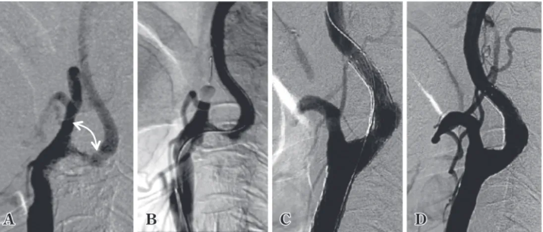
Fig. 1 Large CCA ICA angle(≧60 )as anatomical risk for carotid artery stenting
A: An illustrative case demonstrating a common carotid artery internal carotid artery with
an angle of 90 degree. In this case, a complicated intervention maneuver is necessary.
B: Patient required a buddy wire technique to introduce a stent to the internal carotid
artery.
C , D:The internal carotid artery was straightened by stiff wire, and a stent was deployed under the proximal balloon protection.
A
症候性病変の脳虚血イベントを 1%/年以下に抑えるこ とが可能になっている観点からも1),CAS 周術期脳虚血 イベントの制御は重要である. CAS の周術期脳虚血イベントは発生時期により,1) ガイディングカテーテル誘導,2)遠位型 EPD1 などのデ バイスの通過,3)バルーンあるいはステント血管拡張, 4)ステント留置後,以上の 4 相に分類できる.1∼4 の 各相でのイベント発生機転と考えられ得る対応策を述べ る. ガイディングカテーテル誘導時のイベント CAS 術者は,術後 MRI で治療頚動脈の灌流外領域の DWI陽性病変を経験する.これは,ガイディングカテー テルの罹患頚動脈誘導前に,上行あるいは大動脈弓部で のカテーテル操作で塞栓症を発症させたことによる病変 で,術者経験が浅い場合に多く経験される.術者選定が 問題とされた EVA 3S では,イベントの約 30%がこの相 で発生していた20).Verzini ら31)の,単一施設における
CAS導入から 6 年間の連続 627 例の learning curve の解
析では,合併症は前半 3 年間に有意に高率で,前半期に 占めるイベントの多くはガイディングカテーテル誘導時 に生じたと報告されている.この相でのイベント低減に は,頭頚部血管のカテーテル手術手技の習熟と,learning curveの克服が必須とされる11).加えて,ガイディング カテーテル誘導時のイベントを回避するには,術前のア クセスルート評価や,大動脈弓の形状,さらには大動脈 弓部のアテローム性硬化の評価も重要となる.それぞ れ,胸・腹部大動脈瘤や高度な粥状動脈硬化,TypeⅢ arch,bovine aortic arch さらには right sided aortic arch な
ど大動脈弓の変異(Fig. 2),上行∼大動脈弓部の高度な アテローム性硬化症を有する症例では,経上腕動脈アプ ローチの採用が対策の 1 つとなる.ただし,経上腕動脈 アプローチにおいてもアクセスルート評価が重要で,右 鎖骨下動脈から右総頚動脈,あるいは,腕頭動脈から左 総頚動脈への進入角度の評価が必要となる.症例ごと に,安全に手技を完遂するためのアプローチ方法を検討 することが重要である. 遠位型EPDなどのデバイスの通過時のイベント 内頚動脈の高度狭窄例や高度屈曲例,病変部に可動性 プラークが存在する場合には,EPD の病変通過時にイベ ントが誘発され得る.1と同様に,術前 CT や MR によ 1 2
Table 3 Guideline changes for carotid revascularization
Classification of recommendations and the level of evidence on American Heart Association(AHA)and American Stroke Association(ASA)guidelines.
Symptom CEArisk Stenosis AHA/ASA2006 AHA/ASA2011 AHA/ASA2014
Yes Normal
50 69 CEA(class Ⅰ/LOE A)†* CEA(class Ⅰ/LOE B)†*
CAS(class Ⅰ/LOE B) CEA(class Ⅰ/LOE B)
†*∫
CAS(class Ⅰ/LOE B)†*∫
70%≦ CEA(class Ⅰ/LOE A)†* CEA(class Ⅰ/LOE A)†*
CAS(class Ⅰ/LOE B) CEA(class Ⅰ/LOE A)
†*∫
CAS(class Ⅱa/LOE B)†*∫
High 70%≦ CAS(class Ⅱb/LOE B)‡ CAS(class Ⅱb/LOE B)‡ CAS(class Ⅱa/LOE B)†
No Normal 60≦ (DSA) 70%≦ (US)
CEA(class Ⅰ/LOE A)†
CEA(class Ⅱa/LOE A)†
CAS(class Ⅱb/LOE B) CEA(class Ⅱa/LOE A)
† CAS(class Ⅱb/LOE B) CEA/CAS adavantage
compared with medical treatment is not unclear
Effectiveness of CEA/CAS is not established compared with medical treatment alone
High
60≦ (DSA)
70%≦ (US)
CAS(class Ⅱb/LOE B) Usefulness of CAS forhigh risk CEA is uncertain
(class Ⅱb/LOE C)
Effectiveness of CEA/CAS is not well established compared with medical therapy alone
(class Ⅱb/LOE B)
LOE:level of evidence, DSA:digital subtraction angiography, US:ultrasound sonography
†:Treatment is recommended if perioperative stroke, myocardial infarction and death is <3% in asymptomatic lesion.
<6% in symptomatic lesion.
‡:Reasonable by operator with established periprocedural morbidity and mortality rates of 4 6%.
*:Reasonable to perform the procedure within 2 weeks of the index event.
∫: CEA may be associate with improved outcome for older patients than 70 years, particularly with unfavorable arterial
Fig. 2 Unfavorable aortic arch anatomic variants for carotid artery stenting via the trans femoral approach
A
A:Bovine aortic arch.
B:Type Ⅲ aortic arch.
C:Right sided aortic arch.
B C
Fig. 3 Free floating thrombi of the carotid artery detected on carotid ultrasound in patients with cerebral
infarcts
A: Diffusion weighted images demonstrating multiple high intensity spots in the deep border and cortical border
zone.
B:MR angiography showing 60% stenosis of the right internal carotid artery.
C: A T1 weighted with fat suppression image showing the high intensity plaque in the common and internal carotid
artery that suggested intraplaque hemorrhage.
D: Longitudinal B mode sonography detecting a free floating thrombus(arrows)attached to the mobile plaque with
A B C CCA ICA * D CCA ICA * E
る病変の形状および性状評価,さらには頚動脈超音波に よる病変の動的評価を行うことも重要である(Fig. 3). このような症例では,近位バルーンEPDがリスク低減に 有効である. バルーンあるいはステント血管拡張時のイベ ント SAPPHIRE,CREST,ACT 1 では,フィルター型 EPD 使用が規定されていた.フィルター型 EPD は,開発当初 から,①フィルターと血管壁の間隙をすり抜ける栓子, ②フィルター自体をすり抜ける栓子が注目されていた.
古典的な in vitro の研究として,①Müller Hülsbeck ら18)
は,屍体から採取した頚動脈プラークから500∼1,500μm の栓子を作成し,4 種類の異なるフィルター型 EPD での 粒子補足を検討した.いずれのデバイスにおいてもフィ ルターと血管の間に生じる間隙からの栓子のすり抜けが 確認され,デバイスによって血栓捕捉能力の差も大き く,最大 4.4%の粒子がすり抜けることを報告した.②現 行のフィルターのマイクロポア(孔)は 100μ程度に作 成されているが,Rapp ら23)は,CEA で採取したプラー クにバルン拡張を行って飛散デブリスを検討した結果, 100μm 以下のデブリスが平均 70 万個発生することを明 らかにした.そこから抽出された 60∼100μm のプラー ク粒子 100 個を,ラットの頚動脈から注入すると,脳梗 塞が生じることを組織学的に示した.臨床では,フィル ターをすり抜けるデブリスが,CAS 手技中の経頭蓋超音 波(transcranial Doppler:TCD)モニタリングや術後の MRI DWI陽性病変から解析されている.53 病変をラン ダムに近位バルーン EPD とフィルター EPD に割り付け し,術中に生じる栓子を検討した研究では,手技の開始 から終了までに TCD で観察される microembolic signals (MES)は,バルーンで 27%,フィルターでは 100%に 発生し,術後 MRI DWI 陽性病変は,それぞれ 14.3%と 42.8%に発生していたことが報告された17).MRI DWI 陽 性病変の発生に関し,近位バルーンとフィルターEPDに よる CAS を比較した 5 つの RCT と 3 つの non RCT 試験 のメタ解析28)では,近位バルーンEPDが術中塞栓保護に 有利であることが示された〔効果量(ES)=−0.50(95% 信頼区間:−0.97∼−0.27)〕.脆弱な症候性プラークや, 術前評価で lipid rich プラークと評価される症例では,完 全拡張に固執しない治療手技12)に加え,EPD の選択は, 近位バルーン,さらには近位バルーンをフィルター EPD のバックアップとして併用するなどが効果的となろう. ステント留置後のイベント 治療経験数の多い術者では,前述の 1∼3 相でのイベ ントが少なく,4 相でのイベントが相対的に高くなるこ とが報告されている.Bosiers ら3)は,ヨーロッパ 4 施設 の熟練した CAS 術者による 3,179 例の CAS 治療成績を解 析した.彼らの報告では,症候性 1,317 例中に脳虚血イ ベントは 48 例(3.6%)発生し,このうちの 75%にあた る 36 例がステント留置後に発生していた.無症候性病 変 1,862 例では,脳虚血イベント発生は 42 例(2.2%) で,このうちの 60%(25 例)がステント留置後であっ た.彼らは,症候性病変に対して,open cell type のステ ントを使用した際に,ステント留置後イベントが起こり やすいことを報告した.ステント留置以降の遅発性イベ ントの発生機序は,ステントがプラークを十分に被覆し
3
4
Fig. 4 Carotid ultrasonography of a stented vessel on postprocedural Day 3, and photograph
simulat-ing plaque protrusion in an in vitro experiment
A A
A:B mode ultrasonography suggesting plaque protrusion in the stented left carotid artery.
B:Color flow image of ultrasonography demonstrating plaque prolapsing through the stent.
C:In vitro experiment simulating plaque protrusion through a stent.
ていないことが主要因と考えられており3)25)(Fig. 4),ス テント内突出プラークの遊離,突出プラークを核とする 血栓形成が原因と推測されている.ステント内プラーク 突出は,治療中の DSA では診断されにくいが,IVUS で は,7.8%に診断されるとの報告がある25).Kotsugi ら10) は,連続 354 病変の自験例を解析し,ステント内プラー ク突出は 9 病変(2.6%)に発生し,このうちの 6 例が脳 梗塞(major stroke 1 例,minor stroke 5 例)を発症し, リスク因子は,open cell stent(p<0.034),不安定プラー ク(p<0.004)と報告している.
現行のベアメタルステントによるステント内プラーク 突出を低減する目的で,近年 micro mesh covered stent
が開発され,臨床試験が進んでいる(Table 4).本デバ イスは,細かなメッシュ構造をナイチノールステントに 重合させ,プラーク表面のステントの接着面積を増やす ことで,プラークを効率的に被覆する新しいタイプのス テントである.そのうちの 1 つである CGUARDTM EPS の臨床効果については前向き研究の結果が報告され,
101病変に対して,周術期の death or major stroke:0%,
minor stroke 0.9%,治療 30 日以内の新たなイベント発症
0%と報告された(PARADIGM 試験)19).Dual antiplatelet
therapy(DAPT)によっても制御されない,ステント留
結 語
今日までの治療経験の集積によって明らかにされた CASの課題は,周術期脳梗塞発症のさらなる低減であ る.CAS が,合併症を抑え,十分な治療効果を発揮する には,一定の術者経験と CAS のリスク要因の十分な理解 が必須である.そしてさらに治療成績を向上させるため には,症例に応じた適切な EPD 選択と,ステント留置後 イベント回避を可能とする新規ステントの開発と導入が 必要である. 利益相反 著者全員は日本脳神経外科学会への COI 自己申告の登録を 完了しています.本論文に関して開示すべき COI はありませ ん. 文 献1) Abbott AL, Adelman MA, Alexandrov AV, Barber PA, Barnett HJ, Beard J, Bell P, Björck M, Blacker D, Bonati LH, Brown MM, Buckley CJ, Cambria RP, Castaldo JE, Comerota AJ, Connolly ES Jr, Dalman RL, Davies AH, Eckstein HH, Faruqi R, Feasby TE, Fraedrich G, Gloviczki P, Hankey GJ, Harbaugh RE, Heldenberg E, Hennerici MG, Hill MD, Kle-inig TJ, Mikhailidis DP, Moore WS, Naylor R, Nicolaides A, Paraskevas KI, Pelz DM, Prichard JW, Purdie G, Ricco JB, Ringleb PA, Riles T, Rothwell PM, Sandercock P, Sillesen H, Table 4 Micro mesh covered stents
Three kinds of micro mesh covered stent are listed. Photographs of their whole aspect and hyper magnified stent structures are demonstrated, and each structure and cell size is defined.
Roadsaver® (Terumo) GORE ® Carotid Stent (GORE) CGUARD TM EPS (InspireMD)
Closed cell nitinol frame +
Inside nitinol micromesh
Open cell nitinol frame +
Outside ePTFE lattice
Open cell nitinol frame +
Outside PET micronet
Cell size:375 500μm Cell size:500μm Cell size:150 180μm
tispecialty, expert review and position statement. Stroke
44:1186 1190, 2013.
2) Bergeon P, Rudondy P, Benichou H, Raybaud G, Pellati R, Guennaoui T, Courbier R:Transluminal angioplasty for recurrent stenosis after carotid endarterectomy. Prognostic factors and indications. Int Angiol 12:256 259, 1993. 3) Bosiers M, de Donato G, Deloose K, Verbist J, Peeters P,
Castriota F, Cremonesi A, Setacci C:Does free cell area influence the outcome in carotid artery stenting? Eur J Vasc Endovasc Surg 33:135 143, 2007.
4) Brott TG, Hobson RW 2nd, Howard G, Roubin GS, Clark WM, Brooks W, Mackey A, Hill MD, Leimgruber PP, Sheffet AJ, Howard VJ, Moore WS, Voeks JH, Hopkins LN, Cutlip DE, Cohen DJ, Popma JJ, Ferguson RD, Cohen SN, Blacks-hear JL, Silver FL, Mohr JP, Lal BK, Meschia JF;CREST Investigators:Stenting versus endarterectomy for treat-ment of carotid artery stenosis. N Engl J Med 363:11 23, 2010.
5) Calvet D, Mas JL, Algra A, Becquemin JP, Bonati LH, Dob-son J, Fraedrich G, Jansen O, Mali WP, Ringleb PA, Chatel-lier G, Brown MM, Calvet D, Mas JL, Algra A, Becquemin JP, Bonati LH, Dobson J, Fraedrich G, Jansen O, Mali WP, Ringleb PA, Chatellier G, Brown MM, Algra A, Becquemin JP, Chatellier G, Mas JL, Fraedrich G, Ringleb PA, Jansen O, Brown MM;Carotid Stenting Trialists Collaboration: Carotid stenting:is there an operator effect? A pooled anal-ysis from the carotid stenting trialists collaboration. Stroke
45:527 232, 2014.
6) Diethrich EB, Ndiaye M, Reid DB:Stenting in the carotid artery:initial experience in 110 patients. J Endovasc Surg
3:42 62, 1996.
7) Endarterectomy for asymptomatic carotid artery stenosis. Executive Committee for the Asymptomatic Carotid Ath-erosclerosis Study. JAMA 273:1421 1428, 1995. 8) International Carotid Stenting Study investigators, Ederle J,
Dobson J, Featherstone RL, Bonati LH, van der Worp HB, de Borst GJ, Lo TH, Gaines P, Dorman PJ, Macdonald S, Lyrer PA, Hendriks JM, McCollum C, Nederkoorn PJ, Brown MM:Carotid artery stenting compared with endar-terectomy in patients with symptomatic carotid stenosis (International Carotid Stenting Study):an interim analysis
of a randomised controlled trial. Lancet 375:985 997, 2010.
9) Kernan WN, Ovbiagele B, Black HR, Bravata DM, Chimow-itz MI, EzekowChimow-itz MD, Fang MC, Fisher M, Furie KL, Heck DV, Johnston SC, Kasner SE, Kittner SJ, Mitchell PH, Rich MW, Richardson D, Schwamm LH, Wilson JA;American Heart Association Stroke Council, Council on Cardiovascu-lar and Stroke Nursing, Council on Clinical Cardiology, and Council on Peripheral Vascular Disease. Guidelines for the prevention of stroke in patients with stroke and transient ischemic attack:a guideline for healthcare professionals from the American Heart Association/American Stroke Association. Stroke 45:2160 2236, 2014.
10) Kotsugi M, Takayama K, Myouchin K, Wada T, Nakagawa I, Nakagawa H, Taoka T, Kurokawa S, Nakase H, Kichikawa K:Carotid artery stenting:investigation of plaque protru-sion incidence and prognosis. JACC Cardiovasc Interv 10: 824 831, 2017.
11) Lin PH, Bush RL, Peden EK, Zhou W, Guerrero M, Henao EA, Kougias P, Mohiuddin I, Lumsden AB:Carotid artery stenting with neuroprotection:assessing the learning curve
and treatment outcome. Am J Surg 190:850 857. 2005. 12) Malik RK, Vouyouka A, Salloum A, Marin ML, Faries PL:
Tips and techniques in carotid artery stenting. J Vasc Surg
50:216 220, 2009.
13) Markus HS, King A, Shipley M, Topakian R, Cullinane M, Reihill S, Bornstein NM, Schaafsma A:Asymptomatic embolisation for prediction of stroke in the Asymptomatic Carotid Emboli Study(ACES):a prospective observational study. Lancet Neurol 9:663 671, 2010.
14) Marquardt L, Geraghty OC, Mehta Z, Rothwell PM:Low risk of ipsilateral stroke in patients with asymptomatic carotid stenosis on best medical treatment:a prospective, population based study. Stroke 41:e11 e17, 2010. 15) Mas JL, Chatellier G, Beyssen B, Branchereau A, Moulin T,
Becquemin JP, Larrue V, Lièvre M, Leys D, Bonneville JF, Watelet J, Pruvo JP, Albucher JF, Viguier A, Piquet P, Gar-nier P, Viader F, Touzé E, Giroud M, Hosseini H, Pillet JC, Favrole P, Neau JP, Ducrocq X;EVA 3S Investigators: Endarterectomy versus stenting in patients with symptom-atic severe carotid stenosis. N Engl J Med 355:1660 1671, 2006.
16) Meschia JF, Bushnell C, Boden Albala B, Braun LT, Bravata DM, Chaturvedi S, Creager MA, Eckel RH, Elkind MS, For-nage M, Goldstein LB, Greenberg SM, Horvath SE, Iadecola C, Jauch EC, Moore WS, Wilson JA;American Heart Asso-ciation Stroke Council;Council on Cardiovascular and Stroke Nursing;Council on Clinical Cardiology;Council on Functional Genomics and Translational Biology;Council on Hypertension:Guidelines for the primary prevention of stroke:a statement for healthcare professionals from the American Heart Association/American Stroke Association. Stroke 45:3754 3832, 2014.
17) Montorsi P, Caputi L, Galli S, Ciceri E, Ballerini G, Agrifoglio M, Ravagnani P, Trabattoni D, Pontone G, Fabbiocchi F, Loaldi A, Parati E, Andreini D, Veglia F, Bartorelli AL: Microembolization during carotid artery stenting in patients with high risk, lipid rich plaque. A randomized trial of proximal versus distal cerebral protection. J Am Coll Cardiol
58:1656 1663, 2011.
18) Müller Hülsbeck S, Jahnke T, Liess C, Glass C, Paulsen F, Grimm J, Heller M:In vitro comparison of four cerebral protection filters for preventing human plaque embolization during carotid interventions. J Endovasc Ther 9:793 802, 2002.
19) Musialek P, Mazurek A, Trystula M, Borratynska A, Lesn-iak Sobelga A, Urbanczyk M, Banys RP, Brzychczy A, Zajdel W, Partyka L, Zmudka K, Podolec P:Novel PARADIGM in carotid revascularisation:Prospective evaluation of All comer peRcutaneous cArotiD revascularisation in symptom-atic and Increased risk asymptomsymptom-atic carotid artery steno-sis using CGuardTM MicroNet covered embolic prevention
stent system. EuroIntervention 12:e658 670, 2016. 20) Naggara O, Touzé E, Beyssen B, Trinquart L, Chatellier G,
Meder JF, Mas JL;EVA 3S Investigators:Anatomical and technical factors associated with stroke or death during carotid angioplasty and stenting:results from the endarter-ectomy versus angioplasty in patients with symptomatic severe carotid stenosis(EVA 3S)trial and systematic review. Stroke 42:380 388, 2011.
21) North American Symptomatic Carotid Endarterectomy Trial Collaborators, Barnett HJM, Taylor DW, Haynes RB, Sack-ett DL, Peerless SJ, Ferguson GG, Fox AJ, Rankin RN,
Hachinski VC, Wiebers DO, Eliasziw M:Beneficial effect of carotid endarterectomy in symptomatic patients with high grade carotid stenosis. N Engl J Med 325:445 453, 1991. 22) Rantner B, Goebel G, Bonati LH, Ringleb PA, Mas JL, Frae-drich G;Carotid Stenting Trialists Collaboration:The risk of carotid artery stenting compared with carotid endarterec-tomy is greatest in patients treated within 7 days of symp-toms. J Vasc Surg 57:619 626, 2013.
23) Rapp JH, Pan XM, Yu B, Swanson RA, Higashida RT, Simp-son P, Saloner D:Cerebral ischemia and infarction from atheroemboli <100 microm in size. Stroke 34:1976 1980, 2003.
24) Rosenfield K, Matsumura JS, Chaturvedi S, Riles T, Ansel GM, Metzger DC, Wechsler L, Jaff MR, Gray W;ACT I Investigators:Randomized trial of stent versus surgery for asymptomatic carotid stenosis. N Engl J Med 374:1011 1020, 2016.
25) Shinozaki N, Ogata N, Ikari Y:Plaque protrusion detected by intravascular ultrasound during carotid artery stenting. J Stroke Cerebrovasc Dis 23:2622 2625, 2014.
26) Sigwart U, Puel J, Mirkovitch V, Joffre F, Kappenberger L: Intravascular stents to prevent occlusion and restenosis after transluminal angioplasty. N Engl J Med 316:701 706, 1987.
27) SPACE Collaborative Group, Ringleb PA, Allenberg J, Brückmann H, Eckstein HH, Fraedrich G, Hartmann M, Hennerici M, Jansen O, Klein G, Kunze A, Marx P, Nieder-korn K, Schmiedt W, Solymosi L, Stingele R, Zeumer H, Hacke W:30 day results from the SPACE trial of stent protected angioplasty versus carotid endarterectomy in symptomatic patients:a randomised non inferiority trial.
Lancet 368:1239 1247, 2006.
28) Stabile E, Sannino A, Schiattarella GG, Gargiulo G, Toscano E, Brevetti L, Scudiero F, Giugliano G, Perrino C, Trimarco B, Esposito G:Cerebral embolic lesions detected with dif-fusion weighted magnetic resonance imaging following carotid artery stenting:a meta analysis of 8 studies com-paring filter cerebral protection and proximal balloon occlu-sion. JACC Cardiovasc Interv 7:1177 1183, 2014. 29) Stingele R, Berger J, Alfke K, Eckstein HH, Fraedrich G,
Allenberg J, Hartmann M, Ringleb PA, Fiehler J;SPACE investigators, Bruckmann H, Hennerici M, Jansen O, Klein G, Kunze A, Marx P, Niederkorn K, Schmiedt W, Solymosi L, Zeumer H, Hacke W:Clinical and angiographic risk fac-tors for stroke and death within 30 days after carotid endar-terectomy and stent protected angioplasty:a subanalysis of the SPACE study. Lancet Neurol 7:216 222, 2008. 30) Theron JG, Payelle GG, Coskun O, Huet HF, Guimaraens
L:Carotid artery stenosis:treatment with protected bal-loon angioplasty and stent placement. Radiology 201: 627 636, 1996.
31) Verzini F, Cao P, De Rango P, Parlani G, Maselli A, Romano L, Norgiolini L, Giordano G:Appropriateness of learning curve for carotid artery stenting:an analysis of periproce-dural complications. J Vasc Surg 44:1205 1212, 2006. 32) Yadav JS, Wholey MH, Kuntz RE, Fayad P, Katzen BT,
Mish-kel GJ, Bajwa TK, Whitlow P, Strickman NE, Jaff MR, Popma JJ, Snead DB, Cutlip DE, Firth BG, Ouriel K ;Stent-ing and Angioplasty with Protection in Patients at High Risk for Endarterectomy Investigators:Protected carotid artery stenting versus endarterectomy in high risk patients. N Engl J Med 351:1493 1501, 2004. 頚動脈狭窄症に対する血管内治療 ―今日までの治療経験の集積と次世代に向けて― 秋山 恭彦 宮嵜 健史 萩原 伸哉 中右 博也 神原 瑞樹 吉金 努 将大 藤原 勇太 内村 昌裕 永井 秀政 頚動脈ステント留置術は,全身性動脈疾患であるアテローム性動脈硬化症に対し,頚部局所のアテ ロームを制御する治療で,低侵襲であるために,周術期の心筋梗塞をはじめとする全身性合併症を低 く抑えることが可能である.しかし,治療本来の目的は脳梗塞予防であるため,現行の治療は,周術 期の脳虚血イベントを十分に制御できているとはいえない.本稿では,これまでの頚動脈ステント留 置術と頚動脈内膜剝離術のランダム化比較試験および,そのサブ解析の結果を概説し,本治療法の現 状と課題を周術期脳虚血合併症に焦点をあて整理し,次世代への治療の進歩を探る. 脳外誌 26:728⊖737,2017 要 旨