Nanoplasmonics for Real‑Time and Label‑Free Monitoring of Microbial Biofilm Formation
Author Riccardo Funari, Nikhil Bhalla, Kang‑Yu Chu, Bill Soderstrom, Amy Q. Shen
journal or
publication title
ACS Sensors
volume 3
number 8
page range 1499‑1509
year 2018‑07‑31
Publisher American Chemical Society
Rights (C) 2018 American Chemical Society Author's flag publisher
URL http://id.nii.ac.jp/1394/00000775/
doi: info:doi/10.1021/acssensors.8b00287
ACS AuthorChoice (https://pubs.acs.org/page/policy/authorchoice̲termsofuse.html)
Nanoplasmonics for Real-Time and Label-Free Monitoring of Microbial Bio fi lm Formation
Riccardo Funari,*
,†,¶Nikhil Bhalla,
†,¶Kang-Yu Chu,
†Bill Söderström,
‡and Amy Q. Shen*
,††
Micro/Bio/Nano fl uidics Unit and
‡Structural Cellular Biology Unit, Okinawa Institute of Science and Technology Graduate University, 1919-1 Tancha, Onna-son, Okinawa 904-0495, Japan
*
S Supporting InformationABSTRACT: Microbial bio fi lms possess intrinsic resistance against conventional antibiotics and cleaning procedures; thus, a better understanding of their complex biological structures is crucial in both medical and industrial applications. Existing laboratory methodologies have focused on macroscopic and mostly indirect characterization of mechanical and micro- biological properties of bio fi lms adhered on a given substrate.
However, the kinetics underlying the bio fi lm formation is not well understood, while such information is critical to understanding how drugs and chemicals in fl uence the bio fi lm formation. Herein, we report the use of localized surface plasmon resonance (LSPR) for real-time, label-free monitor- ing of E. coli bio fi lm assembly on a nanoplasmonic substrate consisting of gold mushroom-like structures. Our LSPR sensor
is able to capture the signatures of bio fi lm formation in real-time by measuring the wavelength shift in the LSPR resonance peak with high temporal resolution. We employ this sensor feature to elucidate how bio fi lm formation is a ff ected by di ff erent drugs, including conventional antibiotics (kanamycin and ampicillin) as well as rifapentine, a molecule preventing cell adhesion yet barely a ff ecting bacterial viability and vitality. Due to its fl exibility and simplicity, our LSPR based platform can be used on a wide variety of clinically relevant bacteria, thus representing a valuable tool in bio fi lm characterization and drug screening.
KEYWORDS: bio fi lms, real-time monitoring, LSPR, antibiotics, drug screening, E. coli
B acteria exhibit two types of growth regimes: planktonic, where cells are freely moving in a bulk solution, and sessile aggregates known as bio fi lms.
1,2In the latter form, the microorganisms are closely packed on a solid surface within self-produced matrix of extracellular polymeric substances (EPS). This structure provides many structural and functional bene fi ts such as improved resource capture, adhesion to surfaces, digestive capacity, protection against external agents, and inhibition of bacterial dehydration. Furthermore, the EPS sca ff old facilitates intercellular interactions and horizontal gene transfer.
3Another key feature of bacterial bio fi lms concerns the development of their peculiar resistance against antimicrobial agents.
4Some of their possible protection strategies involve poor antibiotic penetration, formation of gradients of nutrients or cell products, and phenotypic di ff erentiation induced by the EPS matrix.
5,6In addition, the lack of nutrients and the high concentrations of bacterial metabolites in bio fi lms result in areas where cells are in a stationary phase (a slow or nongrowth phase of bacterial growth cycle), which become mostly immune to conventional antibiotic drugs since they target speci fi cally to the metabolically active microorganisms.
7Because of their unique properties, bacterial bio fi lms have been actively studied across diverse fi elds, ranging from industrial processes to medicine. For example, bio fi lms have
been used in bioremediation, waste treatment, and production of fi ne chemicals and biofuels.
8On the other hand, bio fi lms are associated with serious health issues stemming from persistent infections due to the contamination of medical devices (e.g., intravenous and urinary catheters), arti fi cial implants,
9and pollution of drinking water.
10Similar concerns have been recently raised by the world health organization (WHO) through their fi rst global antimicrobial resistance surveillance system (GLASS) report.
11In this context, bacterial detection, antibiotic susceptibility, and the rise of antibiotic resistant microorganisms are pressing issues requiring novel and sensitive detection strategies which can also be adopted to screen new drugs. For instance, Jo et al.
12recently developed a capacitive aptamer-based biosensor to monitor the bacterial growth and antibiotic susceptibility in real-time. In another study, Brosel-Oliu et al.
13detected pathogenic E. coli using impedance spectroscopy. However, both approaches focused on the detection of single planktonic bacteria instead of microbial bio fi lm formation. In contrast, bio fi lm assembly requires the direct adhesion of the bacterial cells onto the
Received: April 9, 2018 Accepted: July 31, 2018 Published: July 31, 2018
Article pubs.acs.org/acssensors Cite This:ACS Sens.2018, 3, 1499−1509
See https://pubs.acs.org/sharingguidelines for options on how to legitimately share published articles.
Table 1. Overview of the Latest Techniques for Bacterial Bio fi lm Characterization
real-timekinetics methodtemporal resolutioncostofsystemassaytypethroughputdrug screeninglimitationskey references BiofilmRingTestYesLowLabeledLowNo Allthesetechniquesarelaboratorybased,unsuitableforon-sitetestingofbacterialbiofilmsandrequireuserto havehighlyspecifictechnicalskillstoperformmeasurements14 ≲1h MicrotiterplatesNoLowLabeledHighYes15,16 N/A StainingAssays(e.g., CrystalViolet)NoLowLabeledLowYes17 N/A DropFlowReactorsYesHighLabeledLowNo18 N/A RheometersYesHighLabel-freeLowNoOnlysuitabletostudymechanicalpropertiesofbiofilm19 Tunable(max1s) AtomicForce MicroscopyNoHighLabel-freeLowNoEventhoughAFMcanprovidedetailedinformationontheadhesiveandstructuralpropertiesofbiofilm,the complexityoftheimagingprocedurestronglylimitstheuseofthismethodologyforhighthroughputtests.20−23 Tunable(max 0.1s) QuartzCrystal MicrobalanceYesLowLabel-freeLowYesCrosssensitivityagainstdensityandviscoelasticpropertiesinthebulkmedia24 Tunable(max 0.1s) Electrochemical Impedance Spectroscopy
YesHighLabel-freeLowYesCrosssensitivitytomassandchargetransportofanalytewhichaffectssensorstability13,25−27 ≲30sperscan Capacitivesensors basedonaptamersYesHighLabeledHighYesTheuseofaptamersforcatchingspecificbacteriapreventsstudyingbacterialbiofilmsinnativeconditions12 ≲3minperscan FieldEffectSensorsYesLowLabel-freeLowYesUnstablemeasurementsduetoelectricalnoiseandmaterialbiocompatibilityissuesatnanoscale28 Tunable(max 0.1s) SurfacePlasmon ResonanceYesHighLabel-freeLowYesPronetochangesinbulksolutionandhencelargenoisyinturbidmedia29 Tunable(max1s) PCRYesHighLabeledHighYesCrosssensitivityandselectivityissueswithnonspecificDNA30 ≲1min FTIRspectroscopyYesHighLabel-freeLowNoRequirestime-consumingpostmeasurementanalysistoidentifypeaksrelatedtosignaturesofbiofilmformation31 ≲5s MicrofluidicsYesHighLabeled/ Label- free HighYesSmallvolumesmakeitdifficulttoanalyzeturbidsamples.Microfluidicsoftenrequireintegrationofsensorsor microscopytoolstovalidatebiofilms.32 ≲1s LocalizedSurface Plasmon Resonance
YesLow(≲200USD)Label-freeHighYesSensitivity/biocompatibilitytradeoffThiswork Tunable(max1 s)
ACS Sensors
substrate because the use of a capturing element (e.g., aptamers or antibodies) prevents the bio fi lm detection under its native condition. Moreover, since bacteria in bio fi lms are extremely resistant to conventional drugs, there is a tremendous interest in discovering new bio fi lm speci fi c antibiotics that can penetrate the polymeric matrix, thus reaching bacterial cells, and/or a ff ecting the EPS structure to destabilize the bio fi lm so that standard antibiotics can subsequently attack the microorganisms.
Motivations described above underpin the research on developing reliable characterization tools to provide the real- time monitoring of biofilm formation under different drugs and chemicals. This activity is promising since detailed information on the bio fi lm growth kinetics is extremely valuable in discovering novel treatments and drugs to combat bio fi lm related infections (e.g., types and concentrations of antibiotics to use). A comparative overview of the latest technologies reported for the characterization of bacterial bio fi lm is
summarized in Table 1 while more details about these methodologies can be found in the Supporting Information.
Recently, nanomaterial based label-free photonic biosensors
have revealed unprecedented information on DNA and protein
molecular interactions, fi nding wide applications in medical
diagnostics, food safety, and environmental monitoring.
33,34However, few attempts have been made to apply label-free
photonic biosensors to cellular assays, as it is challenging to
develop nanostructured substrates with large surface areas that
promote both sensing and long-term cell survival. Localized
surface plasmon resonance (LSPR) is the coherent oscillation
of the surface electrons of metal nanostructures due to
interactions between the incident light and the conduction
band electrons of the metal.
35,36This technology has been
utilized to perform highly sensitive label-free detection of
biomolecular interactions in real time, an essential feature for
the early detection of diseases and point-of-care (POC) clinical
evaluations. However, most recent uses of plasmonic materials
Figure 1.LSPR biochip and microscopic characterization of the plasmonic nanostructures. (a) Snapshot of the device. (b) Schematic of the Au NM LSPR substrate. In order to monitor the bacterial biofilm formation, the nanostructured glass substrate bonds with a PDMS slab containing multiwell structures, where each well can accommodate up to 150μL of cell suspensions. The AFM scan of the Au NM substrate with (c) a 2μm× 2μm scan area; (d) magnification of a 500 nm×500 nm area. This set of measurements shows the uniformity of the Au NM, with an average roughness of 2.0±0.1 nm. SEM images of Au NMs acquired at 100 000×magnification: (e) top; and (f) tilted (40°) images.on bio fi lms exploited the local temperature increase induced by the LSPR e ff ect to prevent bio fi lm formation.
37−39We have recently demonstrated a biocompatible nano- plasmonic substrate by developing a gold-based nanoplasmonic material for long-term monitoring of eukaryotic cell prolifer- ation.
40In this work, by using a similar nanofabrication protocol, we report the use of LSPR for characterizing bio fi lm formation on a highly sensitive, large-scale, and biocompatible nanoplasmonic substrate containing high density gold nano- mushroom structures. Our LSPR substrates consist of gold mushroom-like structures, with stems of silicon dioxide and caps of gold on the order of 30 nm in diameter, to achieve the LSPR e ff ect. Speci fi cally, the model organism Escherichia coli ( E. coli) is used for all studies. The localized surface plasmons on the nanomushroom caps are exploited to monitor bio fi lm formation without any labeling procedure. In addition, we have developed an automated high resolution system allowing the real-time monitoring of bio fi lm formation by continuously illuminating the LSPR biochip in a Faraday cage and recording the resonance peak shift every minute for 24 h. Since LSPR e ff ect is very sensitive to changes of a few tens of nanometers from the sensor surface, our methodology enables precise monitoring of the activities in the bottom layer of the adhering bio fi lm, which is di ffi cult to achieve by using conventional techniques. This information is of paramount importance for fi ghting persistent infections in bio fi lms, since the microbial cells in direct contact with the substrate are mostly protected from the external environment, thus guarded against antibiotic treatment. As a proof of concept study, we further apply our LSPR chips to investigate how three di ff erent types of antibiotics a ff ect the E. coli growth as well as the bio fi lm formation. We demonstrate that our biochip serves as a powerful characterization tool for investigating real-time bio fi lm formation, screening of new drugs, and evaluating alternative cleaning procedures.
■ EXPERIMENTAL SECTION
Bacterial Growth.Precultures of wild type strain MC4100 (K12 derivative) are grown overnight in a 20 mL lysogeny broth (LB) (rich media), shaken at 200 rpm at 37°C. The cultures are back-diluted to 1:200 in a fresh LB media after overnight culture. This roughly corresponds to 2×10⁷CFU mL−1, as estimated from counting single colonies from serial dilutions on LB-agarosefilled Petri dishes. The cultures are gently vortexed before a volume of 125μL of the sample is directly aliquoted into the microwells of the LSPR biochip.
LSPR Biochip Fabrication. The gold (Au) nanomushroom (NM) based LSPR chip is fabricated by a 3-step process. First, we deposit 5 nm Aufilm onto a SiO2substrate at 0.1−0.2 Å s−1using an e-beam evaporator (KE604TT1-TKF1, Kawasaki Science) in a class 1000 clean room. Prior to deposition, SiO2 substrates were cleaned with acetone and isopropanol. Next we anneal the 5 nm goldfilm at 560°C for 3 h to generate a distribution of Au nanoislands on the SiO2substrate. Finally, we selectively etch the Au nanoislands on SiO2 to generate mushroom like structures using reactive ions of SF6. The reactive ion etching (RIE) is performed by inductively coupled plasma (ICP) chemical vapor deposition equipment (Plasmalab 100, Oxford Instruments). SF6 gas is introduced inside the RIE-ICP chamber, maintained at an inside pressure of 10 mTorr and aflow rate of 45 sccm (standard cubic centimeters per minute). The RF power coil and the RF bias coils arefixed to 150 and 10 W, respectively, and the temperature inside the plasma chamber is maintained at 5°C. The total duration of RIE is 5 min. More details on the fabrication techniques can be found in our recent work.41
To fabricate PDMS wells, wefirst make slabs of PDMS by pouring 10:1 polydimethylsiloxane (PDMS) (Dow Corning, Japan) in a Petri dish and then cure the prepolymer for 3.5 h at 60°C after degassing
to remove air bubbles. We then punch holes of 8 mm in diameter by using disposable biopsy punchers (Kai Medical, Japan) to create PDMS wells. Both PDMS and the NM substrate are exposed to oxygen plasma (Harrick Plasma, USA) at 30 W for 1 min. After plasma treatment, PDMS well and NM based LSPR substrate are immediately brought in close contact to ensure strong bonding (Figure 1a). This treatment ensures proper confinement of the sample solution without any leakage. Thereafter the developed LSPR biochips are used for bacterial biofilm sensing.
Morphological Characterization of the LSPR Substrate.
AFM imaging is performed in tapping mode using a Dimension Icon3 (Bruker, Japan) microscope equipped with an aluminum back-coated, antimony-doped Si cantilever from Bruker (TESPA-V2), with typical values of a nominal tip radius∼8 nm, spring constantk≈42 N m−1, and resonance frequencyf0≈320 kHz. Areas of 4μm2and 0.25μm2 have been scanned under a scanning speed of 1 Hz, with a resolution of 512 pixels per line, and a relatively high amplitude set-point ratio (Asp/Afree≈0.85;Afree≈23 nm;Asp≈17 nm). All the measurements have been repeated three times and the experimental results have been processed using NanoScope Analysis 1.8 software (Bruker, USA).
The scanning electron microscopy (SEM) is carried out by using a high performance scanning electron microscope (FEI Quanta 250 FEG, Thermo Fisher Scientific). Images are acquired at 20 kV, with a magnification of 100 000×inside a vacuum chamber maintained at a pressure of 10−4 Pa. To avoid charging of SiO2 surfaces during imaging, the NM substrates are coated with Pt/Pd at a few angstroms thickness. The top view of the NM substrate is captured with the electron gun placed normal to the substrate. In contrast, the side view of the NM substrate is captured by tilting the electron gun at 40°. The SEM images are processed with ImageJ software to analyze detailed morphological features such as the size and gaps between the NM structures.
Sensing Procedure.The instrument involving LSPR consists of two fiber optics patch cords, one connected with a halogen light source (LS-1-LL) and the other connected to the spectroscope (USB4000-UV-vis-ES). All discrete components (spectrometer, light source, and patch chords) are purchased from Ocean Optics (Japan).
The LSPR signals are acquired in T-LSPR (Transmission) mode.
Before taking any signal from the spectroscope, the system is calibrated for dark and light spectrum modes. The LSPR signal is then recorded in absorption mode by observing the wavelength depend- ence of the light absorbed by Au NM via the Spectrasuite software (cross-platform spectroscopy operating software from Ocean Optics).
For noncontinuous experiments, the biochip containing the bacteria grows in a Taitec BR-23PF incubator (Japan) under 37°C, but the biochip is removed from the incubator periodically for the absorption spectrum acquisition under room temperature. The real-time measurements are performed in a Faraday cage under continuous illumination for 24 h. The real-time measurements are acquired by processing the spectrometer data through a homemade graphics user interface on a Matlab platform. The data are then analyzed using OriginPro 2017 (OriginLab, USA).
Crystal Violet Staining. Crystal violet (CV) assay is a well established methodology to evaluate biofilm formation under various growth conditions (e.g., drug screening, strain comparison).42,43For this reason, the CV assay is used to validate the measurements from our LSPR biochip system. The detailed procedure is reported in the Supporting Information.
Viability Staining.LIVE/DEAD BacLight Bacterial Viability Kit (L13152) from Invitrogen has been used to evaluate cell viability on Au NM LSPR substrate and investigate the effect of the continuous white light illumination on E. coli cells. The detailed protocol is reported in theSupporting Information.
■ RESULTS AND DISCUSSION
Characterization of the Nanoplasmonic Chip. An
integrated LSPR biochip (Au NM LSPR substrate bonded
with PDMS wells) is shown in Figure 1a. The nanostructured
ACS Sensors
SiO
2substrate is optically translucent and pink in color due to the presence of plasmonic Au nanostructures. The Au nanostructures have a characteristic pillar shape, with the schematic shown in Figure 1b. The morphological features of the substrate such as the size of the Au NM, gap size between NMs, and their surface density is characterized by using tapping mode atomic force microscopy (AFM) and scanning electron microscopy (SEM), which provide detailed nano- metric features of the LSPR substrate (Figure 1). Based on the AFM images (Figure 1c and d), the Au NMs are quasi- uniformly distributed on the SiO
2surface ( ∼ 10
6− 10
8NM cm
−2) with a root-mean-square roughness of 2.0 ± 0.1 nm.
Based on the high contrast images between Au NMs and the glass substrate obtained by SEM (Figure 1e and f), the average diameter of NMs is 29 ± 0.3 nm (standard error of mean, SE) and 29 ± 14 nm (standard deviation, SD). The average spacing among the Au NMs is 9 ± 1 nm (SE) and 9 ± 6 nm (SD).
For LSPR sensing applications, the average refractive index (RI) sensitivity of our NM LSPR substrate is estimated as 98.6 nm RIU
−1(see more details in the Supporting Information).
The sensitivity of the Au NM can be tuned by changing the size and gaps of the NM structures (discussed in our previous work
40). Note that the size and morphology of Au nanostructures are also crucial for the biocompatibility of the LSPR substrate when it is in direct contact with live entities
such as bacteria, virus, and eukaryotic cells.
40In later sections we will discuss the e ff ects of the continuous exposure of LSPR illumination on the bacterial growth.
LSPR Monitoring of Bacteria Bio fi lm Formation. The local sensitivity and the reliability of our Au NM LSPR substrate have been exploited to develop a new character- ization tool to monitor the E. coli growth and bio fi lm formation. E. coli strains can be both nonpathogenic and pathogenic. The nonpathogenic ones are harmless and easy to manipulate, and hence will be used as our model micro- organism to optimize the sensing procedure. However, the pathogenic E. coli strains are important since they are responsible for a wide range of diseases, e.g., urogenital infections where bacterial cells form bio fi lms.
44Nevertheless, our studies can be easily expanded to other bacterial systems.
Below we will illustrate both noncontinuous and real-time LSPR monitoring of bacterial bio fi lm formation by using a model nonpathogenic E. coli system.
Noncontinuous Monitoring. To study the bio fi lm formation, an inoculum of E. coli (125 μ L, 2 × 10
7CFU mL
−1) is loaded into our LSPR biochip and the bacterial growth is then monitored for about 30 h. For the non- continuous monitoring, the LSPR biochip is removed from an incubator at 37 ° C only periodically when it is required to collect the absorbance spectrum ( ∼ 10 s).
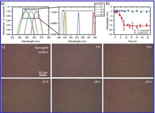
Figure 2.Noncontinuous LSPR monitoring the bacterial adhesion and biofilm formation in the LB culture media. While the bacterial growth is performed at 37°C in an incubator, the absorbance data are obtained at room temperature and take less than 10 s for each measurement. (a) Normalized absorbance spectra for the LSPR biochip incubated with anE. colisample in the LB medium, with zoomed-in details in the wavelength of 546−564 nm region. The peak position shifts to shorter wavelengths during biofilm assembly. The black arrow highlights the progressive blue shift due to microbial biofilm assembly. (b) Absorbance peak position as a function of time. The control experiment performed using only LB culture media produces a negligible shift in the signal (blue circles) when compared with the sample containingE. coli(red squares). Each data point corresponds to at least 3 repetitive experiments (n≥3), with the error bars denoting the standard error of mean (SE). (c) Bacterial growth monitored at 0, 1, 6, 12, 18, and 24 h, captured by bright-field microscopy (Eclipse Ti−U, Nikon Instruments Inc.). The series of images illustrate the progress of the biofilm formation in a LSPR biochip.
As shown in Figure 2a and b, bacterial growth and adhesion on the Au NM substrate produce a signi fi cant blue shift in the absorbance peak of the Au NM, which can be attributed to the charge changes in the overall system upon bio fi lm formation.
As the bacterial fi lm assembly progresses, the charge on the NM substrate increases with the increasing amount of bacteria, protein, and polysaccharide secretion,
45,46which eventually leads to the formation of bio fi lm. Recall that bio fi lm is a slimy matrix composed of mostly proteins and polysaccharides, the latter being negatively charged at physiological pH.
47The increase in the surface charge of Au NM during the bio fi lm formation further leads to an enhanced frequency of plasmonic resonances because the frequency of plasmons is directly proportional to the amount of the charge on the plasmonic material. The increase in the frequency also causes a decrease in the LSPR wavelength, suggesting that the observed blue shifts in the wavelength peak correspond to the bacterial bio fi lm formation. Red square symbols in Figure 2b illustrate that the LSPR signal saturates around 8 h, correlated with the completion of the bio fi lm assembly. For comparison, by incubating the LSPR substrate with the culture medium (LB) for 30 h, only negligible wavelength shift is observed (blue circles in Figure 2b).
The LSPR resonance wavelength versus time data (Figure 2b, red squares) is fitted by using a modified logistic-type function:
Δ =λ + − + − *
b a b
1 e
k t t( ), (1)
where Δλ is the wavelength shift in nanometer, a and b are the initial and the fi nal wavelength values, respectively, k is a rate parameter, and t * corresponds to the time upon which the growth and bio fi lm formation rates are maximized and half of the wavelength shift (a − b) is achieved. By fixing the initial value (a = 0), the best fi t of the experimental responses due to the bacterial growth yields b= − 15.6 ± 0.5 nm, k = 0.93 ± 0.18 h
−1, and t * =5.8 ± 0.3 h.
The bio fi lm formation is also con fi rmed by bright- fi eld microscopy (Figure 2c), which reveals that bacterial growth is barely a ff ected by the nature of the plasmonic substrate nor the LSPR sensing setup. This observation is promising since LSPR substrate has been reported to induce local temperature jump and cause cell death.
48Our results demonstrate that our Au NM LSPR substrate is biocompatible and enables the bacteria to form bio fi lm with sustained viability for more than 30 h.
This simple LSPR platform also allows us to easily evaluate detailed kinetics underlying the bio fi lm formation and can be used to compare di ff erent types of microorganisms or testing the e ff ect of drugs on the bacterial growth.
However, to obtain each measurement point at a speci fi c time (shown in Figure 2b), the LSPR biochip has to be removed from the 37 ° C incubator periodically to capture LSPR signals; hence, some inevitable changes in the LSPR biochip position through the manual alignment and the environmental light have led to some random fl uctuations in the resonance peak wavelength (the error bars in Figure 2b represent the standard error of mean, n ≥ 3). To reduce the contribution of these random fl uctuations, we present the real- time continuous monitoring procedure below.
Real-Time Continuous Monitoring. To enhance the sampling frequency and overcome the noise issues from the noncontinuous monitoring, we have developed an automatized
system to provide the real-time monitoring of the local refractive index changes (hence the LSPR signals) as soon as the E. coli is deposited in the PDMS well of the LSPR biochip.
To prevent sample evaporation, the LSPR biochip is con fi ned in a transparent chamber with a water reservoir. Both bacterial growth and the absorbance spectrum are recorded in a Faraday cage to eliminate the noise caused by the environmental light.
Once the chip is orthogonally aligned between the two optical fi bers, we use a custom-made software to record the absorbance spectrum every minute for a duration of 24 h, thus achieving a much better resolution than those from the noncontinuous measurements. Sensor signals obtained during the fi rst 24 h when an LSPR chip is incubated with an E. coli sample in the LB medium, are shown in the Supporting Information, consistent with the noncontinuous measure- ments.
The stability of the LSPR reading and any nonspeci fi c response resulting from the continuous illumination of the LSPR biochip is fi rst evaluated by recording the transmitted light of a bare Au NM LSPR sensor, in contact with air, for 24 h. The LSPR signals appear to be quite stable for the whole duration of the experiment (purple triangles in Figure 3a).
Thereafter, the same real-time monitoring LSPR procedure is used to compare responses from di ff erent samples: plain
Figure 3. Real-time monitoring of bacterial adhesion and biofilm formation. (a) Resonance wavelength as a function of time for the control (LB),E. coli, andE. coligrowing in the presence of various antibiotics and antibiotic mixture (kanamycin: 100 μg mL−1, ampicillin: 100μg mL−1, rifapentine: 1 μg mL−1). The stability of the sensing platform is evaluated by measuring the resonance peak of the LSPR biochip for 24 h when exposed to air (purple triangles).
Peak positions are estimated byfitting the spectra in the 520−600 nm region using a spline function. (b) Magnification of the sensor responses for thefirst 5 h where the experimental data are shown with the best curvefits.
ACS Sensors
culture medium (LB), E. coli in LB medium, and E. coli treated with di ff erent antibiotics (kanamycin at 100 μ g mL
−1, ampicillin at 100 μ g mL
−1, rifapentine at 1 μ g mL
−1, and a mixture of the three drugs (kanamycin at 100 μ g mL
−1; ampicillin at 100 μ g mL
−1; rifapentine at 1 μ g mL
−1)). These antimicrobial agents attack di ff erent aspects of bacteria metabolisms. Kanamycin and ampicillin are standard anti- biotics: kanamycin interacts with the ribosomes a ff ecting protein synthesis while ampicillin inhibits the transpeptidase, an enzyme involved in cell wall assembling. On the other hand, rifapentine a ff ects the RNA polymerase and has been shown to inhibit curli-dependent bio fi lm formation without killing the bacteria at a concentration of a few μ g mL
−1.
49Bio fi lm growth is controlled by a variety of physical, chemical, and biological phenomena, resulting in cell-to- substrate adhesion and cell-to-cell cohesion events.
19Di ff erent from the noncontinuous measurements (data only captured every 1 − 2 h), a slight red shift in the wavelength is observed during the fi rst 2 h of the real-time monitoring experiments (see Figure 3). The LSPR substrate is first modified by an initial conditioning layer, which promotes the bacteria anchorage and irreversible adhesion. The observed red shift is likely due to the increase in the local dielectric constant of the Au NM structures from the bacterial and protein adhesion on the Au NM substrates since LSPR signal is sensitive to the variation in the local dielectric constant of the sample volume, which extends by ∼ 30 nm from the top of the Au NM surface.
50After 2 h, the Au NM LSPR substrate is completely covered with either bacteria or conditioning biomolecules, preventing further shifts in the local dielectric constant, and thus the LSPR signal starts to exhibit blue shifts. This change of regime is evident in Figure 3b (see the vertical dashed line), where the sensor response from E. coli in LB without any antibiotics (red curve) displays a clear peak and a slope change at about 2 h.
Figure 3a also shows some scattering in the LSPR signal for E. coli and E. coli treated with rifapentine. They are likely due to the high turbidity of the sample, caused by large number of cells in the LSPR-PDMS wells, which makes it di ffi cult to clearly identify the resonance peak. However, this e ff ect does not alter the overall wavelength shift in the resonance peak.
The recorded absorbance spectra captured every minute for all experiments are fi tted using a spline function, providing the resonance wavelength shift data shown in Figure 3a. The experimental results show that bio fi lms treated with conven- tional antibiotics (kanamycin (orange symbols) and ampicillin (green symbols)) lead to signals similar to the one obtained by the control experiment (LB in blue symbols); see Figure 3. On the other hand, rifapentine (gray symbols) at an e ff ective concentration of 1 μ g mL
−149prevents the cell adhesion but
does not eradicate the bacteria, resulting in a smaller wavelength shift than the one obtained by nontreated E. coli (red symbols).
Interestingly, the response of the E. coli treated by the mixed drug system is similar to the one treated with rifapentine during the fi rst 2 h, with the black and the gray curves almost overlapping (see Figure 3b). However, after 2 h, the resonance peak from the mixed drug system shifts to a larger wavelength, reaching similar saturation values as those of the E. coli treated with ampicillin and kanamycin.
The wavelength shifts from all these cases are fi tted by an exponential model:
Δ =λ A(1+e−Bt), (2)
where A and B are the amplitude of the exponential and a rate parameter, respectively.
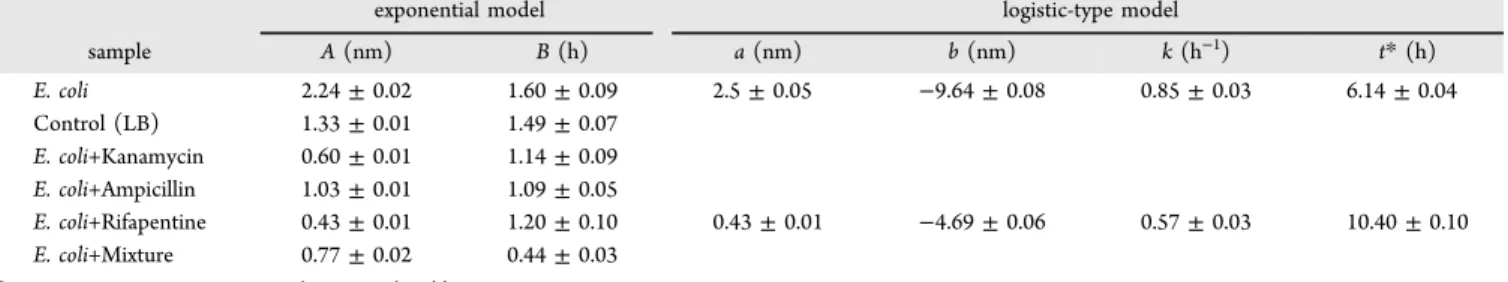
The comparison of the fi tted responses of the tested samples is shown in Figure 3b. Table 2 shows values of fi tted parameters based on the best fi t of the experimental data using the exponential (eq 2) or/and the logistic (eq 1) type models.
The rate parameters k and t * , the time at which the bio fi lm formation rate is maximized, can be correlated with the dampening e ff ect on the bio fi lm formation due to an adhesion inhibiting drug like rifapentine. When rifapentine is added to the culture medium, surface coating rate (k) is signi fi cantly reduced (0.85 ± 0.03 h
−1versus 0.57 ± 0.03 h
−1for E. coli and E. coli treated with rifapentine, respectively), while the time t * to achieve half of the wavelength shift (b − a) increases (6.14 ± 0.04 h versus 10.39 ± 0.11 h, respectively). These results can be very valuable for drug screening studies, since the LSPR signals can be utilized to identify new molecules, which can be combined with conventional antibiotics to either prevent the formation of the microbial superstructure or attack the existing bio fi lm.
Figure 4 provides a general summary of the bio fi lm LSPR responses under di ff erent experimental conditions (non- continuous versus real-time), showing wavelength shifts at 24 h. Note both E. coli - Noncontinuous and E. coli - Real-time tests shown are performed without any drugs or chemicals. In the noncontinuous case, the LSPR biochip is mostly kept in an incubator at 37 ° C for the entire experiment, while in the real- time experiment, the biochip is illuminated with a white light for 24 h, which results in a slightly smaller wavelength shift ( − 11.8 ± 3.4 nm), compared to the noncontinuous test (−14.8 ± 1.3 nm). Among the real-time experiments, the control sample (LB), E. coli treated with kanamycin, ampicillin, and the antibiotic mixture exhibit similar results in the wavelength shift. This is due to the antimicrobial properties of the two conventional drugs which kill mostly all the bacteria Table 2. Parameters Resulting from the Best Fit of the Absorption Maximum Position Data Shown in Figure 3, from the Real- Time Monitoring
aexponential model logistic-type model
sample A(nm) B(h) a(nm) b(nm) k(h−1) t*(h)
E. coli 2.24±0.02 1.60±0.09 2.5±0.05 −9.64±0.08 0.85±0.03 6.14±0.04
Control (LB) 1.33±0.01 1.49±0.07
E. coli+Kanamycin 0.60±0.01 1.14±0.09
E. coli+Ampicillin 1.03±0.01 1.09±0.05
E. coli+Rifapentine 0.43±0.01 1.20±0.10 0.43±0.01 −4.69±0.06 0.57±0.03 10.40±0.10
E. coli+Mixture 0.77±0.02 0.44±0.03
aThe naked LSPR substrate (control (Air)) shows negligible wavelength shift, hence data not included in this table.
before they are able to adhere onto the sensor surface and form the bio fi lm. On the other hand, the LSPR biochip incubated with rifapentine-treated E. coli produces a wavelength shift of
− 4.8 ± 1.1 nm, which is about 50% of those with nontreated bacteria cells. These results are consistent with the intended drug mechanisms of the three antibiotics used: antimicrobial activity versus bacterial adhesion prevention. Indeed, using a drug (like refapentine) to attack bio fi lm formation is not su ffi cient to fi ght the bacterial infection. As evidenced by the response of the LSPR biochip of E. coli treated with a mixture of antibiotics (E. coli + Mixture series of data in Figure 3 and Figure 4), a more e ff ective strategy to fi ght bacterial bio fi lm is to combine conventional bacterial killing antibiotics with drugs that target speci fi cally to the bio fi lm structure.
Since the wavelength shift in the adsorption peaks at 24 h is related to the amount of bacterial cells and EPS absorbed onto the LSPR substrate, these LSPR signals can be compared with the standard end-point microbiology assays such as crystal violet staining assay for the bio fi lm mass evaluation, see section below (see protocol details in Experimental Section).
LSPR Sensor Response versus Crystal Violet Staining and Viability Test. In order to validate the results of our LSPR-based methodology, two independent standard staining procedures have been used to compare the mass and cell viability of bacterial bio fi lms. Crystal violet (CV) assay is the most common methodology to evaluate bio fi lm formation and is based on the direct measurement of microbial biomass. This technique is commonly performed by comparing the absorbance at 570 nm of di ff erent samples after 24 h bacterial growth with CV staining, in a 96-well plate. Since this is a disruptive assay, CV staining does not allow investigations of
dynamic phenomena. After subtracting the contribution of the culture media (LB), the absorbance intensities at 570 nm for stained samples of E. coli treated with kanamycin, ampicillin, and rifapentine are normalized with respect to the intensity value of nontreated E. coli, which is considered as 100% of the response of the assay. In Figure 5a, the results provided by CV staining (blue bars) are compared with the wavelength shifts (normalized by the 24 h value) captured by our LSPR biochip (red bars).
Consistent with the results illustrated by our LSPR-based biosensor, the treatment with kanamycin and ampicillin provides minor signals since most of the bacteria are killed by these two antibiotics. On the other hand, the treatment with rifapentine is less aggressive since it a ff ects only the adhesion properties of the microbes. This produces an absorbance value at 570 nm, which is about 50% of the value from the nontreated E. coli, thus being in excellent agreement with the measurements provided by our LSPR based biochip.
As previously reported, the oscillation of the electrons related to the LSPR sensing can lead to local temperature increase, which can kill bacterial cells.
48As a precaution, we next evaluate how our LSPR based detection system a ff ects the cell vitality. For this reason we use the LIVE/DEAD Baclight Bacterial Viability Kit, which allows us to discriminate between dead (red dye) and living (green dye) cells by fl uorescence microscopy. This test has been performed directly on the LSPR Au NM substrate after 24 h of bacterial growth; see images shown in Figure 5b (I, II, III).
The portion of living cells on a glass slide (Figure 5b panel I), being used as a control study, yields about 95%. For a non- illuminated LSPR substrate (Figure 5b panel II), this value is about 85%. On the other hand, the survival percentage of the bacteria reduces to 65% on the LSPR biochip used for real- time measurements (Figure 5b panel III), likely due to the local stress induced by the continuous light irradiation. This decrease in vitality for continuously illuminated sample matches the di ff erence observed in the LSPR response between the noncontinuous and real-time LSPR measurements (in Figure 4). Nevertheless, despite the substrate and stress in fl uence on the bacterial cell vitality, the bacterial cells are still able to multiply and adhere onto the substrate with bio fi lm formation. For future research, we plan to include a shutter to illuminate the LSPR biochip only brie fl y (few seconds) to collect the transmitted light. This will reduce the stress applied to the microbial cells, thus improving their vitality.
■ CONCLUSIONS
We report the application of an LSPR-based sensor for the direct, real-time, and nondisruptive label-free characterization of bacterial bio fi lm in a liquid environment. Our LSPR sensor platform is able to capture the bio fi lm assembling kinetics and illustrate how such process is a ff ected by di ff erent antibiotics.
The sensing device is based on Au nanoplasmonic structures realized by a cost-e ff ective nanofabrication process. The quanti fi cation of E. coli bio fi lm formation by our LSPR-based device shows excellent agreement with the measurements performed by conventional microbiology techniques (e.g., crystal violet assay and viability staining), thus proving the reliability and the robustness of our approach and paving the way to its utility as a promising tool for characterizing bacterial bio fi lms. Moreover, we demonstrate how e ffi cacy of drugs can be tested using our LSPR biochips, which lays the foundation to apply LSPR technology in antibiotic drug discovery. Our
Figure 4.Comparison at 24 h between nontreated E. coli, culturemedia LB, and bacteria under different antibiotic treatments. Real- time measurements (E. coli - Real-time) require the continuous illumination of the sample with a white light, inducing additional stress on bacteria than noncontinuous measurements (E. coli - Noncontinuous), which results in a slightly smaller wavelength shift (−14.8±1.3 nm and−11.8±3.4 nm forE. coli- Noncontinuous and E. coli - Real-time, respectively). While LB, E. coli treated with kanamycin, and ampicillin are mostly comparable, the rifapentine- treated sample produces a signal of−4.8± 1.1 nm, which is about 50% of the one due to nontreated cells.Note: The error bars represent standard error of the mean. Each error bar corresponds to at least 3 sets of experiments (n≥3).
ACS Sensors
detection methodology can be easily used on other bacteria systems and can also be adapted to study mixed-species bio fi lm formation. In addition, our real-time setup is extremely fl exible since it can be easily integrated with a temperature control system and can acquire data for several days without any action from the operator. One potential future application of our LSPR system is miniaturized portable version, which can be valuable for in situ monitoring of bio fi lm formation in sensitive areas such as hospitals or food industries where bacterial contamination is a looming concern.
■ ASSOCIATED CONTENT
*
S Supporting InformationThe Supporting Information is available free of charge on the ACS Publications website at DOI: 10.1021/acssen- sors.8b00287.
Detection and interrogation techniques of microbial bio fi lms, Size characterization of Gold nanomushrooms, Sensitivity characterization of the LSPR biochip, Wave- length shift due to microbial bio fi lm formation on an LSPR substrate, Crystal violet staining protocol, Viability staining protocol. (PDF)
Figure 5.(a) Biofilm biomass estimation using CV staining (blue bars) and normalized LSPR sensor responses for various samples (red bars) where the output of nontreatedE. colirepresents 100% of the response. The errors bars represent SE. Each error bar corresponds to at least three sets of experiments. (b) LIVE/DEAD BacLight Bacterial Viability staining to estimate the effect of the nanoplasmonic structures on cells viability.
The experiment is performed using two differentfluorescent dyes (SYTO 9 and propidium iodide), emitting in the green and red spectral regions, being specific for living and dead cells, respectively. Fluorescence images of bacteria growing on glass (I), on LSPR Au NM substrate without light exposure (II), and on the LSPR biochip at the end of the 24 h real-time measurement (III). (IV) Fractions of living and dead cells in (I), (II), and (III) images.
■ AUTHOR INFORMATION Corresponding Authors
* E-mail: riccardo.funari@oist.jp.
* E-mail: amy.shen@oist.jp.
ORCID
Amy Q. Shen:
0000-0002-1222-6264 Author Contributions¶
Riccardo Funari and Nikhil Bhalla contributed equally.
Notes
The authors declare no competing fi nancial interest.
■ ACKNOWLEDGMENTS
We gratefully acknowledge the support of the Okinawa Institute of Science and Technology Graduate University (OIST) with subsidy funding from the Cabinet O ffi ce, Government of Japan. A.Q.S. also acknowledges funding from the Japan Society for the Promotion of Science (Grants-in-Aid for Scienti fi c Research (C), Grant No.
17K06173).
■
(1) Flemming, H.-C.; Wingender, J.; Szewzyk, U.; Steinberg, P.;REFERENCES
Rice, S. A.; Kjelleberg, S. Biofilms: an emergent form of bacterial life.Nat. Rev. Microbiol.2016,14, 563−575.
(2) Donlan, R. M. Biofilms: microbial life on surfaces. Emerging Infect. Dis.2002,8, 881−890.
(3) Flemming, H.-C.; Neu, T. R.; Wozniak, D. J. The EPS matrix:
the″house of biofilm cells″.Journal of bacteriology2007,189, 7945−
7947.
(4) Davies, D. Understanding biofilm resistance to antibacterial agents.Nat. Rev. Drug Discovery2003,2, 114−122.
(5) Stewart, P. S.; Costerton, J. W. Antibiotic resistance of bacteria in biofilms.Lancet2001,358, 135−138.
(6) Stewart, P. S. Mechanisms of antibiotic resistance in bacterial biofilms.Int. J. Med. Microbiol.2002,292, 107−113.
(7) Høiby, N.; Bjarnsholt, T.; Givskov, M.; Molin, S.; Ciofu, O.
Antibiotic resistance of bacterial biofilms. Int. J. Antimicrob. Agents 2010,35, 322−332.
(8) Halan, B.; Buehler, K.; Schmid, A. Biofilms as living catalysts in continuous chemical syntheses.Trends Biotechnol.2012,30, 453−465.
(9) Kostakioti, M.; Hadjifrangiskou, M.; Hultgren, S. J. Bacterial biofilms: development, dispersal, and therapeutic strategies in the dawn of the postantibiotic era. Cold Spring Harbor Perspect. Med.
2013,3, a010306.
(10) Wingender, J.; Flemming, H.-C. Biofilms in drinking water and their role as reservoir for pathogens.Int. J. Hyg. Environ. Health2011, 214, 417−423.
(11) World Health Organization Global antimicrobial resistance surveillance system (GLASS) report - Early implementation 2016−2017;
World Health Organization, 2017.
(12) Jo, N.; Kim, B.; Lee, S.-M.; Oh, J.; Park, I. H.; Lim, K. J.; Shin, J.-S.; Yoo, K.-H. Aptamer-functionalized capacitance sensors for real- time monitoring of bacterial growth and antibiotic susceptibility.
Biosens. Bioelectron.2018,102, 164−170.
(13) Brosel-Oliu, S.; Ferreira, R.; Uria, N.; Abramova, N.; Gargallo, R.; Muñoz-Pascual, F.-X.; Bratov, A. Novel impedimetric aptasensor for label-free detection of Escherichia coli O157: H7.Sens. Actuators, B2018,255, 2988−2995.
(14) Olivares, E.; Badel-Berchoux, S.; Provot, C.; Jaulhac, B.;
Prévost, G.; Bernardi, T.; Jehl, F. The BioFilm Ring Test: a rapid method for routine analysis of Pseudomonas aeruginosa biofilm formation kinetics.J. Clin. Microbiol.2016,54, 657−661.
(15) Peeters, E.; Nelis, H. J.; Coenye, T. Comparison of multiple methods for quantification of microbial biofilms grown in microtiter plates.J. Microbiol. Methods2008,72, 157−165.
(16) Stepanović, S.; Vuković, D.; Hola, V.; Di Bonaventura, G.;
Djukić, S.; Ćirković, I.; Ruzicka, F. Quantification of biofilm in microtiter plates: overview of testing conditions and practical recommendations for assessment of biofilm production by staph- ylococci.Apmis2007,115, 891−899.
(17) Hoffman, L. R.; D’argenio, D. A.; MacCoss, M. J.; Zhang, Z.;
Jones, R. A.; Miller, S. I. Aminoglycoside antibiotics induce bacterial biofilm formation.Nature2005,436, 1171.
(18) Goeres, D. M.; Hamilton, M. A.; Beck, N. A.; Buckingham- Meyer, K.; Hilyard, J. D.; Loetterle, L. R.; Lorenz, L. A.; Walker, D.
K.; Stewart, P. S. A method for growing a biofilm under low shear at the air−liquid interface using the drip flow biofilm reactor. Nat.
Protoc.2009,4, 783−788.
(19) Garrett, T. R.; Bhakoo, M.; Zhang, Z. Bacterial adhesion and biofilms on surfaces.Prog. Nat. Sci.2008,18, 1049−1056.
(20) Bremer, P. J.; Geese, G. G.; Drake, B. Atomic force microscopy examination of the topography of a hydrated bacterial biofilm on a copper surface.Curr. Microbiol.1992,24, 223−230.
(21) Ansari, M. J.; Al-Ghamdi, A.; Usmani, S.; Al-Waili, N. S.;
Sharma, D.; Nuru, A.; Al-Attal, Y. Effect of jujube honey on Candida albicans growth and biofilm formation. Arch. Med. Res. 2013, 44, 352−360.
(22) Potthoff, E.; Ossola, D.; Zambelli, T.; Vorholt, J. A. Bacterial adhesion force quantification by fluidic force microscopy.Nanoscale 2015,7, 4070−4079.
(23) Huang, Q.; Wu, H.; Cai, P.; Fein, J. B.; Chen, W. Atomic force microscopy measurements of bacterial adhesion and biofilm formation onto clay-sized particles.Sci. Rep.2015,5, 16857.
(24) Schofield, A. L.; Rudd, T. R.; Martin, D. S.; Fernig, D. G.;
Edwards, C. Real-time monitoring of the development and stability of biofilms of Streptococcus mutans using the quartz crystal micro- balance with dissipation monitoring.Biosens. Bioelectron. 2007, 23, 407−413.
(25) Gutiérrez, D.; Hidalgo-Cantabrana, C.; Rodríguez, A.; García, P.; Ruas-Madiedo, P. Monitoring in real time the formation and removal of biofilms from clinical related pathogens using an impedance-based technology.PLoS One2016,11, e0163966.
(26) Pires, L.; Sachsenheimer, K.; Kleintschek, T.; Waldbaur, A.;
Schwartz, T.; Rapp, B. E. Online monitoring of biofilm growth and activity using a combined multi-channel impedimetric and ampero- metric sensor.Biosens. Bioelectron.2013,47, 157−163.
(27) Goikoetxea, E.; Routkevitch, D.; De Weerdt, A.; Green, J. J.;
Steenackers, H.; Braeken, D. Impedimetric fingerprinting and structural analysis of isogenic E. coli biofilms using multielectrode arrays.Sens. Actuators, B2018,263, 319−326.
(28) Matsuura, K.; Asano, Y.; Yamada, A.; Naruse, K. Detection of Micrococcus Luteus biofilm formation in microfluidic environments by pH measurement using an ion-sensitive field-effect transistor.
Sensors2013,13, 2484−2493.
(29) Filion-Côté, S.; Melaine, F.; Kirk, A. G.; Tabrizian, M.
Monitoring of bacterial film formation and its breakdown with an angular-based surface plasmon resonance biosensor. Analyst 2017, 142, 2386−2394.
(30) Suzuki, N.; Yoshida, A.; Nakano, Y. Quantitative analysis of multi-species oral biofilms by TaqMan Real-Time PCR.Clin. Med.
Res.2005,3, 176−185.
(31) Donlan, R.; Piede, J.; Heyes, C.; Sanii, L.; Murga, R.; Edmonds, P.; El-Sayed, I.; El-Sayed, M. Model system for growing and quantifying Streptococcus pneumoniae biofilms in situ and in real time.Applied and environmental microbiology2004,70, 4980−4988.
(32) Bruchmann, J.; Sachsenheimer, K.; Rapp, B. E.; Schwartz, T.
Multi-channel microfluidic biosensor platform applied for online monitoring and screening of biofilm formation and activity.PLoS One 2015,10, e0117300.
(33) Anker, J. N.; Hall, W. P.; Lyandres, O.; Shah, N. C.; Zhao, J.;
Van Duyne, R. P. Biosensing with plasmonic nanosensors.Nat. Mater.
2008,7, 442−453.
ACS Sensors
(34) Špačková, B.; Wrobel, P.; Bocková, M.; Homola, J. Optical biosensors based on plasmonic nanostructures: a review.Proc. IEEE 2016,104, 2380−2408.
(35) Willets, K. A.; Van Duyne, R. P. Localized surface plasmon resonance spectroscopy and sensing.Annu. Rev. Phys. Chem.2007,58, 267−297.
(36) Hammond, J. L.; Bhalla, N.; Rafiee, S. D.; Estrela, P. Localized surface plasmon resonance as a biosensing platform for developing countries.Biosensors2014,4, 172−188.
(37) Meeker, D. G.; Jenkins, S. V.; Miller, E. K.; Beenken, K. E.;
Loughran, A. J.; Powless, A.; Muldoon, T. J.; Galanzha, E. I.; Zharov, V. P.; Smeltzer, M. S.; Chen, J. Synergistic photothermal and antibiotic killing of biofilm-associated Staphylococcus aureus using targeted antibiotic-loaded gold nanoconstructs.ACS Infect. Dis.2016, 2, 241−250.
(38) Ding, X.; Yuan, P.; Gao, N.; Zhu, H.; Yang, Y. Y.; Xu, Q.-H. Au- Ag core-shell nanoparticles for simultaneous bacterial imaging and synergistic antibacterial activity.Nanomedicine2017,13, 297−305.
(39) Pallavicini, P.; Dona, A.; Taglietti, A.; Minzioni, P.; Patrini, M.;
Dacarro, G.; Chirico, G.; Sironi, L.; Bloise, N.; Visai, L.; Scarabelli, L.
Self-assembled monolayers of gold nanostars: a convenient tool for near-IR photothermal biofilm eradication.Chem. Commun.2014,50, 1969−1971.
(40) Bhalla, N.; Sathish, S.; Sinha, A.; Shen, A. Q. Large-Scale Nanophotonic Structures for Long-Term Monitoring of Cell Proliferation.Advanced Biosystems2018,2, 1700258.
(41) Bhalla, N.; Sathish, S.; Galvin, C. J.; Campbell, R. A.; Sinha, A.;
Shen, A. Q. Plasma assisted large-scale nanoassembly of metal- insulator bioplasmonic mushrooms.ACS Appl. Mater. Interfaces2018, 10, 219−226.
(42) O’Toole, G. A. Microtiter dish biofilm formation assay. J.
Visualized Exp.2011, No. e2437,DOI: 10.3791/2437.
(43) Feoktistova, M.; Geserick, P.; Leverkus, M. Crystal violet assay for determining viability of cultured cells.Cold Spring Harbor Protocols 2016,2016, pdb.prot087379.
(44) Kaper, J. B.; Nataro, J. P.; Mobley, H. L. Pathogenic escherichia coli.Nat. Rev. Microbiol.2004,2, 123.
(45) Kang, F.; Alvarez, P. J.; Zhu, D. Microbial extracellular polymeric substances reduce Ag+ to silver nanoparticles and antagonize bactericidal activity. Environ. Sci. Technol. 2014, 48, 316−322.
(46) Kroll, A.; Behra, R.; Kaegi, R.; Sigg, L. Extracellular polymeric substances (EPS) of freshwater biofilms stabilize and modify CeO2 and Ag nanoparticles.PLoS One2014,9, e110709.
(47) Wingender, J.; Neu, T. R.; Flemming, H.-C. Microbial extracellular polymeric substances; Springer, 1999; pp 1−19.
(48) Pihl, M.; Bruzell, E.; Andersson, M. Bacterial biofilm elimination using gold nanorod localised surface plasmon resonance generated heat.Mater. Sci. Eng., C2017,80, 54−58.
(49) Maher, M. C.; Lim, J. Y.; Gunawan, C.; Cegelski, L. Cell-based high-throughput screening identifies rifapentine as an inhibitor of amyloid and biofilm formation in Escherichia coli.ACS Infect. Dis.
2015,1, 460−468.
(50) Zalyubovskiy, S. J.; Bogdanova, M.; Deinega, A.; Lozovik, Y.;
Pris, A. D.; An, K. H.; Hall, W. P.; Potyrailo, R. A. Theoretical limit of localized surface plasmon resonance sensitivity to local refractive index change and its comparison to conventional surface plasmon resonance sensor.J. Opt. Soc. Am. A2012,29, 994−1002.