Self‑Assembly‑Directed Cancer Cell Membrane Insertion of Synthetic Analogues for
Permeability Alteration
Author Enming Du, Xunwu Hu, Guanying Li, Shijin Zhang, Dingze Mang, Sona Roy, Toshio Sasaki, Ye Zhang
journal or
publication title
Langmuir
volume 35
number 23
page range 7376‑7382
year 2018‑08‑09
Publisher American Chemical Society
Rights (C) 2018 American Chemical Society This document is the Accepted Manuscript version of a Published Work that appeared in final form in Self‑Assembly‑Directed Cancer Cell Membrane Insertion of Synthetic Analogues for Permeability Alteration, copyright (C) American Chemical Society after peer review and technical editing by the publisher. To access the final edited and published work see [insert ACS Articles on Request
author‑directed link to Published Work, see https://pubs.acs.org/doi/10.1021/acs.langmuir.
8b02107.
Author's flag author
URL http://id.nii.ac.jp/1394/00001108/
doi: info:doi/10.1021/acs.langmuir.8b02107
Self-assembly-Directed Cancer Cell Membrane Insertion of Synthetic Analogues for Permeability Alteration
Enming Du,
†Xunwu Hu,
†Guanying Li,
†Shijin Zhang,
†Dingze Mang,
†Sona Roy,
†Toshio Sasaki,
‡and Ye Zhang*
††
Bioinspired Soft Matter Unit,
‡Imaging Section, Okinawa Institute of Science and Technology Graduate University, 1919-1 Tancha, Onna-son, Okinawa, 904-0495, Japan
Self-assembly, Cancer cell, Membrane Insertion, rigid molecule, permeability alteration
ABSTRACT
Inspired by the metamorphosis of pore-forming toxins (PFTs) from soluble inactive monomers to cytolytic transmembrane assemblies, we developed self-assembly-directed membrane insertion of synthetic analogues for permeability alteration. An expanded π-conjugation based molecular precursor with extremely high rigidity, long hydrophobic length that comparable to the hydrophobic width of plasma membrane, is synthesized for membrane inserted self-assembly.
Guided by the cancer biomarker expression in vitro, the soluble precursors transform into
hydrophobic monomers for self-assembly structure inserting into the fluid phase of membrane
exclusively. Membrane insertion of rigid synthetic analogue destroys the selective permeability of the plasma membrane gradually. It eventually leads to cancer cell death, including drug resistant cancer cells.
INTRODUCTION
Selective permeability is one of the key features of cell membrane to maintain its functionality as active barrier between life and death. Altering the membrane permeability not just leads to cell death, but also potentially leads to subtle manipulation of cellular functions. The mechanistic insights of membrane targeting proteins enable the development of novel therapeutic strategies.
Unfortunately, the application of membrane modification and manipulation as part of cancer therapy is lagging.
1-2As a major class of pore-forming proteins, PFTs have been well known for altering the plasma membrane permeability of their target cells.
3The targeted surface binding leads to a drastic increase in a local concentration triggering the oligomerization of PFTs. Upon the exposure of hydrophobic surfaces in the transition process, PFTs insert into membrane leading to cell death.
These mechanistic insights inspired us to design a potential cancer therapeutic strategy using molecular self-assembly facilitated membrane insertion of synthetic analogues on cancer cells.
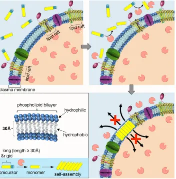
As shown in Figure 1, non-toxic water-soluble molecular precursors are constructed to mimic the
inactive hydrophilic PFT monomers distributed in extracellular fluid. Under tumor
microenvironment, the alkaline phosphatases (ALPs) that over expressed by cancerous cells
dephosphorylate the precursors into hydrophobic building blocks upon cell membrane triggering
self-assembly into rigid aggregates.
4-8The hydrophobic surface of the aggregate facilitates its
insertion into the hydrocarbon core of membrane constructing nanoarchitectonics among the
phospholipid bilayer.
9-11The porosity binding effect with molecules of the inserted synthetic analogues lead to membrane permeability alteration by blocking the transportation of molecules.
Severe permeability alteration eventually leads to cancer cell death.
Figure 1. Schematic illustration of self-assembly directed membrane insertion of long and rigid synthetic analogue causing permeability alteration.
Regarding the complexity and dynamic features of membrane structure, the live cell membrane insertion of synthetic analogues, without the help of external force, is difficult and challenging.
Except single molecular synthetic peptides derived from natural proteins or peptides,
12live cell
membrane insertion of sub-micron synthetic structures was rarely reported.
13Computational
simulations indicate that besides intrinsic rigidity of the synthetic analogue, a proper length of
the purely hydrophobic portion match to the hydrophobic core of the membrane to maximize the
interactions
13is also critical to the successful insertion.
14Therefore, we select the hydrophobic
domain of 'muscle molecule',
15-16the expanded π-conjugation system with extremely high rigidity,
long linear length, for the construction of desired molecular precursor leading to self-assembly- guided membrane insertion.
EXPERIMENTAL SECTION
Materials and Instruments. Ammonium acetate was purchased from Nacalai Tesque, INC.
and purified by sublimation before usage. Other chemical reagents were purchased from Sigma, Nacalai and Wako and used without further purification. Mass spectra were recorded using a Thermo LTQ-ETD mass spectrometer (ESI) and high resolution mass spectra were measured with a Thermo LTQ-Orbitrap Classic mass spectrometer (ESI).
1H,
13C and
31P NMR spectra were recorded on a Bruker Ascend 400 (400, 100 and 162 MHz, respectively) spectrometer. Time- dependent
31P NMR for dephosphorylation process was recorded on a JEOL 600 MHz NMR (243 MHz). TEM micrographs were obtained on a JEM-1230R Transmission Electron Microscope.
SEM micrographs were measured on a FEI Quanta 250 FEG Scanning Electron Microscope.
Confocal images were obtained on a Zeiss LSM780 Confocal Microscope. The fluorescence intensity of PI was detected using imaging flow cytometer (ImageStream X Mark, Germany).
LDH release was determined using a Tecan Infinite M1000 PRO microplate reader.
Synthesis. Synthetic procedures and characterizations of surface ligands applied in this study are described in Supporting Information.
TEM imaging. Aliquots (10 μL) of sample solution were added into a glow discharge copper
grid (400 mesh) coated with thin carbon film and incubated for 30 s at room temperature. After
removing excess solution, the grid was washed with deionized water three times and then stained
with 2.0% (w/v) uranyl acetate (UA) by exposing the grid in three drops of UA solution for 30 s.
TEM images were captured at high vacuum on transmission electron microscope JEM-1230R (JEOL, Japan).
Molecular dynamics simulations and polymorph prediction. Molecular mechanics calculations were performed using Materials Studio. As the crystal structure prediction method uses a rigid body approximation in the initial search for crystal packing alternatives, it is necessary to perform an analysis to determine low energy geometry to be used as input for the packing calculations. The molecules were drawn and geometrical energy minimization scans were performed using Forcite module of Materials Studio. The optimized low energy conformations were used as the starting points for crystal structure prediction using the Materials Studio Polymorph Predictor (PP).
Cell viability assay. Cells in exponential growth phase were seeded in a 96 well plate at a concentration of 7×10
3cells/well for HeLa, A375, and 1×10
4cells/well for HS5, OVCAR3, cell lines. The cells were allowed to attach to the wells for 12 h at 37ºC, 5% CO
2. The culture medium was removed followed by addition of 100 μL culture medium containing different concentrations (0.1, 1, 10, 100 and 200 μM) of compound 2 (immediately diluted from 10 mM stock solution in PBS buffer). After the desired time of exposure, 10 μL MTT solution (5 mg/mL) was added to each well and incubated at 37°C for another 4 h, and then 100 μL of SDS solution (10% in Milli-Q water) was added to stop the reduction reaction and dissolve the purple formazan. The absorbance of each well at 570 nm was measured by a Tecan microplate reader.
All experiments were conducted triplicate. The results were calculated as means, which are expressed as cell viability (%).
Live Cell imaging. Molecular probes were purchased from Life Technologies (Thermo Fisher
Scientific, USA). Cells in exponential growth phase were seeded into a glass bottomed culture
dish at 2×10
5cells per dish. The cells were allowed for attachment for 24 h at 37 °C under 5%
CO
2. Culture medium was removed and fresh medium containing different concentrations of compounds was added. After incubation for desired time, cells were washed with live cell imaging solution for three times, and further stained with commercial cell labels. Cells were then washed two times with fresh live-cell imaging solution and visualized by laser confocal microscopy (LSM 780, Carl Zeiss) immediately (λ
ex: 405 nm for compound 2, 488 nm for Lysotracker Green DND-26, 561 nm for Actinred 555 and Alexa Fluor 555, 633 nm for CellMask deep red; λ
em: 420-480 nm for compound 2, 508-570 nm for Lysotracker Green DND- 26, 580-720 nm for Actinred 555, 570-606 nm for Alexa Fluor 555, 650-750 nm for CellMask deep red).
SEM imaging. After taking confocal images, the samples were washed with PBS and then the cells were cross-linked with 2.5% glutaraldehyde in 0.1 M cacodylate buffer for 10 min. The samples were washed with 0.1 M cacodylate buffer for 5 min (×3) and then further fixed with 1% osmium tetroxide in 0.1 M cacodylate buffer for 30 min, followed by washing with water for 5 min (×3) and then progressive dehydration in a graded series of ethanols (70%, 80%, 90%, 95% and twice in 100%, 3 minutes at each concentration). The cells were rinsed with t-butanol for 3 min (×3) and then dried by freeze-dryer overnight. SEM images of the dried samples were captured before platinum coating.
LDH release. LDH release activity was measured by pierce LDH cytotoxicity assay kit (Thermo Scientific) according to the manufacturers’ instructions. HeLa, A375 and OVCAR3 cells were seeded at 1.5 ×10
4cells/well in 100 μL of medium in a 96 well plate, respectively.
After incubation with compound 2 (10, 50, 100 μM) for 12 h, the release of LDH in the
supernatant was measured with a microplate reader at 490 nm. Cells treated with 10 μL of water
(12 h) or Lysis buffer (10X, 45 min) was used as negative and positive controls, respectively. All experiments were carried out in triplicates. LDH activity was calculated as follows:
LDH release (%) = Compound treated activity − Negative activity Positive activity − Negative activity × 100
RESULTS AND DISCUSSION
Molecular design and synthesis.1,10-phenanthroline analogous containing two bidentates were reported as optimal ligands in the preparation of multinuclear cyclic pseudorotaxanes as
¢muscle molecules¢ through metal(I)-mediated assembly because of their long linear length and
highly rigidity. By replacing the polyethylene glycol macrocycle with two carboxyl groups, we were able to introduce enzyme responsive amino acids obtaining the target molecule 2 (Scheme 1A). The synthesis commences with 2,9-dibromo-1,10-phenanthroline and 3,8-dibromo-1,10- phenanthroline both prepared from 1,10-phenanthroline through different paths (Scheme S1 and S2). Following Sauvage’s method, we were able to apply 2,9-dibromo-1,10-phenanthroline and methyl 4-(5,5-dimethyl-1,3,2-dioxaborinan-2-yl)benzoate for Suzuki-coupling obtaining the dione building block in elevated yield (84%). The linear aldehyde building block was prepared in 5 steps from 3,8-dibromo-1,10-phenanthroline. A condensation of both building blocks at the presence of ammonium acetate in a sealed tube at 105
oC for 24 h constructed an oxazole ring as rigid spacer leading to molecule 1 identified by high-resolution mass spectroscopy (HRMS).
Two carboxyl groups of the rigid molecule 1 were activated with diisopropylcarbodiimide (DIC)
and N-hydroxysuccinimide (NHS), following the conjugation with two o-phospho-
L-tyrosine
(Yp) forming the soluble precursor 2. Upon the expression of alkaline phosphatase (ALP), the
precursor 2 transforms into 3, the hydrophobic monomer ready for self-assembly.
The geometrical energy minimized structures of 2 and 3 (Scheme 1B) indicate both molecules adopt Y-shape with linear length of hydrophobic domain beyond 30 Å,
1longer than the thickness of the hydrophobic core of cell membrane. The hydrophobic domains of both molecules remain static without deformation during ALP induced dephosphorylation due to the extreme rigidity, which may lead to efficient membrane insertion.
Scheme 1. (A) The synthetic path of precursor 2, and the enzyme triggered transformation of 2
into monomer of self-assembly 3. (B) The stick models of geometrical energy minimized
ALP induced molecular self-assembly. The ALP instructed transformation of 2 into 3 was monitored by
31P NMR (Figure 2A).
17The time-lapse spectra indicated that after 90 min incubation with ALP (Figure S1B), a complete conversion of 2 to 3 is reached. Time-lapse emission spectroscopies (Figure 2B and S1) indicate the decrease of emission intensity during the first 60-min incubation induced by self-quenching. After that, the intensity gradually becomes stable. The two-stage migration of maximum emission peak, red shift until 60 min and blue shift afterwards, suggests a dynamic adjustment of molecular packing following the molecular transformation from 2 to 3. Regarding the rigidity of the molecule, the intermolecular π–π stacking is suggested as the main driving force for self-assembly resulting in the overall red
shift (from 457nm to 475 nm) of emission spectra. Optical images taken at the end-point of the transformation exhibit clear solutions with neither precipitation nor gel-formation observed.
Under the UV light, the solution emits light blue fluorescence (Figure 2C and S2). The
transmission electron microscopy (TEM) (Figure 2D and S2) confirms the self-assembly of 3
into irregular nanoscale aggregates distributed in PBS buffer. The Polymorph Predictor module
18predicts molecular self-assembly is driven by strong intermolecular π-π stacking (Figure 2E and
Figure S3) forming close packed nanostructure with hydrophobic surface, which is consistent to
the emission spectra variations.
Figure 2. (A) Time-lapse
31P NMR spectroscopies of 2 (400 μM ) in Tris-HCl D
2O buffer (50mM, pH = 8.5) reacting with ALP (1 U/mL). Na
2HPO
4(6mM) is used as internal standard.
(B) Time-lapse normalized maximum emission intensity and wavelength of 2 (100 μM) in PBS buffer reacted with ALP (1 U/mL). Optical images under the normal light and UV light (365 nm) (C) and TEM image (D) of end-point of 2 (100 μM) reacted with ALP (1U/mL) in PBS buffer.
(E) The predicted molecular stacking of 3 in CPK model correlated to the self-assemblies in TEM image.
Membrane insertion induced toxicity in vitro. Three cancer cell lines, the cervical cancer
cell (HeLa), the skin malignant melanoma cell (A375), the drug resistant ovarian cancer cell
(OVCAR-3), and stromal cell line HS-5 cell with ALP isozymes in different expression patterns
cultured in different concentrations of exogenous ALP (the ALP expression profiles are
summarized in Table S1), are applied in cytotoxicity tests of 2 (Figure 3A). With no toxicity on
HS-5 cells, cancerous cell targeted cytotoxicity of 2 is revealed. Cancerous cell viability profiles
also suggested concentration dependent/cell type dependent toxicity of 2. 2 started showing
toxicity on HeLa cell beyond 10 μM. But it started showing toxicity on both A375 cell and OVCAR3 cell beyond 100 μM. At 200 μM, 2 had similar toxicity on both HeLa and A375 cells.
And it killed 40% of the OVCAR3 cells after 3-day incubation.
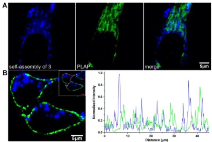
Live cell imaging by confocal microscopy exhibits toxicity correlated selective subcellular targeting. Towards cancerous cells, under compatible condition ([2] = 10 μM), we observe blue fluorescence distributed mainly in lysosomes (Figure S4A). Accompanied with cytotoxicity, self- assemblies start accumulating on cell membranes with stochastic distribution. Following the rising of toxicity, the population of targeted cell by self-assemblies rises (Figure S4B and S4C).
Comparing to normal cell (HS-5)’s lysosome targeted accumulation of self-assemblies (Figure
S5), precursor 2 selectively target cancerous cell membranes inducing toxicity. Fluorescent
imaging along the z-axis (Figure 3B, 3C 3D and S6) reveals cancerous cell membrane insertion
of self-assemblies. The fluorescent intensity profile along the cell membrane exhibiting
enhancement of self-assemblies’ fluorescent intensity (blue) accompanied with decline of cell
membrane’s fluorescent intensity (red) vice versa, confirms the membrane insertion of self-
assemblies on three types of cancerous cells. Under the same incubation condition, the
membrane insertion rate in order from the highest to the lowest is HeLa cell, A375 cell and
OVCAR-3 cell, consistent to the cytotoxicity profile of 2 towards these cancer cells. SEM
images indicated microscale synthetic analogues inserting on membrane close to the cell margin
of all three types of cancerous cells (Figure S7). By increasing the incubation concentration of 2,
expansion of assembled analogues on membrane is observed (Figure S4B and S4C). Scientists
predicted the phospholipids exaction from membrane to inserted analogue due to the van der
Waals and hydrophobic interactions during the late stage of insertion.
19We observed the same
phenomenon in the experiment that the membrane shifted toward self-assembled analogues (Figure S8).
ALP guided membrane insertion in vitro. The non-selective membrane insertion on all three types of cancer cells with (HeLa) and without (A375 and OVCAR3) placental alkaline phosphatase (PLAP) expression suggests that overexpression of PLAP that locate on the membrane is not critical to self-assembly-facilitated membrane insertion here. After treatment of PLAP inhibitor
L-phenylalanine, HeLa cells showed unaffected membrane insertion of assembled analogues (Figure S9). HeLa cell membrane insertions show no overlap with PLAP (Figure 4A) suggest no physical binding interactions between the PLAP and self-assemblies, which is different from PLAP guided self-assembly on cancer cell membrane showing co-localizations.
4-5, 7-8,20-21