Cwh43 regulates G0 quiescence, nutrient
metabolism and lipid homeostasis in fission yeast
Author Norihiko Nakazawa, Takayuki Teruya, Kenichi Sajiki, Kazuki Kumada, Alejandro
Villar‑Briones, Orie Arakawa, Junko Takada, Shigeaki Saitoh, Mitsuhiro Yanagida
journal or
publication title
Journal of Cell Science
volume 131
number 16
page range jcs217331
year 2018‑08‑21
Publisher The Company of Biologists Ltd.
Rights (C) 2018 The Author(s).
Author's flag publisher
URL http://id.nii.ac.jp/1394/00000824/
doi: info:doi/10.1242/jcs.217331
RESEARCH ARTICLE
The putative ceramide-conjugation protein Cwh43 regulates G0 quiescence, nutrient metabolism and lipid homeostasis in fission yeast
Norihiko Nakazawa
1,‡, Takayuki Teruya
1, Kenichi Sajiki
1, Kazuki Kumada
1, Alejandro Villar-Briones
1,*, Orie Arakawa
1, Junko Takada
1, Shigeaki Saitoh
2and Mitsuhiro Yanagida
1,‡ABSTRACT
Cellular nutrient states control whether cells proliferate, or whether they enter or exit quiescence. Here, we report characterizations of fission yeast temperature-sensitive (ts) mutants of the evolutionarily conserved transmembrane protein Cwh43, and explore its relevance to utilization of glucose, nitrogen source and lipids. GFP-tagged Cwh43 localizes at ER associated with the nuclear envelope and the plasma membrane, as in budding yeast. We found that cwh43 mutants failed to divide in low glucose and lost viability during quiescence under nitrogen starvation. In cwh43 mutants, comprehensive metabolome analysis demonstrated dramatic changes in marker metabolites that altered under low glucose and/
or nitrogen starvation, althoughcwh43cells apparently consumed glucose in the culture medium. Furthermore, we found thatcwh43 mutant cells had elevated levels of triacylglycerols (TGs) and coenzyme A, and that they accumulated lipid droplets. Notably, TG biosynthesis was required to maintain cell division in the cwh43 mutant. Thus, Cwh43 affects utilization of glucose and nitrogen sources, as well as storage lipid metabolism. These results may fit a notion developed in budding yeast stating that Cwh43 conjugates ceramide to glycosylphosphatidylinositol (GPI)-anchored proteins and maintains integrity of membrane organization.
KEY WORDS: Cwh43, Ceramide, Fission yeast, Low glucose, Nitrogen starvation, Lipid droplet
INTRODUCTION
Nutrition is a major determinant of whether cells divide or undergo cell cycle arrest to enter quiescence. Principal nutrients include carbohydrates, proteins and lipids, although vitamins and minerals are also essential. Carbohydrates are hydrolyzed to monosaccharides, such as glucose, the essential carbon and energy source for all organisms. Amino acids are a critical nitrogen source for synthesis of numerous biological substances, such as proteins, nucleotides and neurotransmitters, and are also necessary for gluconeogenesis under low-glucose conditions. Under environmental conditions in which carbon and/or nitrogen sources
are not available, cells have the ability to utilize lipids as an alternative energy source. Most eukaryotic cells are capable of storing lipid droplets (LDs), comprised primarily of triacylglycerols (TGs) and sterols, in specialized intracellular organelles (Meyers et al., 2017; Pol et al., 2014; Thiam et al., 2013). Remarkably, changes in the cellular regulation of LDs and LD-associated proteins are linked to human diseases including obesity, diabetes and atherosclerosis (Krahmer et al., 2013). Therefore, metabolic processes involving principal nutrients are tightly integrated.
Mechanisms controlling metabolism of principal nutrients are assumed to be evolutionarily conserved. Because the fission yeast Schizosaccharomyces pombe is a suitable model organism for the study of cell division and quiescence, we investigated genetically controlled mechanisms in this organism that respond to fasting or starvation. Nitrogen starvation induces entry into the quiescent G0 phase, in which cells remain alive for months without growth or division (Su et al., 1996; Yanagida, 2009), demonstrating S. pombe G0 quiescence provides a model system to study chronological lifespan. Using a collection of temperature-sensitive (ts) mutants, we identified more than 80
‘super housekeeping genes
’required for both proliferation and nitrogen starvation-induced G0 quiescence (Sajiki et al., 2009 and our unpublished data). For example, the stress- responsive mitogen-activated protein kinase (MAPK) Sty1 is required for G0 entry. The 26S proteasome maintains G0 quiescence by minimizing reactive oxygen species (ROS) in collaboration with autophagy (Takeda et al., 2010). By contrast, the quiescent state has also been studied under limited glucose conditions. In media containing low glucose (4.4 mM, 0.08%), S. pombe wild-type cells are able to divide as well as in normal glucose media (111 mM, 2%). It is reported that the critical glucose transporter Ght5, the zinc-finger transcription factor Scr1, the calmodulin kinase kinase (CaMKK)-like Ssp1, the PP2A phosphatase inhibitor Sds23 and the TORC2- mediated signaling pathway are essential for cell division under low- glucose conditions (Hanyu et al., 2009; Saitoh et al., 2015). In response to a wide range of environmental stresses, a limited number of key signaling pathways including protein kinase C (PKC) (Levin et al., 1990; Toda et al., 1993), MAPKs (Chen and Thorner, 2007;
Perez and Cansado, 2010; Shiozaki and Russell, 1995; Sugiura et al., 1999, 1998) and the Ca
2+/calmodulin-dependent phosphatase calcineurin (Cyert, 2003; Sugiura et al., 2002; Yoshida et al., 1994), become activated. Thus, identification of factors involved in utilization of both carbon and nitrogen sources should help to illuminate common signaling pathways of principal nutrients.
In this study, we isolated eight temperature-sensitive (ts) mutant alleles of the fission yeast cwh43 gene from our ts mutant collections, and found that seven of these mutants showed sensitivity to both nitrogen starvation and glucose limitation.
Cwh43 encodes a conserved transmembrane protein that is thought
Received 1 March 2018; Accepted 20 July 2018
1G0 Cell Unit, Okinawa Institute of Science and Technology Graduate University, Tancha 1919-1, Onna-son, Okinawa 904-0495, Japan.2Institute of Life Science, Kurume University, Hyakunen-Kohen 1-1, Kurume, Fukuoka 839-0864, Japan.
*Present address: Okinawa Institute of Science and Technology Graduate University, Instrumental Analysis Section, Tancha 1919-1, Onna-son, Okinawa, Japan 904-0495.
‡Authors for correspondence ([email protected]; [email protected]) N.N., 0000-0001-8386-0812; M.Y., 0000-0003-0293-5654
Journal of Cell Science
to incorporate the sphingolipid, ceramide, into a lipid moiety of glycosylphosphatidylinositol (GPI)-anchored proteins (GPI-APs) in budding yeast plasma membranes (Ghugtyal et al., 2007; Martin- Yken et al., 2001; Reggiori et al., 1997; Umemura et al., 2007;
Yoko-o et al., 2013). Ceramide is present in all eukaryotic cells and serves structural functions in lipid bilayers (Chaurasia and Summers, 2015). In addition, ceramide acts as a signaling molecule in response to stresses such as UV irradiation and chemotherapy, which induce cell cycle arrest, apoptosis and cell senescence (Hannun, 1996; Hannun and Obeid, 2008). To address the novel, nutrient-related phenotypes in S. pombe cwh43 mutant cells, we comprehensively measured the levels of metabolites and lipids using metabolomic and lipidomic approaches. Strikingly, Cwh43-deficient cells had changed levels of nutrient stress- inducible metabolic compounds and they also over-accumulated TGs (neutral lipids) in LDs. We present evidence that Cwh43, a putative ceramide-conjugation protein, is intimately involved in cellular metabolism of principal nutrients and storage lipids.
RESULTS
Isolation ofS. pombe cwh43temperature-sensitive mutants
We previously developed 1015 ts haploid S. pombe strains (Hayashi et al., 2004), and identified genes responsible for their ts phenotypes (Hanyu et al., 2009; Hayashi et al., 2004; Nakamura et al., 2012). To
identify the mutant gene, plasmids that rescued the ts phenotype of the mutant strain were isolated by transformation, using an S. pombe genomic DNA library. Plasmid DNAs recovered from Ts
+transformants were then subcloned and sequenced. Nevertheless, the colony number of obtained transformants was occasionally insufficient to identify the high-copy suppressor gene owing to low plasmid transformation efficiency. To overcome this technical difficulty for determination of mutation sites in eight ts mutant strains (ts200, ts202, ts285, ts609, ts724, ts824, ts833, ts941), we performed whole-genome sequencing of the mutant strains. Upon completion, all mutation sites responsible for these eight ts phenotypes turned out to be located in cwh43
+(SPAC589.12), encoding the putative glycosylceramide biosynthesis protein Cwh43 (PomBase, https://www.pombase.org/) (Fig. 1A). We backcrossed these ts strains with the wild-type multiple times and confirmed that the amino acid changes co-segregated with the ts phenotypes.
Cwh43 is a conserved eukaryotic transmembrane protein. It contains the Frag1/DRAM/Sfk1 family domain at its N-terminus and an endonuclease/exonuclease/phosphatase superfamily domain at its C-terminus (Dlakic
́, 2000; Ghugtyal et al., 2007; Martin-Yken et al., 2001; Umemura et al., 2007) (Fig. 1A). Nineteen transmembrane helix motifs occur in the N-terminal 700 amino acids. The mutation sites of four cwh43 ts mutants, K273stop (ts609), G300E (ts824), N418K (ts285) and W605stop (ts202), are
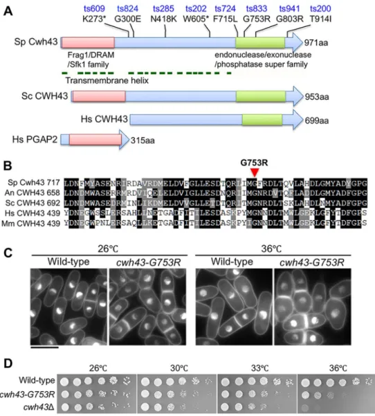
Fig. 1. Isolation ofS. pombe temperature-sensitive mutants of the gene encoding the conserved transmembrane protein Cwh43.
(A)S. pombeCwh43 (Sp Cwh43) is drawn schematically along withS. cerevisiae CWH43 (Sc CWH43) protein. Mutation sites of eightS. pombe cwh43ts mutants are indicated with amino acid changes.
Asterisks indicate stop codons. The N- and C-terminal conserved regions of the Frag1/DRAM/Sfk1 and endonuclease/
exonuclease/phosphatase superfamilies are indicated with red and green boxes, respectively. Horizontal green lines show positions of transmembrane helices in Sp Cwh43 protein. Cwh43 comprises two distinct proteins in mammalian cells.
Human PGAP2 and CWH43/FLJ21511 are homologous to the N- and C-terminal regions of yeast Cwh43, respectively (Ghugtyal et al., 2007; Umemura et al., 2007). (B) Amino acid sequences of the conserved C-terminal regions that surround the G753R mutation in the S. pombe cwh43-833ts strain. Amino acid sequences of Cwh43 orthologs in S. pombe(Sp),Aspergillus niger(An), Saccharomyces cerevisiae(Sc),Homo sapiens(Hs), andMus musculus(Mm) are aligned. (C) DAPI-stained wild-type andcwh43-G753Rmutant cells are shown. Mild and severe defects in cell morphology were observed incwh43- G753Rmutant cells at 26°C and 36°C for 6 h, respectively. Scale bar: 10μm.
(D) Wild type, thecwh43-G753Rmutant and the deletion mutant lacking the cwh43+gene (cwh43Δ) were spotted on EMM2 solid medium.
Journal of Cell Science
located in the region that contains transmembrane motifs. In the C-terminal domain, the evolutionarily conserved F715, G753 and G803 were mutated to leucine (ts724), arginine (ts833) and arginine (ts941) residues, respectively (Fig. 1B; Fig. S1). T914I (ts200) is located just behind the C-terminal domain. In contrast, none of our mutants resulted from mutations in the conserved N-terminal domain of the Frag1/DRAM/Sfk1 family. Thus, the transmembrane region and conserved C-terminal domain appear to be indispensable for S. pombe Cwh43 function, although the N-terminal domain may have an additive role, consistent with the more-severe phenotype of the deletion mutant of the full-length cwh43
+gene (see below).
Construction ofcwh43-G753Randcwh43Δmutant strains
All eight cwh43 mutants produced partly elongated, swollen cell shapes at the permissive temperature (26°C), and this morphological abnormality was more severe at the restrictive temperature (36°C) (Fig. 1C; Fig. S2A). Abnormal cell morphology was indistinguishable among the eight mutant strains. We selected the G753R mutation (ts833) for further phenotypic analyses. It is located in the C-terminally conserved region and loses viability in vegetative and G0 quiescent cells (see below).
To confirm that the ts phenotype is caused by single amino acid changes in the Cwh43 protein, we introduced the G753R mutation into the wild-type strain under the native promoter, along with the hemagglutinin (HA) tag. The resulting transformants, designated Cwh43-WT-HA:KanR and cwh43-G753R-HA:KanR (hereafter, termed wild type and cwh43-G753R, respectively), were used in this study. The HA-tagged wild-type strain showed normal cell growth, as did the non-tagged wild-type; however, the cwh43-G753R mutant and the original cwh43-833 mutant failed to form colonies at the restrictive temperature (36°C) (Fig. 1D; Fig. S2B). After the temperature shift from 26°C to 36°C, the cell count of the cwh43- G753R mutant hardly increased, while viability decreased from 55% to 20% (Fig. S2C). Thus, cell number increase and viability of cwh43-G753R mutant cells were impaired at 36°C. The cell morphological defect was evident even at 26°C, suggesting that the cell morphological abnormality itself was not a lethal attribute. The proportion of cells with septa was significantly higher in the cwh43- G753R mutant at both 26°C and 36°C, while the numbers with multiple septa increased at 36°C, suggesting that this mutant is defective in cytokinesis (Fig. S2D). These phenotypes of the integrated strain were indistinguishable from those of the original cwh43-833 mutant.
Next, we constructed a deletion mutant of the cwh43
+gene by replacing it with the hygromycin-resistance gene. The resulting cwh43 deletion mutant (cwh43
Δ) failed to form colonies at 36°C, as did the cwh43-G753R ts mutant (Fig. 1D), indicating that deletion of cwh43
+is indispensable for cell proliferation at high temperature.
The colony-forming capacity of the cwh43
Δmutant is less than that of the cwh43-G753R mutant at all tested temperatures. Consistent with this, the morphological abnormality of the cwh43
Δmutant was more severe than that of the cwh43-G753R mutant at both 26°C and 36°C (Fig. S2E). Thus, cwh43-G753R mutant cells may be not fully devoid of Cwh43 function.
Monitoring defective cytokinesis in livingcwh43 mutant cells
To examine cell division of cwh43-G753R mutant cells in detail, we made time-lapse observations of living cells. Wild-type and cwh43- G753R cells were cultivated at 26°C and then observed using a microfluidic perfusion system at 26°C or 36°C (Materials and Methods). At 26°C, DIC images showed that most wild-type cells
divided within 4 h, and septum formation occurred in one or two movie frames (15
–30 min) just before cell division (Fig. 2A, top;
Movie 1). However, in the cwh43 mutant, cell division required 2 to 8 h and septum formation took >2 h, demonstrating that completion of cytokinesis was significantly delayed (Fig. 2A, bottom; Movie 2).
After the temperature shift to 36°C, division of cwh43-G753R mutant cells was blocked but this did not occur in wild-type cells (Fig. 2B; Movies 3,4). Mutant cells gradually became elongated and swelled without cell division, resulting in multiple septa and protrusion of cytosol. In addition, Aniline Blue staining of cwh43- G753R mutant cells showed abnormally high concentrations of the cell wall component 1,3-
β-glucan, particularly at the septum, compared with dividing wild-type cells (Fig. 2C). This abnormal accumulation of 1,3-
β-glucan was obvious at 36°C. As reported in a large number of mutant strains defective in cell wall integrity, the ts phenotype of cwh43 mutant cells was suppressed in the presence of the osmotic stabilizer sorbitol (1.2 M) (Fig. S2F). Concomitantly, the glucan accumulation phenotype was slightly alleviated by sorbitol treatment (Fig. S2G), implying a correlation between cell wall defects and excessive glucan deposition. Taken together, these data indicate that impaired Cwh43 delayed completion of cytokinesis, presumably by causing excess 1,3-
β-glucan to accumulate at the septum.
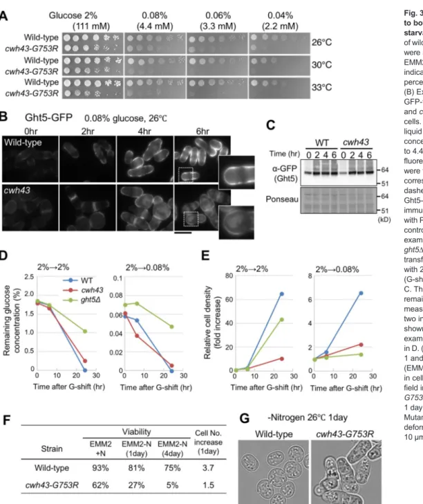
cwh43mutant shows low-glucose sensitivity and improper localization of glucose transporters, in spite of glucose consumption from the culture medium
To test whether the cwh43
+gene is required for cell proliferation under nutrient deficiency, we examined the colony-forming ability of cwh43-G753R mutant cells on solid EMM2 medium containing 0.04
–2% glucose (2.2-111 mM; low to high levels of glucose). As previously described for the authentic low-glucose-sensitive mutant strains defective in Ssp1 kinase (Hanyu et al., 2009) and for a deletion mutant of hexose transporter Ght5 (Saitoh et al., 2015), the cwh43-G753R mutant failed to form colonies on low-glucose media (0.04
–0.08%) at 26
–33°C (Fig. 3A). Seven of the eight cwh43 original ts mutant strains failed to divide under low-glucose conditions, exhibiting the low-glucose sensitivity (LGS) phenotype (Table S1).
Under low-glucose conditions, expression of specific hexose transporters is induced, and these transporters localize at the cell surface (Saitoh et al., 2015). We examined intracellular localization of the GFP-tagged hexose transporter Ght5 in cwh43 mutant cells. After a shift from 2% to 0.08% glucose at 26°C, Ght5
–GFP localization at the cell surface was not intense and showed cytoplasmic signals in the mutant (Fig. 3B). Expression of Ght5
–GFP itself was properly induced in cwh43 mutant cells, as well as in wild-type cells (Fig. 3C).
In wild-type cells, another GFP-tagged hexose transporter, Ght8, was located at the plasma membrane exclusively at the poles of the cells (Saitoh et al., 2015) (Fig. S3). By contrast, cwh43 mutant cells showed cytoplasmic accumulations of Ght8
–GFP similar to those of Ght5-GFP, suggesting that Cwh43 affects the localization of hexose transporters under glucose limitation.
To examine glucose consumption by wild-type and cwh43
mutant cells, we measured glucose concentrations in the culture
medium. Cells initially cultivated in high-glucose (2%) medium
were transferred to high (2%)- or low (0.08%)-glucose medium and
incubated at 26°C, along with ght5 deletion mutant cells (ght5
Δ) as
a control (Saitoh et al., 2015). Unexpectedly, glucose consumption
was not significantly decreased in cwh43 mutant cultures compared
with wild-type, in either high or low glucose (Fig. 3D), although the
cell number increase of cwh43 mutant was decidedly slow (Fig. 3E). Journal of Cell Science
These data suggest that the cwh43 mutant consumes glucose in culture media, even if cell division is declined. Although subcellular localization of Ght5 and Ght8 were affected in the cwh43 mutant, these transporters, which were partially retained at the plasma membrane, may be sufficient for glucose uptake.
Cwh43 is required to maintain cell viability during G0 quiescence induced by nitrogen starvation
We next determined whether the cwh43
+gene is required for maintaining viability during G0 quiescence under nitrogen starvation.
Wild-type and cwh43-G753R mutant cells grown in EMM2 medium (EMM2+N) were transferred to nitrogen-deficient EMM2-N at 26°C for 1 and 4 days, and the resulting quiescent G0 cells were spotted onto YPD solid medium in order to count the cells that were able to form colonies. In comparison with the wild type, under nitrogen starvation, the viability of cwh43-G753R mutants plunged to 27%
after 1 day and to only 5% after 4 days, indicating severe defects in
regeneration capacity, even after replenishment of the nitrogen source (Fig. 3F). The cell number of wild-type cells increased 3.7× after 1 day under nitrogen starvation, indicating that two rounds of cell division occurred. In contrast, numbers of cwh43 mutant cells increased only 1.5×. Wild-type S. pombe cells have a spherical shape during G0 quiescence (Sajiki et al., 2009; Su et al., 1996); however, cwh43- G753R mutant cells presented non-spherical, deformed shapes (Fig. 3G). Six of the seven remaining cwh43 ts mutant strains also decreased in viability under nitrogen starvation (Table S1). These results suggest that cwh43 mutant cells failed to enter a normal and viable G0 quiescence in response to nitrogen deficiency.
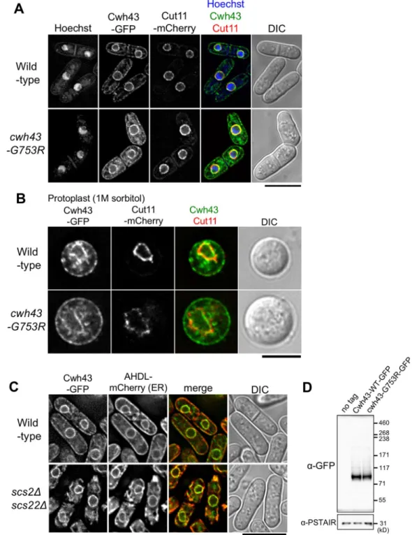
Intracellular localization of Cwh43
To observe the intracellular localization of Cwh43 protein, the C-terminus of cwh43
+or cwh43-G753R mutant genes was tagged with GFP. At 26°C, Cwh43-WT
–GFP apparently localized at the nuclear membrane and the plasma membrane, accompanying
Fig. 2. Monitoring defective cytokinesis and excessiveβ-glucan accumulation at cell septa incwh43 mutant cells.(A,B) Left, time-lapse images of wild-type andcwh43-G753R strains. Cells were first cultivated at 26°C, and then either incubated further at 26°C (A) or shifted to 36°C (B) in a microfluidic perfusion chamber with a continuous supply of EMM2 medium. Differential interference contrast (DIC) images were presented at 2-h intervals (also see Movies 1–4). Arrows and arrowheads in B indicate the positions of multiple septa and a protruded cytosolic structure in cwh43-G753Rcells, respectively. Right, division time and duration of septum formation were monitored for individual cells. (C) Left, fluorescence and bright- field images of wild-type andcwh43- G753Rcells were captured after staining for the cell wall component 1,3-β-glucan using the fluorescent dye Aniline Blue.
Cells were cultured at 26°C or 36°C for 6 h in liquid medium. Right, Aniline Blue- stained cells were spotted on filter paper.
Scale bars: 10μm.
Journal of Cell Science
granule-like cytoplasmic signals (Fig. 4A, top). At 36°C, localization of Cwh43
–GFP was similar to that at 26°C, although GFP-tagged Cwh43 proteins were possibly unstable in high temperature, unlike the HA-tagged proteins (Fig. S4A,B).
GFP-tagged Cwh43-G753R mutant proteins also showed similar localization as observed in wild type at both temperatures, implying that the G753R mutation does not affect Cwh43 localization itself (Fig. 4A, bottom; Fig. S4A). We also demonstrated that Cwh43
–GFP localized in close proximity to plasma membranes by performing a protoplast preparation with enzymatic digestion of cell walls (Fig. 4B).
In budding yeast, CWH43 protein is reported to localize at the endoplasmic reticulum (ER) (Umemura et al., 2007). Considering that the ER is continuous with the nuclear envelope and is associated with the plasma membrane (Pidoux and Armstrong, 1993; Zhang et al., 2010), S. pombe Cwh43 proteins are also
predicted to enrich in the ER. To test this possibility, we observed Cwh43
–GFP localization in an scs2
Δscs22
Δdouble-deletion mutant strain in which the cortical ER dissociates from plasma membrane (Zhang et al., 2012). In wild-type cells, the Cwh43
–GFP signals colocalized with the artificial luminal ER marker AHDL
–mCherry (Zhang et al., 2010) at the plasma membrane and nuclear periphery (Fig. 4C, top). However, in scs2
Δscs22
Δdouble deletion mutant cells, localization of both Cwh43
–GFP and AHDL
–mCherry at the plasma membrane disappeared, and, instead, accumulated in the cytoplasm (Fig. 4C, bottom). This result indicates that Cwh43, as in budding yeast CWH43, enriches at ER associated with the plasma membrane.
Immunodetection of Cwh43 protein
Protein extracts of exponentially growing wild-type and cwh43- G753R strains were examined by immunoblotting using antibodies
Fig. 3.cwh43mutants are sensitive to both low glucose and nitrogen starvation.(A) Aliquots (5×10⁴cells) of wild-type andcwh43-G753Rstrains were serially diluted 5×, spotted onto EMM2 medium containing the indicated concentrations (mM and percentage) of glucose at 26–33°C.
(B) Expression and localization of GFP-tagged Ght5 in wild-type (top) andcwh43-G753R(bottom) mutant cells. Cells were cultivated in EMM2 liquid medium at 26°C and the glucose concentration was switched from 111 to 4.4 mM at time 0 h. GFP fluorescence microscopy images were taken at 2-h intervals. Inserts correspond to the areas of the white dashed boxes. (C) The protein level of Ght5–GFP was measured by immunoblotting. Total protein stained with Ponceau S is shown as a loading control. (D) Glucose consumption was examined in the wild-type,cwh43and ght5Δmutant strains. Cells were transferred to fresh EMM2 supplied with 2% (left) or 0.08% (right) glucose (G-shift) at time 0 and cultivated at 26°
C. The glucose concentration remaining in the medium was measured after 0, 6 and 24 h. Mean of two independent experiments are shown. (E) Cell proliferation was examined for the same conditions as in D. (F) Cell viability was measured at 1 and 4 days after nitrogen starvation (EMM2-N) at 26°C along with changes in cell number after 1 day. (G) Bright- field images of wild-type andcwh43- G753Rmutant cells cultured after 1 day in EMM2-N medium at 26°C.
Mutant cells showed non-spherical, deformed cell shapes. Scale bars:
10μm.
Journal of Cell Science
against HA or GFP. GFP- or HA-tagged Cwh43-WT and -G753R bands were observed at
∼71 kDa, which is less than their molecular masses (
∼114 kDa for HA;
∼137 kDa for GFP) (Fig. 4D; Fig. S4C).
Overproduction of chromosomally integrated Cwh43-WT protein tagged with HA at its N-terminus also showed bands at
∼71 kD (Fig. S4D, nmt-HA-Cwh43). In addition, overproduced Cwh43-
Fig. 4. GFP-tagged Cwh43 proteins localize at ER associated with the nuclear envelope and the plasma membrane.(A) Intracellular localization of GFP- tagged Cwh43-WT and -G753R proteins. Wild-typecwh43+orcwh43-G753Rmutant genes were tagged with GFP and integrated into the chromosome under the native promoter with a kanamycin-resistance gene. The GFP-taggedcwh43+gene was functional, because the resulting integrant strain Cwh43-WT–GFP grew normally. These cells were cultivated at 26°C in EMM2 medium and fluorescence images were captured immediately without fixation. The nuclear envelope was visualized via mCherry-tagged Cut11 protein, a spindle pole body (SPB)-anchoring nuclear membrane protein (Broek et al., 1991; West et al., 1998), along with Hoechst 33342 staining for DNA and differential interference contrast (DIC) images. A single focal plane is shown. (B) Localization of Cwh43-WT–GFP or -G753R–GFP proteins in protoplasts. Cells were treated with a zymolyase and lysing enzyme mixture for 1 h in the presence of 1 M sorbitol and cells bearing both Cwh43–GFP and Cut11–mCherry were observed. A single focal plane is shown. (C) Localization of Cwh43–GFP and the artificial ER marker AHDL–mCherry in wild-type orscs2Δscs22Δdouble deletion mutant cells at 26°C. Scs2 and Scs22 link the cortical ER to the plasma membrane (Zhang et al., 2012).
(D) Immunoblotting for GFP-tagged Cwh43-WT and -G753R proteins. Cells were cultivated at 26°C in EMM2 medium, and total protein extracts were prepared and subjected to SDS-PAGE. Levels of Cwh43-WT-GFP or -G753R–GFP proteins were examined by immunoblotting using an antibody against GFP, as well as a non-tagged control. Anti-PSTAIR antibody (against amino acid sequence of PSTAIR in Cdc2) was used as a loading control. Scale bars: 10μm (A,C); 5μm (B).
Journal of Cell Science
WT
–HA protein derived from cwh43
+cDNA migrated faster than its expected molecular mass (Fig. S4D, pCwh43-HA). Hence, S.
pombe Cwh43 presents a fast-migrating band in SDS-PAGE, a property often reported in ER- or membrane-associated proteins (Shirai et al., 2008).
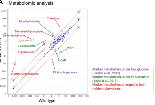
Metabolic changes of biomarkers for nutrient stress in cwh43mutant cells
To further characterize the cwh43 mutant phenotype, we applied our established metabolomic approach to cwh43-G753R mutant cells. Since cell viability of the cwh43-G753R mutant at 36°C and of the the cwh43
Δdeletion mutant at both 26°C and 36°C was remarkably low, the cwh43-G753R strain was cultivated at 26°C in liquid EMM2 medium to mid-log phase (5×10
6cells/ml) along with wild-type cells, and these exponentially growing vegetative cells were collected. Then, cellular metabolites were extracted in 50% methanol and metabolites were analyzed using an LC-MS (LTQ Orbitrap mass spectrometer), as described previously (Pluskal et al., 2010b; Pluskal et al., 2011; see Materials and Methods). 119 metabolites were detected in wild-type and cwh43- G753R mutant strains (Fig. 5A; Table S2). In cwh43 mutant cells, the levels of 26 metabolites increased more than 3-fold, while ten decreased more than 3-fold compared with wild-type cells (Fig. 5B, C; Table S3 and S4). Most of the compounds displaying significantly altered abundances in the mutant have been previously reported as marker metabolites that change under nitrogen starvation or low-glucose conditions (Fig. 5A) (Pluskal et al., 2011; Sajiki et al., 2013). The cwh43 mutant also accumulated compounds that commonly change in both types of nutritional starvation.
The most strikingly accumulated compound in cwh43 mutant cells was deferriferrichrome, which increased 19,000-fold relative to wild-type cells (Fig. 5B). Deferriferrichrome is an iron storage compound that increases with ferrichrome under low-glucose conditions (Pluskal et al., 2011). The iron-free form (deferri-form) of ferrichrome is produced under iron deficiency (Labbé et al., 2007; Schrettl et al., 2004), suggesting that cwh43 mutant cells were deficient in iron.
Another group of drastically increased compounds includes ergothioneine and S-methyl-ergothioneine. Ergothioneine acts as a physiological antioxidant (Cheah and Halliwell, 2012) that has been reported to increase under both low-glucose and nitrogen starvation conditions (Pluskal et al., 2011; Sajiki et al., 2013).
Hercynylcysteine sulfoxide is a precursor of ergothioneine.
Consistent with the fact that ergothioneine is a sulfur-containing derivative of trimethyl histidine, tri- and di-methylated histidine also accumulated in cwh43 mutant cells. Additionally, trehalose increased over 60-fold in the mutant. This compound is produced to stabilize proteins and membranes under various physiologic stresses, including heat shock, low glucose and nitrogen starvation (Cansado et al., 1998; Elbein et al., 2003; Pluskal et al., 2010b).
2-Oxoglutarate and succinate, intermediates of the TCA cycle, increased 89-fold and 6-fold, respectively, in cwh43-G753R mutant cells (Fig. 5B; Table S3). These compounds are specific biomarkers that increase during the nitrogen starvation response (Sajiki et al., 2013). By contrast, the purine biosynthesis pathway intermediates, 5-phospho-ribose 1-diphosphate (PRPP), aminoimidazole-4-carboxamide ribonucleotide (AICAR), N- succinocarboxamide-5-aminoimidazole ribonucleotide (SAICAR) and N-formylglycinamide ribonucleotide (FGAR) decreased remarkably in mutant cells (Fig. 5C; Table S4). These compounds have also been reported as marker metabolites that decrease
immediately under nitrogen starvation (Sajiki et al., 2013). Taken together, the metabolomic analysis indicated that the cwh43 mutant is apparently defective in utilization of both carbon and nitrogen sources, producing massive nutritional stresses, even when these nutrients are present in the culture medium.
In wild-type S. pombe cells, the level of free-form coenzyme A (CoA) is normally extremely low relative to other metabolites (Nakamura et al., 2012). However, in cwh43 mutant cells, we found that the level of CoA increased more than 4000-fold compared to wild-type cells (Fig. 5A,B). This drastic increase in CoA content in the absence of Cwh43 function is a unique metabolic property that has not been reported under low glucose or nitrogen starvation. CoA is an essential cofactor in the metabolism of carboxylic acids and lipids, implying that cwh43 mutant cells have altered lipid abundance.
cwh43mutant cells overproduce triacylglycerols
To examine whether Cwh43 influences cellular lipid metabolism, we performed lipidomic analysis in wild-type and cwh43-G753R mutant cells. Cells were cultivated in EMM2 at 26°C and total lipids were extracted with tert-butylmethylether (Matyash et al., 2008).
Lipid extracts were then subjected to LC/MS analysis and compounds were identified using Lipidomics Gateway Software (see Materials and Methods). We identified 124 lipids in both strains (Fig. 6A; Table S5). 15 lipids increased
≥3-fold in abundance while nine others diminished
≥3-fold in cwh43-G753R mutant cells compared to the wild type (Table S6). Notably, in mutant cells, 14 of 15 increased lipids were triacylglycerols (triglycerides, TGs) and the levels of 14 TG species were
∼3
–5-fold higher than in wild-type cells (Fig. 6B). TG species containing unsaturated fatty acids increased, although TGs composed of only saturated fatty acids decreased in cwh43 mutant (Fig. 6C).
Thin-layer chromatography (TLC) confirmed that TGs were enriched in cwh43 mutant cells compared to wild-type cells (Fig. 6D). In dga1
Δplh1
Δdouble-mutant cells lacking the TG biosynthetic enzymes Dga1 and Plh1 (Meyers et al., 2016) (see below), TGs were not detected, as expected. Sterol esters also increased in cwh43 mutant, though levels of diacylglycerols (DGs) were comparable to those in wild-type and dga1
Δplh1
Δmutant cells. Taken together, these results provide clear evidence that defects in Cwh43 function raise levels of TGs along with levels of sterol esters.
Lipid droplets accumulate incwh43-G753Rmutant cells
To further validate elevated levels of TGs in cwh43 mutant cells, we stained them with BODIPY 493/503, a fluorescent dye that specifically binds to TGs, and we observed LDs, consisting of TGs and cholesterol (Long et al., 2012; Pol et al., 2014; Thiam et al., 2013). In cwh43 mutants, the number and size of BODIPY-stained LDs was substantially greater than in wild-type cells (Fig. 6E
–G), confirming that impaired function of Cwh43 causes excess accumulation of storage lipids in vegetative cells.
Next, we constructed deletion mutant strains of two TG synthesis
enzymes, Dga1 and Plh1, to determine whether the increased
number of LDs in cwh43 mutant cells depends on TG synthetic
pathways (Meyers et al., 2016). Both Dga1 (a diacylglycerol
O-acyltransferase) and Plh1 (a phospholipid-diacylglycerol
acyltransferase) conjugate acyl chains to diacylglycerol, using
distinct acyl chain donors, acyl-CoA and glycerophospholipid,
respectively (Oelkers et al., 2002, 2000; Zhang et al., 2003). As
previously reported (Meyers et al., 2016), dga1
Δplh1
Δdouble
mutant cells barely produce LDs (Fig. 6E,F). Furthermore, cwh43- Journal of Cell Science
Fig. 5. Metabolic changes in nutritional starvation biomarkers in vegetativecwh43mutant cells.(A) Scatter plot comparing normalized peak areas of all 119 metabolites detected in wild-type andcwh43-G753Rcells. Metabolites were extracted from exponentially growing vegetative cells at 26°C and analyzed using LC/MS. Compounds that increased or decreased >10-fold incwh43mutant cells relative to wild-type are indicated by arrows. Marker metabolites specific for low glucose (Pluskal et al., 2011) and nitrogen starvation (Sajiki et al., 2013) are shown in purple and green, respectively. Metabolic compounds that changed in both low glucose and nitrogen starvation (Pluskal et al., 2010b; Pluskal et al., 2011; Sajiki et al., 2013) are indicated in red. CoA has not been reported as marker metabolite under both nutrient deficiencies previously (orange). (B,C) 26 metabolites increased more than 3-fold (B) and ten metabolites decreased more than 3-fold (C) incwh43mutant cells compared with wild-type cells. The degree of increase or decrease in the ratio of each compound is shown with asterisks in the ternary logarithm. Compounds previously reported to increase (upward arrows) or decrease (downward arrows) under low glucose (LG) or nitrogen starvation (−N) are indicated. Incwh43mutant cells, antioxidants, deferriferrichrome, trehalose, TCA cycle intermediates, methylated amino acids and the free form of coenzyme A (CoA) substantially increased; however, purine biosynthesis intermediates, diphosphoglycerate and biotin, which reflect a high-energy state,
decreased.
Journal of Cell Science
Fig. 6. Increased triacylglycerols and LDs incwh43mutant cells.(A) Lipidomic analysis of cellular extracts in wild-type andcwh43-G753Rcells. Normalized peak areas of 124 lipid compounds are presented as a scatterplot. Cells were cultivated in EMM2 medium at 26°C and extracted lipid samples were analyzed by LC/MS. Plots for TGs are marked in orange. (B) Levels of 14 TG species increased∼3–5-fold in mutant cells compared to wild-type cells. MIPC is a complex sphingolipid (mannosylinositol phosphorylceramide). (C) TG species composed of unsaturated fatty acids were increased incwh43mutant cells. The degrees of change (the ratios ofcwh43to WT) of triacylglycerols (TG), diacylglycerols (DG), phosphatidylcholine (PC), phosphatidylethanolamine (PE), phosphatidylinositol (PI), phosphatidylserine (PS) and phytosphingosine (Sph) are shown with numbers of unsaturated bonds. The sum of detected lipids incwh43mutant was compared with wild-type in each category. (D) Thecwh43-G753Rmutant accumulated TGs whiledga1Δplh1Δdouble-mutant failed to produce them, as shown via TLC of total lipids. Positions of TGs, DGs and sterol esters are indicated with the origin of sample spot and the solvent front. (E) LDs were stained with the fluorescent dye BODIPY 493/503 in wild-type,cwh43-G753R,dga1Δplh1Δandcwh43-G753R dga1Δplh1Δmutant strains at 26°C. Dga1 and Plh1 catalyze production of TGs from DGs through distinct reactions (Meyers et al., 2016). Differential interference contrast (DIC) images were merged with fluorescence images. Inserts correspond to the area of the white dashed boxes. Scale bar: 10μm. (F) Mean±s.d. (n=81, 85, 62 and 67 cells for wild type,cwh43-G753R, dga1Δ plh1Δandcwh43-G753R dga1Δplh1Δstrains, respectively) of the LD number in a cell. (G) Distribution of LD size in wild-type andcwh43mutant cells. The mean LD size in each cell is shown in arbitrary units. (H) Severe synthetic growth defect of triple mutants betweencwh43-G753Randdga1Δplh1Δdouble mutants.
Journal of Cell Science
G753R dga1
Δplh1
Δtriple mutant cells also scarcely showed LDs, demonstrating that elevated LD formation in cwh43 mutant cells requires the TG biosynthesis pathway.
The dga1
Δplh1
Δdouble mutant grew normally; however, cwh43-G753R and dga1
Δplh1
Δmutants were additively defective in colony formation at 26
–36°C (Fig. 6H). This negative genetic interaction was striking and indicated that LD formation is indispensable for cell division in Cwh43-deficient cells. Since this synthetic phenotype remained in the presence of 1.2 M sorbitol (Fig. S5A), the positive effect of TG synthesis on cwh43 mutant cells may be independent of morphological recovery. Simultaneous depletion of dga1
+and plh1
+genes in cwh43 mutant apparently abolished sensitivity to glucose limitation, implying that the LGS phenotype of cwh43 mutant depends on TG synthesis (Fig. S5B).
DISCUSSION
In the present study, we identified multiple ts mutant strains defective in S. pombe Cwh43 and characterized their phenotypes.
Among the eight ts strains, we identified seven cwh43 ts mutants that showed sensitivities to both nitrogen starvation and glucose- limited conditions. Identification of these mutant phenotypes is unique in suggesting that Cwh43 contributes to coordinated utilization of distinct major nutrients. Metabolomic analysis indicates that Cwh43-defective cells suffer from nutrient stresses and decline in metabolic activity required for cell division, even in the presence of nutrients. Our results provide the first evidence that Cwh43, which is a proposed ceramide-conjugating protein, strongly affects cellular metabolism of principal nutrients and maintains storage lipid homeostasis.
What is the physiological significance of TG accumulation and its requirement for cell division in cwh43 mutant cells? First, these results suggest that TGs may function as a critical energy reserve under nutritional stress caused by carbon and nitrogen starvation in cells with defective Cwh43. In fact, wild-type S. pombe cells under nitrogen starvation significantly increase the cellular level of TGs accompanying formation of massive LDs (Fig. S6A,B). Consistent with this, both the protein and mRNA levels of two TG synthetic enzymes, Dga1 and Plh1, were upregulated in nitrogen-starved G0 quiescent cells (Fig. S6C,D,E). These levels were not significantly changed between wild-type and the cwh43 mutant, suggesting that the mechanism of TG accumulation in cwh43 mutant cells differs from that in wild-type G0 cells.
An alternative possibility for the significance of TG accumulation is that TGs preserve altered plasma membranes in cwh43 mutant cells. The activity of storage lipid synthesis affects membrane lipid metabolism in budding and fission yeast (Gaspar et al., 2011; He et al., 2014; Péter et al., 2017). Considering that dga1
Δplh1
Δdouble mutants grew as well as wild-type cells under normal culture conditions, TG synthesis is deduced to be non-essential for cell growth in the presence of functional Cwh43. In sharp contrast, with impaired Cwh43, TGs may reinforce the disorganized membranes, in which ceramide incorporation is probably defective, compensating for the loss of cell division ability. The increased level of TGs containing unsaturated fatty acids may confer fluidity on the plasma membrane in cwh43 mutant cells. However, because LDs originate at the ER, where Dga1 and Plh1 proteins localize (Meyers et al., 2016; Thiam et al., 2013), we cannot exclude the possibility that Cwh43 directly affects formation of LDs at the ER.
cwh43 mutant cells are defective in cell division under low glucose and alter the level of metabolic markers for glucose limitation, despite the fact that mutant cells consume glucose from the culture medium. These facts appear inconsistent, but suggest the
possibility that cwh43 mutants may fail to utilize glucose properly after its uptake. Furthermore, we presented genetic evidence that cwh43 mutant shows sensitivity to glucose limitation only in the presence of dga1
+and plh1
+genes (Fig. S5B). Thus, we hypothesize that cwh43 mutant cells preferentially consume the principal nutrients for TG production. In nutritionally rich media, carbon and nitrogen sources may be converted into TGs for maintaining cell division in cwh43 mutant; however, TG production may be decreased in mutant cells under nutritionally limiting conditions.
The drastic increase of the iron-storage compounds, ferrichrome and deferriferrichrome, in cwh43 mutant implies cellular iron deficiency. S. pombe has two separate iron uptake pathways. The first is reductive iron transport, employing Frp1 (an Fe
3+reductase), Fio1 (an Fe
2+oxidase) and Fip1 (an Fe
3+permease). The second is non-reductive transport, which acquires ferrichrome-bound iron via the specific transporters Str1, Str2 and Str3. Hence, one possibility is that localization of these iron transporters at the plasma membrane might be adversely affected in cwh43 mutant cells. Alternatively, we assume that the transcriptional response to iron deficiency may be coupled with glucose limitation. Transcriptional repressors for genes encoding iron transporters and ferrichrome synthetases are actually shared with those of genes involved in the response to low glucose (Janoo et al., 2001; Labbé et al., 2007; Mercier and Labbe, 2010).
We found that a drastically increased CoA level is characteristic of the metabolic phenotype of Cwh43-defective cells. CoA is biosynthesized from pantothenate in five reactions and acts as an acyl carrier involved in numerous metabolic reactions, including fatty acid synthesis (Leonardi et al., 2005; Srinivasan and Sibon, 2014). As an initial reaction, CoA is utilized for production of acetyl-CoA. Therefore, our results raise the possibility that elevated levels of CoA in cwh43 mutants accelerate acetyl-CoA synthesis.
Generally, excessive acetyl-CoA enables fatty acid synthesis and increases triacylglycerol production. In contrast, the S. pombe ppc1- 537 mutant, defective in phosphopantothenoylcysteine synthetase, which catalyzes the second step of CoA synthesis from pantothenate, decreases acetyl-CoA levels and suppresses LD formation (Nakamura et al., 2012). Cwh43 deficiency may thus create a cellular environment in which CoA levels increase to a greater extent than in wild-type cells, promoting triacylglycerol accumulation via acetyl-CoA and fatty acid synthesis. Hydrolysis of thioester bonds in CoA-conjugated metabolites releases high free energy (
ΔG°
′=
−31.4 kJ/mol), which is comparable to that of ATP hydrolysis (
ΔG°
′=
−30.5 kJ/mol), implying that cwh43 mutant cells may consume the cellular energy stored in numerous CoA derivatives and maintain their survival under nutrient stresses.
Further study is definitively required to understand metabolic changes implicated in CoA accumulation in Cwh43-deficient cells and its relationship to availability of principal nutrients.
The broad phenotypes in cwh43 mutant cells can be explained by hypothesizing that Cwh43 properly organizes plasma membranes to provide functional platforms for nutrient signaling and also for cytokinesis. S. cerevisiae CWH43 is proposed to be essential for conjugation of ceramide to GPI-anchored proteins (GPI-APs).
Indeed, the S. pombe Cwh43 C-terminal amino acid sequence
around the G753R mutation is highly conserved in both budding
and fission yeasts. GPI-APs are modified at the ER and then
recruited to the plasma membrane, where they act as receptors, cell
adhesion factors or enzymes (Fujita and Jigami, 2008; Kinoshita
and Fujita, 2016). Ceramide-enriched micro-domains at the plasma
membrane, such as lipid rafts, are believed to be centers for signal Journal of Cell Science
transduction in various biological processes (Lingwood and Simons, 2010). Although further analysis is required, we speculate that metabolic and lipidomic alterations in S. pombe cwh43-G753R mutant cells are associated with defects in ceramide conjugation.
MATERIALS AND METHODS Strains and plasmids
S. pombestrains used in this study were derived from haploid wild-type strains 972 (h−) and 975 (h+). A collection of temperature-sensitive (ts) strains made by random mutagenesis was used (Hayashi et al., 2004).
Mutation sites and amino acid substitutions incwh43ts mutant strains were determined by whole-genome sequencing (Illumina) and confirmed by Sanger dideoxy sequencing. Genetic linkage between the ts phenotype and mutation sites was verified by tetrad analysis. Strains expressing a C-terminal 3HA (hemagglutinin antigen)- or GFP-tagged Cwh43-WT or -G753R were made by chromosomal integration under the native promoter with the kanamycin-resistance gene. Strains expressing GFP-tagged Dga1 and Plh1 were constructed in the same manner as Cwh43–GFP with the hygromycin-resistance gene. Strains overproducing HA-tagged Cwh43 proteins were constructed by replacing the native promoter with thenmt1+ promoter using the pFA6a-kanMX6-P3nmt1 plasmid (Bähler et al., 1998).
The plasmid pRep1 carrying thenmt1+promoter and thecwh43+cDNA sequence tagged with HA at its C-terminus was also used for overproduction. Deletion of thecwh43+,dga1+,plh1+,scs2+andscs22+ genes was performed by replacing the entire genomic locus with the hygromycin or nourseothricin (clonNAT)-resistance genes in a haploid strain. C-terminal mCherry-tagged Cut11 strains were made by chromosomal integration under the native promoter. GFP-tagged Ght5 and Ght8 proteins and deletion of the ght5+ were previously described (Saitoh et al., 2015). The strain expressing mCherry-tagged AHDL, an artificial ER marker, was a gift from Dr Snezhana Oliferenko (King’s College London, UK).
Growth conditions
S. pombecells were cultivated in YPD (rich medium) or EMM2 (minimal medium) with modified glucose concentrations as indicated (Moreno et al., 1991). For nitrogen starvation, cells were first cultivated in EMM2 at 26°C and then transferred to nitrogen-deficient EMM2-N medium at 26°C for 24 h (Sajiki et al., 2009). Cell viability was calculated as a percentage of the number of colonies formed versus the number of plated cells. Liquid- cultured cells were counted using a Multisizer 3 (Beckman Coulter).
Fluorescence microscopy and live-cell analysis
DAPI staining was carried out as previously described (Adachi and Yanagida, 1989). Fluorescent staining of 1,3-β-glucan was performed using Aniline Blue, as previously described (Okada and Ohya, 2016). For lipid droplet staining, 107 cells were stained in EMM2 medium containing 100 nM BODIPY 493/503 (Thermo Fisher Scientific, D3922) (Meyers et al., 2016). Procedures for live-cell analysis were carried out using a DeltaVision Elite Microscopy System (GE Healthcare) as described previously (Nakazawa et al., 2016). Oil immersion (60× and 100×; NA 1.4; Olympus) or silicon objective lenses (UPLSAPO 60XS2 and 100XS;
NA 1.3 and 1.35; Olympus) were used. For time-lapse imaging, cells were loaded into an ONIX microfluidic perfusion chamber (CellASIC, Hayward, CA) and cultivated with a continuous medium supply. All-in-one microscopes BZ9000 and BZ-X700 (Keyence, Osaka, Japan), were used to observe fixed cells.
Protoplast preparation
S. pombecells, cultured in 10 ml EMM2 medium at 1×10⁷cells/ml, were collected by centrifugation (1700gfor 3 min) and washed twice with an equal volume of SCS buffer (20 mM sodium citrate pH 5.8, 1 M sorbitol).
Harvested cells were re-suspended in 1 ml SCS containing 2 mg/ml zymolyase 100T (Seikagaku, Tokyo, Japan) and 2 mg/ml lysing enzymes (Sigma, L1412), and incubated for 60 min at 36°C. After cell wall digestion, protoplasts were washed once with SCS buffer.
Immunochemistry
Protein extracts were prepared by cell breakage with trichloroacetic acid (TCA) and glass beads. Cell cultures were harvested by adding a quarter volume of 100% TCA and extracts were prepared in 10% TCA. Precipitated cell extracts were boiled with LDS sample buffer and loaded onto a custom- made 4–12% gradient Bis-Tris gel with MOPS buffer (NuPAGE, Invitrogen). Immunoblotting was performed using antibodies against HA (Roche, catalog number 11 666 606 001, 1:500), GFP (Roche, catalog number 11 814 460 001, 1:500) and PSTAIR (a gift from Dr Yoshitaka Nagahama, National Institute for Basic Biology, Japan, 1:200).
Measurement of glucose consumption
Glucose consumption was measured as previously described (Saitoh et al., 2015). An aliquot of cell culture was obtained and cell pellets were removed by centrifugation. The amount of glucose remaining in the medium was measured using a Glucose HK Assay Kit (Sigma-Aldrich).
RNA extraction and reverse transcription-quantitative PCR Total RNA fromS. pombecells was extracted using a MasterPure yeast RNA purification kit (Epicentre). Purified RNA was reverse-transcribed using a PrimeScript RT reagent kit (TaKaRa) with oligo dT and random primers according to the manufacturer’s instructions. The genomic DNA eraser supplied with the above reagent was used to remove contaminated genomic DNA in the RNA sample. cDNA was quantified using real-time PCR (ExiCycler; Bioneer) with a SYBR Premix Ex Taq II solution (TaKaRa). PCR primer sequences are available upon request.
Metabolomic sample preparation
Metabolomic samples were prepared using procedures described previously (Pluskal et al., 2010b). Briefly, cultured cells (40 ml per sample, 5×10⁶ cell/ml) were harvested by vacuum filtration and immediately quenched in−40°C methanol. After cells were collected by centrifugation, internal standards (10 nmol of HEPES and PIPES) were added to each sample. Cells were disrupted using a Multi-Beads Shocker (Yasui Kikai, Osaka, Japan) in 50% methanol. Proteins were removed by filtration with an Amicon Ultra 10-kDa cut-off filter (Millipore). Samples were then concentrated by vacuum evaporation and re-suspended in 40μl of 50% acetonitrile; 1μl of sample was used for each LC-MS injection.
Lipidomic analysis and thin-layer chromatography
Cultured cells (10 ml per sample, 5×10⁶ cell/ml) were harvested and disrupted in 50% methanol as performed in metabolome sample preparation. Two independentS. pombe cell cultures were prepared for each strain. For lipid extraction ofS. pombecells, non-polar lipids were first extracted with 750μltert-butyl methyl ether (TBME) (Matyash et al., 2008) and 250μl of 40 mM NaCl. Second, polar lipids were extracted with 500μl TBME with water-saturated-1-butanol (3:2) and mixed with non-polar lipid extracts. Samples were then concentrated by vacuum evaporation and re- suspended in 100μl of the mixed solvent (water-saturated-1-butanol/2- Propanol/H2O, 1:1:1); 1μl of sample was used for each LC-MS injection.
For thin-layer chromatography, 40μl of sample were used as described previously (Meyers et al., 2016).
LC-MS analysis
LC-MS data were acquired using a Paradigm MS4 HPLC system (Michrom Bioresources, Auburn, USA) coupled to an LTQ Orbitrap mass spectrometer (Thermo Fisher Scientific), as described previously (Pluskal et al., 2010b). For metabolome analysis, LC separation was performed on a ZIC-pHILIC column (150 mm×2.1 mm, 5μm particle size, Merck, Darmstadt, Germany). Acetonitrile (A) and 10 mM ammonium carbonate buffer ( pH 9.3) (B) were used as the mobile phase, with gradient elution from 80% (A) to 20% (A) in 30 min, at a flow rate of 100μl/min.
Each sample was analyzed twice, once in negative and once in positive ionization mode. Raw LC-MS data were analyzed using MZmine 2 (version 2.21) software. Data analytical procedures and parameters have been described previously (Pluskal et al., 2010a). Compounds were identified using either commercially available standards (STD) or analysis
Journal of Cell Science
of MS/MS spectra (MS/MS) (Table S2). Peak areas were normalized by a weighted contribution of the internal standards (PIPES and HEPES) using MZmine 2. For lipidomic analysis, LC separation was performed on a Hypersil Gold C18column (100 mm×2.1 mm, 3μm particle size, Thermo Fisher Scientific). 60% acetonitrile, 40% water, 10 mM ammonium formate, 0.1% formic acid (A) and 90% isopropanol, 10% acetonitrile, 10 mM ammonium formate and 0.1% formic acid (B) was used as the mobile phase, with gradient elution from 70% (A) to 0% (A) in 20 min followed by isocratic elution for 10 min, at a flow rate of 100μl/
min. The mass spectrometer was operated in full scan mode with a scan range of 200–1200m/z. Peak identification by mass search was performed using Lipidomics Gateway (http://www.lipidmaps.org/) and verified using accurate mass, MS/MS fragmentation patterns and retention times (Table S5). We defined the‘relative peak area’ratio as: relative peak area ( ppm)=raw peak area/total raw peak area×106. Raw LC-MS data in mzML format are accessible via the MetaboLights repository (http://
www.ebi.ac.uk/metabolights). Metabolomic and lipidomic analysis data are available under accession numbers MTBLS577 and MTBLS578, respectively.
Acknowledgements
We are indebted to Dr Yukinobu Nakaseko for analyzing the mutation sites of the originalcwh43ts mutant strains in an initial stage of this work and to Dr Snezhana Oliferenko for providing the mCherry-tagged AHDL strain. We thank Dr Takeshi Hayashi for providing the mCherry-tagged Cut11 strain, Ms Risa Uehara for assistance with glucose consumption assay and Dr Steven D. Aird for editing the manuscript.
Competing interests
The authors declare no competing or financial interests.
Author contributions
Conceptualization: N.N.; Methodology: N.N., T.T., K.S., K.K., A.V., O.A., J.T., S.S.;
Software: T.T.; Validation: N.N., S.S.; Formal analysis: N.N., T.T., K.S., K.K., S.S.;
Investigation: N.N.; Resources: N.N.; Data curation: N.N., T.T.; Writing - original draft: N.N., M.Y.; Writing - review & editing: N.N., T.T., K.S., S.S., M.Y.; Visualization:
N.N.; Supervision: M.Y.; Project administration: N.N., M.Y.
Funding
This study was supported by grants from the Japan Society for the Promotion of Science (KAKENHI grant JP 17K07394 to S.S.) and by the MEXT-Supported Program for the Strategic Research Foundation at Private Universities from the Ministry of Education, Culture, Sports, Science and Technology, Japan (to S.S.).
We are also grateful for the generous support of Okinawa Institute of Science and Technology Graduate University.
Data availability
Metabolomic and lipidomic analysis data are available in the MetaboLights repository (https://www.ebi.ac.uk/metabolights/) under accession numbers MTBLS577 and MTBLS578, respectively.
Supplementary information
Supplementary information available online at
http://jcs.biologists.org/lookup/doi/10.1242/jcs.217331.supplemental
References
Adachi, Y. and Yanagida, M.(1989). Higher order chromosome structure is affected by cold-sensitive mutations in a Schizosaccharomyces pombe gene crm1+ which encodes a 115-kD protein preferentially localized in the nucleus and its periphery.J. Cell Biol.108, 1195-1207.
Bähler, J., Wu, J.-Q., Longtine, M. S., Shah, N. G., McKenzie, A., Steever, A. B., Wach, A., Philippsen, P. and Pringle, J. R.(1998). Heterologous modules for efficient and versatile PCR-based gene targeting in Schizosaccharomyces pombe.Yeast14, 943-951.
Broek, D., Bartlett, R., Crawford, K. and Nurse, P.(1991). Involvement of p34cdc2 in establishing the dependency of S phase on mitosis.Nature349, 388-393.
Cansado, J., Vicente-Soler, J., Soto, T., Fernandez, J. and Gacto, M.(1998).
Trehalose-6P synthase is essential for trehalase activation triggered by glucose, nitrogen source or heat shock, but not by osmostress, in Schizosaccharomyces pombe.Biochim. Biophys. Acta1381, 271-278.
Chaurasia, B. and Summers, S. A. (2015). Ceramides–lipotoxic inducers of metabolic disorders.Trends Endocrinol. Metab.26, 538-550.
Cheah, I. K. and Halliwell, B. (2012). Ergothioneine; antioxidant potential, physiological function and role in disease.Biochim. Biophys. Acta1822, 784-793.
Chen, R. E. and Thorner, J.(2007). Function and regulation in MAPK signaling pathways: lessons learned from the yeast Saccharomyces cerevisiae.Biochim.
Biophys. Acta1773, 1311-1340.
Cyert, M. S.(2003). Calcineurin signaling in Saccharomyces cerevisiae: how yeast go crazy in response to stress. Biochem. Biophys. Res. Commun. 311, 1143-1150.
Dlakić, M.(2000). Functionally unrelated signalling proteins contain a fold similar to Mg2+-dependent endonucleases.Trends Biochem. Sci.25, 272-273.
Elbein, A. D., Pan, Y. T., Pastuszak, I. and Carroll, D.(2003). New insights on trehalose: a multifunctional molecule.Glycobiology13, 17R-27R.
Fujita, M. and Jigami, Y.(2008). Lipid remodeling of GPI-anchored proteins and its function.Biochim. Biophys. Acta1780, 410-420.
Gaspar, M. L., Hofbauer, H. F., Kohlwein, S. D. and Henry, S. A.(2011).
Coordination of storage lipid synthesis and membrane biogenesis.J. Biol. Chem.
286, 1696-1708.
Ghugtyal, V., Vionnet, C., Roubaty, C. and Conzelmann, A.(2007). CWH43 is required for the introduction of ceramides into GPI anchors in Saccharomyces cerevisiae.Mol. Microbiol.65, 1493-1502.
Hannun, Y. A.(1996). Functions of ceramide in coordinating cellular responses to stress.Science274, 1855-1859.
Hannun, Y. A. and Obeid, L. M.(2008). Principles of bioactive lipid signalling:
lessons from sphingolipids.Nat. Rev. Mol. Cell Biol.9, 139-150.
Hanyu, Y., Imai, K. K., Kawasaki, Y., Nakamura, T., Nakaseko, Y., Nagao, K., Kokubu, A., Ebe, M., Fujisawa, A., Hayashi, T. et al.(2009). Schizosaccharomyces pombecell division cycle under limited glucose requires Ssp1 kinase, the putative CaMKK, and Sds23, a PP2A-related phosphatase inhibitor.Genes Cells14, 539-554.
Hayashi, T., Fujita, Y., Iwasaki, O., Adachi, Y., Takahashi, K. and Yanagida, M.
(2004). Mis16 and Mis18 are required for CENP-A loading and histone deacetylation at centromeres.Cell118, 715-729.
He, Y., Yam, C., Pomraning, K., Chin, J. S. R., Yew, J. Y., Freitag, M. and Oliferenko, S.(2014). Increase in cellular triacylglycerol content and emergence of large ER-associated lipid droplets in the absence of CDP-DG synthase function.Mol. Biol. Cell25, 4083-4095.
Janoo, R. T., Neely, L. A., Braun, B. R., Whitehall, S. K. and Hoffman, C. S.
(2001). Transcriptional regulators of the Schizosaccharomyces pombe fbp1 gene include two redundant Tup1p-like corepressors and the CCAAT binding factor activation complex.Genetics157, 1205-1215.
Kinoshita, T. and Fujita, M.(2016). Biosynthesis of GPI-anchored proteins: special emphasis on GPI lipid remodeling.J. Lipid Res.57, 6-24.
Krahmer, N., Farese, R. V. and Walther, T. C.(2013). Balancing the fat: lipid droplets and human disease.EMBO Mol. Med.5, 973-983.
Labbé, S., Pelletier, B. and Mercier, A.(2007). Iron homeostasis in the fission yeast Schizosaccharomyces pombe.Biometals20, 523-537.
Leonardi, R., Zhang, Y.-M., Rock, C. O. and Jackowski, S.(2005). Coenzyme a:
back in action.Prog. Lipid Res.44, 125-153.
Levin, D. E., Fields, F. O., Kunisawa, R., Bishop, J. M. and Thorner, J.(1990). A candidate protein kinase C gene, PKC1, is required for the S. cerevisiae cell cycle.
Cell62, 213-224.
Lingwood, D. and Simons, K. (2010). Lipid rafts as a membrane-organizing principle.Science327, 46-50.
Long, A. P., Manneschmidt, A. K., VerBrugge, B., Dortch, M. R., Minkin, S. C., Prater, K. E., Biggerstaff, J. P., Dunlap, J. R. and Dalhaimer, P.(2012). Lipid droplet de novo formation and fission are linked to the cell cycle in fission yeast.
Traffic13, 705-714.
Martin-Yken, H., Dagkessamanskaia, A., De Groot, P., Ram, A., Klis, F. and Francois, J.(2001). Saccharomyces cerevisiae YCRO17c/CWH43encodes a putative sensor/transporter protein upstream of the BCK2branch of the PKC1- dependent cell wall integrity pathway.Yeast18, 827-840.
Matyash, V., Liebisch, G., Kurzchalia, T. V., Shevchenko, A. and Schwudke, D.
(2008). Lipid extraction by methyl- tert-butyl ether for high-throughput lipidomics.
J. Lipid Res.49, 1137-1146.
Mercier, A. and Labbe, S.(2010). Iron-dependent remodeling of fungal metabolic pathways associated with ferrichrome biosynthesis.Appl. Environ. Microbiol.76, 3806-3817.
Meyers, A., del Rio, Z. P., Beaver, R. A., Morris, R. M., Weiskittel, T. M., Alshibli, A. K., Mannik, J., Morrell-Falvey, J. and Dalhaimer, P.(2016). Lipid droplets form from distinct regions of the cell in the fission yeast Schizosaccharomyces pombe.Traffic17, 657-669.
Meyers, A., Weiskittel, T. M. and Dalhaimer, P.(2017). Lipid droplets: formation to breakdown.Lipids52, 465-475.
Moreno, S., Klar, A. and Nurse, P.(1991). Molecular genetic analysis of fission yeast Schizosaccharomyces pombe.Meth. Enzymol.194, 795-823.
Nakamura, T., Pluskal, T., Nakaseko, Y. and Yanagida, M.(2012). Impaired coenzyme A synthesis in fission yeast causes defective mitosis, quiescence-exit failure, histone hypoacetylation and fragile DNA.Open Biol.2, 120117-120117.
Nakazawa, N., Mehrotra, R., Arakawa, O. and Yanagida, M.(2016). ICRF-193, an anticancer topoisomerase II inhibitor, induces arched telophase spindles that snap, leading to a ploidy increase in fission yeast.Genes Cells21, 978-993.