Introduction
Of the various causes of stroke, carotid atherosclerosis is one of the leading causes of morbidity and mortality world- wide. The most common source of emboli in transient ischemic attacks and embolic stroke originates from atherosclerotic disease at the carotid bifurcation.
1),2)The chemical constituents of the atherosclerotic plaque, as well as the plaque morphology and degree of stenosis, are extremely important in predicting the clinical outcome and in determin- ing the management of the disease.
3) 7)Several investigators have reported the impact of magnetic resonance imaging, as well as Doppler sonography, to evalu-
ate the chemical composition of carotid arteriosclerotic plaques. Vulnerable/soft plaques, which comprise lipid-rich necrotic cores and/or intraplaque hemorrhage can be assessed noninvasively by magnetic resonance plaque imaging. In par- ticular, high signal intensity plaques on T1-weighted (T1W) images have been considered to correspond to vulnerable plaques which correlate with a higher level of ischemic events.
5),8) 10)Two-dimensional (2D), black-blood (BB) imaging utiliz- ing the turbo spin-echo (TSE) with double inversion recovery (DIR) technique is commonly used to evaluate the composi- tion of the carotid plaques in routine clinical examination.
With this method, triggering by using ECG or peripheral pulse unit (PPU) is typically required to suppress the
Correspondence to:Koichi Takano
Department of Radiology, Faculty of Medicine, Fukuoka University, 7 45 1 Nanakuma, Jonan-ku, Fukuoka-shi, Fukuoka 814 0180, Japan.
Tel: 092 801 1011 Fax: 092 864 6652 E-mail: [email protected]
T1-weighted Black-Blood Carotid Plaque Imaging Using Variable Flip-Angle 3D Turbo Spin-Echo: Comparison with
2D Turbo Spin Echo and 3D Turbo Field Echo Sequences
Koichi T
AKANO1), Shinnichi Y
AMASHITA1), Koichiro T
AKEMOTO2), Toru I
NOUE2)and Kengo Y
OSHIMITSU1)1)
Department of Radiology, Faculty of Medicine, Fukuoka University
2)
Department of Neurosurgery, Faculty of Medicine, Fukuoka University
Abstract:Purpose: To compare a T1-weighted, three-dimensional variable-flip-angle (3D-VRFA) turbo spin-echo
(TSE) sequence with both a two-dimensional (2D)-TSE double inversion recovery (DIR) sequence and a 3D turbo field-echo (3D-TFE) DIR sequence in magnetic resonance black-blood carotid plaque imaging. Materials and Methods: Fifteen patients underwent black-blood plaque imaging including pulse-triggered axial T1-weighted (T1W) 2D-TSE, pulse-triggered sagittal 3D-TFE, and non-triggered coronal T1W 3D-VRFA sequences. The imaging quality of each sequence was visually graded on a three-point score. The signal ratio of the carotid plaque to the arterial lumen (RPL), and that of the carotid plaque to the adjacent muscle (RPM) was calculated in each sequence. Results: The score for the 3D-TFE was lower than that of 3D-VRFA. Although no significant differences were observed between the scores of the 2D-TSE and 3D-VRFA sequences, the 3D-VRFA allowed visualization of arteries in arbitrary orien- tations, as well as small plaque compositions such as ulcerations and calcifications not visualized on 2D-TSE. The RPL was highest on 2D-TSE, whereas the RPM was highest on 3D-VRFA. Conclusions: 3D-VRFA is a promising technique for the diagnosis of carotid plaques.
Key words: Carotid plaque, Magnetic resonance imaging, Turbo spin-echo, Variable flip-angle
intraluminal flow signal, and to reduce artifacts related to the blood flow.
11),12)However, the 2D-TSE DIR technique has several limita- tions, such as restriction of imaging direction to the cross- sectional transaxial plane, with a compromise on the examination time-efficiency, and restriction of the contrast setting depending on the cardiac cycle of each patient.
13) 15)In addition, 2D-TSE is subject to partial volume effects, owing to poor spatial resolution in the slice-selecting direction in assessing small plaque compositions.
16)To overcome these limitations of 2D imaging, several 3D imaging techniques have been employed for plaque imaging.
Investigators have reported a carotid BB imaging technique using field-echo-based 3D sequences, such as 3D turbo field echo (TFE) or magnetization-prepared rapid gradient-echo (MPRAGE).
5),8),9),14)However, usually these field-echo- based 3D-BB techniques also require ECG/PPU triggering for intraluminal signal suppression, which may become degraded due to either a complex flow or by difficulty in suppressing the flow within a longer arterial segment.
17)Recently, a variation of the black-blood 3D-TSE technique, which uses non-selective variable refocusing flip angle (VRFA) along the echo train to achieve a pseudosteady state for a low refocusing flip angle, was introduced. With this technique, a longer echo train can be applied while reducing the specific absorption ratio (SAR), image blurring, and deg- radation of image contrast.
18),19)3D-VRFA-TSE, combined with a lower refocus flip angle and a flow-sensitizing gradient (sensitized flow compensation), has been reported to effi- ciently reduce the intravascular signal and provide single-slab 3D-TSE BB imaging without using ECG/PPU- triggering.
20)Hence, it is expected that the 3D-VRFA-TSE sequence may be useful in assessing carotid plaques.
The purpose of this study was to compare a non-triggered, T1-weighted 3D-VRFA-TSE sequence with a PPU-triggered 2D-TSE sequence and a PPU-triggered 3D-TFE sequence in image quality and contrast for carotid plaques.
Materials and Methods
Study Population
A total of fifteen consecutive patients (fourteen males and one female: age range= 67-83 years: mean age= 73 years) with unilateral carotid stenosis greater than fifty percent, which was found on routine time-of flight MR angiography (MRA) were included in this study.
Imaging Protocol
Bilateral carotid MR images were obtained on a 1.5T Philips Achieva whole-body scanner (Philips Medical Systems, Best, the Netherlands) using a SENSE head/neck coil with a quadrate head part with two neck elements. A chemical shift selective fat suppression of spectral presaturation with inversion recovery (SPIR) was applied to all carotid BB sequences. Axial 2D-TSE double-IR (DIR) T1W was acquired using PPU-triggering with the following parameters: TE/TI/echo train length/number of excitations=
7/263-399/7/2, matrix size of 320 x 320, a reconstruction ma- trix of 512 x 512, with a 200mm-field of view and 3mm sec- tion thickness. The TR was 1 cardiac cycle and ranged from 600 to 1000ms. The scan time ranged from 3 minutes 1 sec- ond to 4 minutes 24 seconds to obtain 4 slices.
An oblique-sagittal 3D-TFE DIR sequence with PPU- triggering was acquired centered at the unilateral carotid bi- furcation with stenosis detected on MRA, with the following parameters: TR/TE/flip angle/ TI /number of excitations=
8/4/10/400/2, matrix size of 224 x 214 with reconstruction matrix of 512 x 512 with a 200mm field of view, 35 to 40 overcontiguous slices with the acquired section thickness of 1.2mm and reconstructed section thickness of 0.6mm. The scan time was 4 minutes 6 seconds to 4 minutes 51 seconds.
Finally, non-triggered, 3D-VRFA-TSE T1W was obtained in a coronal direction with the following parameters:
TR/TE/echo train length /number of excitations= 450/16/20/2, matrix size of 225 x 224 with reconstruction matrix of 512 x 512, with a 200mm field of view. A refocus control of 60 de- grees was selected to reduce the intraluminal signal. In addi- tion, flow-sensitizing gradients (sensitized flow compensation) were employed for further suppression of the signal from slowly flowing blood.
20)A total of 50 to 70 overcontiguous slices with an acquired section thickness of 1.2mm and reconstructed section thickness of 0.6mm were obtained with a scan time from 4 minutes 16 seconds to 5 minutes 58 seconds.
Image Analysis
The images obtained in each sequence were independently analyzed by three readers. Each reader was blinded to the re- sults of other imaging sequences. Imaging quality was as- sessed by each reader based on overall image quality, motion/flow artifacts, and plaque-lumen differentiation, using a three-point scale; 3 = good, 2 = appropriate for diagnosis, and 1 = inadequate for diagnosis. Qualitative comparison was made using these scores as indices.
Major segments of the carotid plaque, occupying more than
one-third of the plaque, were quantitatively assessed by signal intensity characterization. The signal intensities of the seg- ments were measured by each reader with polygonal region of interests (ROI) drawn over the segments. The signal ratio of the carotid plaque to the arterial lumen (RPL), as well as that of the carotid plaque to the adjacent sternocleidomastoid or scalenus medius muscle (RPM) was then calculated for each sequence.
A repeated-measures analysis of variance with Bonferroni post hoc comparisons was used to compare image quality, RPLs and the RPMs between the BB sequences. The corre- sponding P values were Bonferroni corrected and thus were considered statistically significant for P < .05. All statistical analyses were performed by using the software program Dr.
SPSS II for Windows, version 11.0.1 J (SPSS, Chicago, Ill).
Results
All fifteen patients successfully underwent the complete set of MR sequences. Representative images of each BB se- quence are presented in Fig. 1. The qualitative assessment of the image quality of each sequence is summarized in Table 1.
The score for 3D-TFE was significantly lower compared with VRFA-TSE (p=0.009). The 2D-TSE and 3D-VRFA-TSE images were acceptable in all fifteen subjects, whereas the 3D-TFE images were inadequate for diagnosis in four sub- jects. In these subjects, there was a pronounced flow artifact, particularly in the distal arterial lumen, and the plaque-lumen interface was indistinct on 3D-TFE (Fig. 2). Although a flow artifact was also observed in the distal lumen in three cases on both 3D-VRFA TSE and 2D-TSE, these sequences were ca-
pable of discriminating the plaque and lumen in all of the sub- jects (Fig. 2).
In contrast to the 2D-TSE, 3D-TFE and 3D-VRFA covered the entire extension of the plaque and enabled visualization of the plaque in arbitrary orientations, particularly in the long- axis views (Figs. 1 and 2). The details in the plaque shape and compositions, such as ulcerations (n=4) and calcifica- tions (n=3) were better visualized on 3D-VRFA than on 2D- TSE (Fig. 1).
The quantitative assessment of the signal ratios acquired on 2D-TSE, 3D-TFE and 3D-VRFA sequences is summarized in Table 2. The RPL on 3D-VRFA was significantly higher than that on 3D-TFE (p=0.013) and lower than 2D-TSE (p=0.048). The RPL on 2D-TSE was also higher than on 3D-TFE (p=0.008). In addition, the RPM on 3D-VRFA was significantly higher than that on 2D-TSE (p=0.001) and 3D- TFE (p<0.001). The RPM on 3D-TFE was also lower than that on 2D-TSE (p=0.03).
Discussion
This study has demonstrated the efficacy of 3D-VRFA im- aging for noninvasive evaluation of carotid plaques. Unlike 2D-TSE, 3D-VRFA allowed for visualization of carotid plaques in arbitrary orientations. In particular, the oblique sagittal view along the long axis of the carotid bifurcation was useful to demonstrate the entire distribution of the plaques (Figs. 1 and 2). In addition, the details about the plaque shape and composition were better visualized on 3D-VRFA than on 2D-TSE (Fig. 1). In contrast to the 3D sequences, 2D-TSE allowed acquisition of only four slices with a thick-
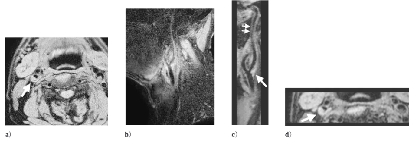
Fig. 1. An atheromatous plaque in a -year-old male.
A high signal intensity plaque at the right carotid bifurcation is shown on transaxial D-TSE a , oblique sagittal D-TFE b , coronal D-VRFA c , and sagittal and transaxial reconstruction images of D-VRFA d and e . The small ulceration is depicted only on D sequences arrows in b and d .
a b c d
e
ness of 3mm and an interslice gap of 4mm in a transaxial di- rection. Small plaque compositions may not be visualized due to low spatial resolution in the slice-select direction on 2D-TSE.
21)Although 3D-TFE with PPU- triggering also allowed image reconstruction in variable orientations, however, the suppres- sion of the intraluminal signal was insufficient, and the im- ages were compromised by plaque-mimicking artifacts in some cases. The residual signal on 3D-TFE reflects the diffi- culty in suppressing the signal from complex flow within the carotid bifurcation throughout the long-axis arterial segment, which is much longer than an axial slice on 2D-TSE.
17),22)In contrast, 3D-VRFA-TSE yielded acceptable suppression of the signal from flowing blood in all of the cases. Park and Kim proposed a T1-weighted 3D-TSE sequence with VRFA for efficiently reducing intravascular signal while retaining the signal from contrast-enhancing brain metastases. In addi- tion to VRFA, adding the proposed flow-sensitizing gradients yielded further suppression of the signal from slowly flowing blood.
20)Likewise, the present study indicated that 3D-TSE with VRFA and flow-sensitizing gradients is also useful in
reducing the signal within the carotid bifurcations to assess atherosclerotic plaques.
The signal ratio of the plaque to the muscle (RPM) on 3D- VRFA was significantly higher than that on conventional 2D- TSE. The difference in the contrast between these sequences may have been caused by the differences in the repetition time (TR). Narumi et al. examined carotid plaques using a non- triggered, self-navigated radial-scan technique, and demon- strated considerable differences in the plaque signal among T1-weighted images with different TRs. In the hyperintensity
Fig. 2. An atheromatous plaque in a -year-old male.An atheromatous plaque arrows is visible on D-TSE a , and on the sagittal and axial reconstruction image of D-VRFA c and d , whereas the plaque-lumen interface is unclear on D-TFE b . Although an intraluminal residual signal double arrows is observed in the distal portion of the internal carotid artery on D-VRFA arrowhead , it does not affect the evaluation of the plaque c .
a b c d
2D-TSE 3D-TFE 3D-VRFA
Image Quality 2.60+/−0.51 2.07+/−0.80 2.80+/−0.41
0 05
+ −
Table 1. Qualitative Comparison of Image Quality Among Three Black Blood Sequences
2D-TSE 3D-TFE 3D-VRFA
RPL 21.33+/−15.7 7.16+/−3.10 11.24+/−4.05
RPM 1.33+/−0.16 1.17+/−0.17 1.60+/−0.31
** *
*
* **
**
Table 2. Quantitative Comparison of RPL and RPM on Three BB Sequences
Data are mean the values +/− standard deviations.
* p<0.05, ** p<0.01
RPL and RPM are the signal ratio of the carotid plaque to the arterial lumen, and that of the carotid plaque to the adjacent muscle, respec- tively.
plaques, a TR-dependent decrease of the plaque signal was evident, whereas the signal remained unchanged regardless of the TR in the isointensity plaques.
15)Hence, conventional triggered 2D T1W images with relatively long and intersubjectively different TRs may be inappropriate for as- sessing vulnerable carotid plaques. In the present study, we observed that a constant and relatively short TR (450 ms), which is independent of the cardiac cycle of the patients, may contribute to the highest signal ratio of the plaques obtained on 3D-VRFA. Hence, it is expected that 3D-VRFA will be useful to detect T1W-high signal carotid plaques.
Although VRFA-TSE provided clinically sufficient BB im- ages, flow artifacts were observed in the distal lumen in three cases (Fig. 2). New blood-suppression techniques, such as motion-sensitized driven-equilibrium (MSDE) prior to TSE imaging may provide more efficient flow signal suppression.
23),24)There were several limitations to this study. One is the lack of pathological evidence of a plaque in all of our patients. We evaluated patients with carotid plaques which may comprise various constituents, including soft and/or hard components, and their signal intensity is therefore expected to vary on T1W images.
4) 10),13) 15)Hence, the detectability of vulner- able/ soft plaques by using 3D-VRFA needs to be evaluated in future studies and correlated with pathological findings.
The second limitation is that we have not conducted sys- tematic experiments to optimize the imaging parameters of the BB pulse sequences used in this study. The 2D-TSE and 3D-TFE in this study were performed according to the meth- ods used in previous studies.
9),13),14)However, 3D-VRFA is a relatively new technique and there are only a few reports re- garding the imaging protocol using this sequence.
20),25)Although Yoneyama et al
25)recommended low refocusing flip angles between 30 and 50 degrees to efficiently decrease flow artifacts in contrast-enhanced brain imaging, we selected a slightly larger refocus flip angle (60 degrees) because a lower refocus flip angle tended to degrade the signal to noise ratio in our experience (unpublished data), and maintaining a higher spatial resolution is desirable in assessing small carotid plaques.
In conclusion, T1-weighted 3D VRFA-TSE is a promising method to assess carotid plaques in terms of imaging cover- age, spatial resolution and lesion contrast. Further studies and pathological validation are needed to confirm the clinical util- ity of this pulse sequence.
References
1) North American symptomatic carotid endarterectomy trial.
Methods, patient characteristics, and progress. Stroke. 22: 711 720, 1991.
2) Golledge J, Greenhalgh RM, Davies AH. The symptomatic carotid plaque. Stroke. 31: 774 781, 2000.
3) Imparato AM, Riles TS, Mintzer R, Baumann FG. The importance of hemorrhage in the relationship between gross morphologic char- acteristics and cerebral symptoms in 376 carotid artery plaques.
Ann Surg 197 (2): 195 203, 1983.
4) Yuan C, Zhang SX, Polissar NL, Echelard D, Ortiz G, Davis JW, Ellington E, Ferguson MS, Hatsukami TS. Identification of fibrous cap rupture with magnetic resonance imaging is highly associated with recent transient ischemic attack or stroke. Circulation. 105:
181 185, 2002.
5) Murphy RE, Moody AR, Morgan PS, Martel AL, Delay GS, Allder S, MacSweeney ST, Tennant WG, Gladman J, Lowe J, Hunt BJ.
Prevalence of complicated carotid atheroma as detected by mag- netic resonance direct thrombus imaging in patients with suspected carotid artery stenosis and previous acute cerebral ischemia.
Circulation. 107: 3053 3058, 2003.
6) Moody AR, Murphy RE, Morgan PS, Martel AL, Delay GS, Allder S, MacSweeney ST, Tennant WG, Gladman J, Lowe J, Hunt BJ.
Characterization of complicated carotid plaque with magnetic reso- nance direct thrombus imaging in patients with cerebral ischemia.
Circulation. 107: 3047 3052, 2003.
7) Ouhlous M, Flach HZ, de Weert TT, Hendriks JM, van Sambeek MR, Dippel DW, Pattynama PM, van der Lugt A. Carotid plaque composition and cerebral infarction: MR imaging study. Am J Neuroradiol. 26: 1044 1049, 2005.
8) Yamada N, Higashi M, Otsubo R, Sakuma T, Oyama N, Tanaka R, Iihara K, Naritomi H, Minematsu K, Naito H. Association between signal hyperintensity on T1-weighted MR imaging of carotid plaques and ipsilateral ischemic events. Am J Neuroradiol. 28:
287 292, 2007.
9) Yoshida K, Endo H, Sadamasa N, Narumi O, Chin M, Inoue K, Mitsudo K, Yamagata S. Evaluation of carotid artery atherosclerotic plaque distribution by using long-axis high- resolution black-blood magnetic resonance imaging. J Neurosurg.
109: 1042 1048, 2008.
10) Bitar R, Moody AR, Leung G, Symons S, Crisp S, Butany J, Rowsell C, Kiss A, Nelson A, Maggisano R. In vivo 3D high- spatial-resolution MR imaging of intraplaque hemorrhage.
Radiology. 249: 259 267, 2008.
11) Edelman RR, Chien D, Kim D. Fast selective black blood MR im- aging. Radiology. 181: 655 660, 1991.
12) Simonetti OP, Finn JP, White RD, Laub G, Henry DA. Black blood" T2-weighted inversion-recovery MR imaging of the heart.
Radiology. 1991: 49 57, 1996.
13) Yoshida K, Narumi O, Chin M, Inoue K, Tabuchi T, Oda K, Nagayama M, Egawa N, Hojo M, Goto Y, Watanabe Y, Yamagata S. Characterization of carotid atherosclerosis and detection of soft plaque with use of black-blood MR imaging. Am J Neuroradiol.
29: 868 874., 2008.
14) Watanabe Y, Nagayama M, Suga T, Yoshida K, Yamagata S, Okumura A, Amoh Y, Nakashita S, Van Cauteren M, Dodo Y.
Characterization of atherosclerotic plaque of carotid arteries with histopathological correlation: vascular wall MR imaging vs. color Doppler ultrasonography (US). J Magn Reson Imaging. 28: 478 485, 2008.
15) Narumi S, Sasaki M, Ohba H, Ogasawara K, Hitomi J, Mori K, Ohura K, Ono A, Terayama Y. Altered carotid plaque signal among different repetition times on T1-weighted magnetic reso- nance plaque imaging with self-navigated radial-scan technique.
Neuroradiology. 52: 285 290, 2010.
16) Fan Z, Zhang Z, Chung YC, Weale P, Zuehlsdorff S, Carr J, Li D.
Carotid arterial wall MRI at 3T using 3D variable-flip-angle turbo spin-echo (TSE) with flow-sensitive dephasing (FSD). J Magn Reson Imaging. 31: 645 654, 2010.
17) Crowe LA, Varghese A, Mohiaddin RH, Yang GZ, Firmin DN.
Elimination of residual blood flow-related signal in 3D volume- selective TSE arterial wall imaging using velocity-sensitive phase reconstruction. J Magn Reson Imaging. 23: 416 421, 2006.
18) Hennig J, Weigel M, Scheffler K. Multiecho sequences with vari- able refocusing flip angles: optimization of signal behavior using smooth transitions between pseudo steady states (TRAPS). Magn Reson Med. 49: 527 535, 2003.
19) Busse RF, Hariharan H, Vu A, Brittain JH. Fast spin echo se- quences with very long echo trains: design of variable refocusing flip angle schedules and generation of clinical T2 contrast. Magn Reson Med. 55: 1030 1037, 2006.
20) Park J, Kim EY. Contrast-enhanced, three-dimensional, whole- brain, black-blood imaging: application to small brain metastases.
Magn Reson Med. 63: 553 561, 2010.
21) Balu N, Chu B, Hatsukami TS, Yuan C, Yarnykh VL. Comparison
between 2D and 3D high-resolution black-blood techniques for ca- rotid artery wall imaging in clinically significant atherosclerosis. J Magn Reson Imaging. 27: 918 924, 2008.
22) Steinman DA, Rutt BK. On the nature and reduction of plaque- mimicking flow artifacts in black blood MRI of the carotid bifurca- tion. Magn Reson Med. 39: 635 641, 1998.
23) Wang J, Yarnykh VL, Hatsukami T, Chu B, Balu N, Yuan C.
Improved suppression of plaque-mimicking artifacts in black-blood carotid atherosclerosis imaging using a multislice motion- sensitized driven-equilibrium (MSDE) turbo spin-echo (TSE) se- quence. Magn Reson Med. 58: 973 81, 2007.
24) Koktzoglou I, Li D. Diffusion-prepared segmented steady-state free precession: Application to 3D black-blood cardiovascular magnetic resonance of the thoracic aorta and carotid artery walls.
J Cardiovasc Magn Reson. 9: 33 42, 2007.
25) Yoneyama M, Nakamura M, Tabuchi T, Takemura A, Ogura J.
Spin-echo like T1 contrast" volumetric black-blood images using 3D LOWRAT: low refocusing flip angle TSE. Proc Intl Soc Mag Reson Med. 18: 3031, 2010.
(Received on January 11, 2011, Accepted on March 11, 2011)