Identification of an HLA class I allele closely involved in the auto‑antigen
presentation in acquired aplastic anemia
著者 材木 義隆
著者別表示 Zaimoku Yoshitaka journal or
publication title
博士論文本文Full 学位授与番号 13301甲第4582号
学位名 博士(医学)
学位授与年月日 2017‑06‑30
URL http://hdl.handle.net/2297/00052027
doi: 10.1182/blood-2016-11-752378
Creative Commons : 表示 ‑ 非営利 ‑ 改変禁止 http://creativecommons.org/licenses/by‑nc‑nd/3.0/deed.ja
1 ZAIMOKU et al
Short title: HLA-B*40:02 MUTATIONS IN APLASTIC ANEMIA Scientific category: IMMUNOBIOLOGY
Identification of an HLA class I allele closely involved in the auto-antigen presentation in acquired aplastic anemia
Yoshitaka Zaimoku,1,2 Hiroyuki Takamatsu,1 Kazuyoshi Hosomichi,3 Tatsuhiko Ozawa,4 Noriharu Nakagawa,1 Tatsuya Imi,1 Hiroyuki Maruyama,1 Takamasa Katagiri,5 Hiroyuki Kishi,4 Atsushi Tajima,3 Atsushi Muraguchi,4 Koichi Kashiwase6 and Shinji Nakao1
YZ, HT and KH contributed equally to this study.
1Department of Hematology, Graduate School of Medical Sciences, Kanazawa University, Kanazawa, Japan;
2Department of Internal Medicine, Keiju Medical Center, Nanao, Japan;
3Department of Bioinformatics and Genomics, Graduate School of Advanced Preventive Medical Sciences, Kanazawa University, Kanazawa, Japan;
4Department of Immunology, Graduate School of Medicine and Pharmaceutical Sciences, University of Toyama, Toyama, Japan;
5Clinical Laboratory Science, Graduate School of Medical Sciences, Kanazawa University, Kanazawa, Japan;
6Department of HLA Laboratory, Japanese Red Cross Kanto-Koshinetsu Block Blood Center, Tokyo, Japan
Keywords: Aplastic anemia; Hematopoiesis; Major histocompatibility complex genes; Responses to self antigens
Text word count: 3944 Abstract word count: 246 Number of figures: 5 Number of tables: 2 Number of references: 37
Blood
2 Abstract
To identify HLA alleles closely involved in the auto-antigen presentation in acquired aplastic anemia (AA), we studied the HLA allelic loss frequencies of 312 AA patients, including 43 patients with loss of
heterozygosity of 6p chromosome (6pLOH). An analysis of the HLA alleles contained in the lost haplotype revealed HLA-B*40:02 to be the most frequently lost allele. When we examined 28 AA (12 6pLOH[+] and 16 6pLOH[-]) patients with HLA-B*40:02 for the presence of leukocytes lacking HLA-B4002 (B4002[-]) using a new monoclonal antibody specific to this allele, B4002(-) granulocytes were detected not only in all 6pLOH(+) patients but also in 9 (56%) of the 16 6pLOH(-) patients. Furthermore, 10 (83%) of the 12 6pLOH(+) patients possessed 1.0%-78% B4002(-) granulocytes that retained the HLA-A allele on the same haplotype (B4002[-]A[+]), suggesting the frequent co-existence of granulocytes that underwent mutations restricted to HLA-B*40:02 with 6pLOH(+) (B4002[-]A[-]) granulocytes. Deep sequencing of the HLA-B*40:02 of sorted B4002(-)A(+) granulocytes revealed various somatic mutations, such as frameshift, nonsense and splice site mutations, in all 15 patients studied. Surprisingly, missense mutations in the α-3 domain of HLA-B*40:02 that is not involved in the antigen presentation were detected
exclusively in the B4002(+) granulocytes of three patients possessing B4002(-) granulocytes. The markedly high prevalence of leukocytes lacking HLA-B4002 as a result of either or both 6pLOH or structural gene mutations suggests that antigen presentation by hematopoietic stem/progenitor cells to cytotoxic T cells via the HLA-B allele plays a critical role in the pathogenesis of AA.
Key Points
1. Somatic mutations of HLA-B*40:02 are highly frequently detected in granulocyte of patients with acquired aplastic anemia.
2. Antigen presentation via HLA-B4002 may play a critical role in the pathophysiology of acquired aplastic anemia.
Introduction
Acquired aplastic anemia (AA) is thought to be a T-cell mediated autoimmune disease based on a good response to immunosuppressive therapy, such as anti-thymocyte globulin (ATG) and/or cyclosporine (CsA).1 A large body of in vitro evidence suggests the essential role of T cells in the development of AA, which includes the presence of T cells with particular T-cell receptor clonotypes,2-7 cytotoxic T-cell clones capable of killing hematopoietic stem/progenitor cells (HSPCs),2,4 and leukocytes that lack HLA class I antigens due to copy-number neutral loss of heterozygosity in the short arm of chromosome 6 (6pLOH).8-12 Among these suggestive findings, the presence of 6pLOH(+) leukocytes is considered to be the most compelling evidence that cytotoxic T cells (CTLs) are involved in the development of bone marrow failure, as it represents the escape of HSPCs with 6pLOH from the attack of CTLs that are specific to
auto-antigens presented by the lacked HLA class I allele. However, the incidence of 6pLOH in AA patients shown by several studies was at most 13%, and it is therefore unclear to what extent the HSPC-specific CTLs contribute to the development of AA and which HLA proteins are the most critically involved in the
3 auto-antigen presentation.
We previously determined the missing frequencies of individual HLA alleles by analyzing the alleles contained in the missing haplotype of 6pLOH(+) AA patients and found that four alleles (HLA-A*02:01, A*02:06, A*31:01, and B*40:02) were significantly more likely to be lost than the other HLA alleles.9 However, it was difficult to determine which allele in the missing haplotype was actually responsible for the antigen presentation because the lost fragment of chromosome 6p usually contained two or more HLA alleles, and allele-specific monoclonal antibodies (mAbs) useful for detecting leukocytes lacking single alleles were not commercially available, except for mAbs specific to some HLA-A alleles.
Among the four HLA class I alleles, an important role of HLA-B*40:02 in auto-antigen presentation in AA has been suggested by other studies. A congress abstract13 reported a markedly high prevalence of HLA-B61, a corresponding serotype to HLA-B*40:02, in Japanese pediatric AA patients. Inaguma et al14 established a CTL clone capable of suppressing HSPCs in a HLA-B*40:02-restricted manner from an AA patient who possessed HLA-B*40:02-lacking leukocytes due to 6pLOH. Osumi et al15 reported a case of AA whose leukocytes had a nonsense mutation in HLA-B*40:02, suggesting the presence of leukocytes lacking only HLA-B4002. Given these findings, a flow cytometry (FCM) analysis using
HLA-B4002-specific mAbs may reveal leukocytes that lack HLA-B4002 due to mechanisms other than 6pLOH in AA patients carrying HLA-B*40:02. Many researchers have tried to generate a mAb specific for HLA-B61 by immunizing transgenic mice, but all attempts have failed for some reason. If anti-HLA-B61 mAbs were generated, it would greatly facilitate the understanding of the mechanisms underlying the lack of HLA-B4002 from leukocytes as well as of the immune mechanisms of AA.
We recently succeeded in generating mAbs specific to HLA-B61, taking advantage of cDNA derived from B lymphocytes from an AA patient possessing anti-HLA-B61 antibody as a result of multiple
transfusions.16 The mAbs successfully identified HLA-B4002-missing leukocytes not only in 6pLOH(+) patients but also in 6pLOH(-) patients in the majority of AA patients possessing HLA-B*40:02, and deep sequencing of HLA-B4002-missing granulocyte-derived DNA revealed various mutations in the structural gene region of HLA-B*40:02, strongly suggesting a critical role of HLA-B4002 in the auto-antigen presentation of AA.
Patients and methods Subjects
A total of 312 Japanese patients with AA (age, 15 to 90 years old, median 65 years old; 132 males and 180 females; 115 severe AA and 197 non-severe AA) were enrolled in an observational study to determine the prevalence of HLA allele-lacking leukocytes (HLA-LLs) between 2011 and 2014 (supplemental Figure 1). The patients’ blood samples were subjected to HLA typing and droplet digital polymerase chain reaction (ddPCR) for detecting 6pLOH. Severe AA was diagnosed when at least two of the following criteria were met; the neutrophil count was < 0.5 × 10⁹/L, the platelet count was < 20 × 109/L and the reticulocyte count was < 20 × 109/L.17 Very severe AA was defined as a neutrophil count < 0.2 × 10⁹/L in addition to the criteria for severe AA.18 The response criteria were as previously described.19 All patients
4
were genotyped for HLA-A, HLA-B, HLA-C, and HLA-DRB1 alleles using the polymerase chain reaction (PCR) sequence-specific oligonucleotide method.20 All patients provided their informed consent to the HLA-typing and genetic analyses. The study protocols were approved by the ethical committee of Kanazawa University Institute of Medical, Pharmaceutical and Health Sciences.
Preparation of anti-HLA-B61 antibody
We developed a new method for generating human mAbs specific to HLA alleles by taking advantage of B cells from patients who had anti-HLA antibodies following repeated transfusions.16 Briefly, mononuclear cells isolated from 20 ml of peripheral blood from an anti-HLA-B61 antibody-positive patient were stimulated to differentiate into antibody-producing cells with a cytokine cocktail containing R848, CpG2006, anti-CD40, recombinant human interleukin (hIL)-2, hIL-4, hIL-17, hIL-21 and human B-cell activating factor in RPMI1640 containing 10% fetal calf serum for 6 days. CD138+ antibody-producing cells were then isolated with anti-CD138 antibody-conjugated microbeads using AutoMACS Pro Separator (Miltenyi Biotec, Bergisch Gladbach, Germany). Anti-HLA-B61 IgG antibody-producing cells were isolated using a microwell array chip coated with purified HLA-B61 proteins and subjected to RNA extraction from single cells as previously described.21,22 The antibody cDNA fragments for heavy and light chain variable domain fragments were amplified using the single-cell 5’-RACE method and inserted into expression vectors. Thereafter, HEK293 cells were transfected with the heavy and light chain vectors and cultured to obtain a supernatant containing complete mAb molecules. The specificity of the mAbs was tested by ELISA and FCM. To prevent nonspecific binding of human IgG to human Fc receptors, the Fc portion of the anti-HLA-B61 mAb was replaced with the constant domain of mouse IgG1.
Detection of HLA-LLs and cell sorting
HLA-LLs were detected using FACS Canto II (BD Biosciences, San Jose, USA) and analyzed with the FlowJo v10.1 software program (Tree star, Ashland, USA). Leukocytes lacking HLA-B4002 were referred to as B4002(-) cells, including both 6pLOH(+) leukocytes that lack both HLA-B4002 and one HLA-A allele on the same haplotype (B4002[-]A[-]) and leukocytes lacking only HLA-B4002 (B4002[-]A[+]).
B4002(-)A(+) granulocytes and HLA-B4002-positive (B4002[+]) granulocytes and/or B4002(+) T cells were sorted using FACS Aria Fusion (BD Biosciences). The mAbs used for this study are provided in supplemental Table 1.
ddPCR to detect 6pLOH involving HLA genes
The presence of 6pLOH(+) leukocytes and their percentages of total leukocytes were determined with ddPCR using a QX200 AutoDG Droplet Digital PCR System (Bio-Rad, Hercules, USA) by comparing the copy number of each HLA allele in individuals heterozygous for the HLA allele. 6pLOH that involves HLA genes gives rise to a copy number imbalance between the two different alleles. The reaction mixtures of ddPCR included 2 TaqMan probes labeled with different fluorochromes (6-FAM or VIC)
complementary to the allele-specific sequences, 2 primers complementary to consensus sequences
5
surrounding the allele-specific sequence, and 40 ng of genomic DNA in 1x ddPCR Supermix for Probes (No dUTP; Bio-Rad). We designed four primer and probe mixture that can detect 6pLOH involving HLA-A (A31/33 mixture), HLA-B (B1 and B2 mixture) and HLA-C (C3 mixture, supplemental Tables 2 and 3).
The detailed protocols for ddPCR were shown in supplemental methods.
When the lacked alleles contained in the haplotype of 6pLOH(+) patients could not be completely determined by ddPCR or FCM, they were estimated using a haplotype database of the Japanese population including 18,604 individuals from 5,824 families.23 An allele was judged to be missing as a result of 6pLOH if the presence of the allele in the lost haplotype was estimated with >95% accuracy.
Detection of HLA-B gene mutations
To identify the somatic mutations of HLA-B*40:02 in B4002(-)A(+) granulocytes, target sequencing of HLA-B in sorted B4002(-)A(+) granulocytes was performed using a next-generation sequencer (NGS, MiSeq; Illumina, San Diego, USA). B4002(+) granulocytes and/or B4002(+) T cells from identical individuals were used as a control. HLA genes were enriched from genomic DNA using sequence capture (SeqCap EZ system; Roche Sequencing, Pleasanton, USA), a hybridization-based gene enrichment method.
Potential mutations responsible for missing HLA-B4002 were identified when variant reads were found only in B4002(-) granulocytes. All of the mutations were validated using deep sequencing of HLA-B locus-specific long-range PCR, as previously described.24 HLA-B alleles carrying those mutations were determined using the nearest allele-specific SNPs. Reference sequences of HLA-B*40:02 were obtained from the IPD-IGMT/HLA database.25
Statistical analyses
The clinical parameters of the patients were compared using Fisher’s exact test for categorical variables and the Mann-Whitney U test for continuous variables with the EZR software package, a graphical user interface for R (The R Foundation for Statistical Computing version 2.13.0).26
Results
Allelic frequency of HLA-genes in this study subjects
When the allelic frequency of HLA genes in 312 AA patients genotyped in this study was compared with those in the Japanese general population,23 HLA-B*40:02 was the fifth-most frequent allele observed in AA patients; the top five alleles were HLA-DRB1*15:02 (22% vs. 11%, OR=2.3, P=3.0×10⁻¹⁵), B*52:01 (19%
vs. 11%, OR=1.9, P=6.0×10⁻⁹), DRB1*15:01 (14% vs. 7.7%, OR=2.0, P=4.5×10⁻⁸), C*12:02 (19% vs.
11%, OR=1.9, P=2.8×10⁻⁸), and B*40:02 (13% vs. 7.9%, OR=1.8, P=9.3×10⁻⁶).
Validation of the 6pLOH(+) cell measurement using ddPCR
The ddPCR generated 18,745±1,553 (mean±2SD) droplets per one reaction mixture, which allowed us to detect at least 3%-4% 6pLOH(+) leukocytes with a specificity of 99%. The specificity of the ddPCR in detecting 6pLOH(+) leukocytes was verified by showing negativity for 50 healthy volunteers or AA
6
patients who were known to be negative for HLA-A-allele lacking leukocytes using FCM. To determine the reliability of estimating the 6pLOH(+) cell percentage by ddPCR, leukocytes from an individual carrying HLA-A2 and Cw3 were mixed with leukocytes from an individual not carrying A2 or Cw3 at various ratios, and the mixed leukocyte populations were examined by FCM and ddPCR. The percentages of 6pLOH(+) cells estimated by ddPCR were almost identical to those determined by FCM, suggesting sufficient reliability of ddPCR in measuring the percentage of 6pLOH(+) cells in the total leukocyte populations (supplemental Figure 2).
Detection of 6pLOH and the frequency of HLA alleles involved in the lost haplotype
Using either of the four different ddPCR mixtures, the presence of 6pLOH was evaluable in 224 (72%) of 312 patients with AA because they were available for ddPCR in at least 1 of the 3 HLA class I loci.
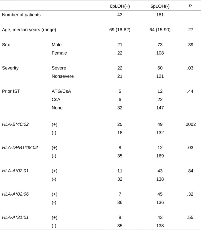
6pLOH(+) leukocytes that accounted for 3.9% to 84% (median, 10%) of the total leukocytes were detected in 43 (19%) of the 224 patients analyzed by the ddPCR. 6pLOH(+) patients had significantly more severe disease (51%) than 6pLOH(-) patients (33%, P=0.03), while age (P=0.27), sex (P=0.39) and prior immunosuppressive therapy (IST, P=0.44) did not differ markedly between the two groups (Table 1).
When the prevalence of 6pLOH was compared between patients carrying and not carrying each
HLA-allele, including alleles in HLA-A, HLA-B, HLA-C and HLA-DRB1 loci, consistent with a previous report,9 HLA-B*40:02 was most strongly involved in 6pLOH (34% [25/74], odds ratio [OR]=3.7, P=0.0002), followed by HLA-DRB1*08:02 (40% [8/20], OR=3.2, P=0.03). Of note, the HLA-B*40:02 allele was included in the lost haplotype in all 25 6pLOH(+) patients possessing this allele. Seven of the 8 patients with HLA-DRB1*08:02 alleles possessed HLA-B*40:02 in the same lost haplotype, while the remaining patient had HLA-DRB1*08:02 in the retained haplotype, suggesting that the higher frequency of HLA-DRB1*08:02 in 6pLOH(+) patients is due to the linkage disequilibrium between HLA-B*40:02 and DRB1*08:02.
Among 150 HLA-B*40:02-negative patients, 6pLOH was detected in 18 (12%) patients. HLA-alleles that were more likely to be possessed by 6pLOH(+) patients were HLA-DRB1*15:01 (23% [9/40], OR=3.3, P=0.02), C*01:02 (24% [8/34], OR=3.3, P=0.03) and B*54:01 (29% [5/17], OR=3.8, P=0.03).
HLA-B*54:01 was missing in all five 6pLOH(+) patients carrying this allele, while 7 of 9
HLA-DRB1*15:01 and 2 of 8 C*01:02 in the 6pLOH(+) patients were in the retained haplotype. Three HLA alleles (HLA-A*02:01, A*02:06 and A*31:01) that had been suggested to be frequently lost due to 6pLOH by our previous study9 were not significantly associated with 6pLOH in this study cohort; each allele was found at similar frequencies between 6pLOH(+) and 6pLOH(-) patients (HLA-A*02:01, 20%
[11/54], OR=1.1, P=0.84; A*02:06, 13% [7/52], OR=0.59, P=0.32; and A*31:01, 16% [8/51], OR=0.73, P=0.55). The trend was consistent in HLA-B*40:02-negative patients (HLA-A*02:01, 11% [4/35], OR=0.93, P=1.0; A*02:06, 14% [5/37], OR=1.2, P=0.77; and A*31:01, 7.5% [3/38], OR=0.55, P=0.56).
B4002(-) granulocytes in patients with HLA-B*40:02
Peripheral blood samples were available for FCM using the anti-HLA-B61 antibody in 28 patients with AA
7
carrying HLA-B*40:02, including 12 6pLOH(+) and 16 6pLOH(-) patients. B4002(-) granulocytes that accounted for 24%-99% of the total granulocytes were detected in all of the 12 6pLOH(+) patients.
Unexpectedly, 1.0%-99% B4002(-) granulocytes were also detected in nine (56%) of the 16 6pLOH(-) patients. None of 12 individuals carrying HLA-B*40:02 (6 healthy individuals and 6 patients with
hematological malignancies) were positive for B4002(-) granulocytes. The 21 patients possessing B4002(-) granulocytes had more severe disease (67%, 14/21), while only 1 of the 7 patients (14%, P=0.03) not possessing B4002(-) granulocytes had severe disease.
Since 10 of the 12 6pLOH(+) patients were heterozygous for the HLA-A allele, the HLA-A allelic expression by their B4002(-) granulocytes was able to be evaluated. As shown in Figure 1 and Table 2, 8.0%-99% granulocytes of all the 10 6pLOH(+) patients lacked both HLA-B4002 and the HLA-A allele (B4002[-]A[-]) on the same haplotype containing HLA-B*40:02 and were therefore thought to represent 6pLOH(+) granulocytes. In contrast, 8 of the 10 patients also had 1.0%-78% granulocytes that lacked HLA-B4002 but retained the HLA-A allele (B4002[-]A[+]), indicating that these B4002(-) cells lacked HLA-B4002 via mechanisms other than 6pLOH.
Two 6pLOH(+) patients (Cases 16 and 18) were homozygous for HLA-A allele, and the percentage of 6pLOH(+) granulocytes was estimated using ddPCR. Only 9% and 66% of the sorted B4002(-) granulocytes were 6pLOH(+), suggesting that these patients also had B4002(-)A(+) granulocytes that accounted for 91% and 34% of B4002(-) granulocytes. None of the 20 (10 6pLOH[+] and 10 6pLOH[-]) patients with HLA-B*40:02 had granulocytes that lacked an HLA-A allele but retained HLA-B4002. The prevalence of missing HLA-B4002 from granulocytes in the 28 patients was 75% (21/28, 9 with
B4002[-]A[+] cells alone, 10 with both B4002[-]A[+] and B4002[-]A[-] [6pLOH] cells, and 2 with B4002[-]A[-] cells alone, Figure 1).
Mutations of HLA-B alleles in B4002(-)A(+) granulocytes
B4002(-)A(+) granulocytes were available for mutation analyses of HLA-B alleles in 15 of the 19 patients who possessed the aberrant granulocytes. The mean coverage of the HLA-B gene was 426× for the capture method and 32,077× for the PCR method. Somatic mutations were judged to be present when the
frequency exceeded 0.01. In total, 59 different somatic mutations of HLA-B were identified in the sorted B4002(-)A(+) granulocytes, all of which were present in HLA-B*40:02 and not in any of the other HLA-B alleles. The median variant allele frequency (VAF) was 3.7% (range, 1.0%-47%), and the number of mutations in each patient ranged from 1 to 9 (median 4, Figure 2 and supplemental Table 4).
Fifty-five mutations were exonic, and four were intronic. The exonic mutations were frameshift duplications (n=12), frameshift deletions (n=23), an inframe deletion (n=1), nonsense mutations (n=14), a missense mutation (n=1), and start losses (n=4). All exonic mutations were expected to result in a lack of HLA-B4002 expression except for one missense mutation in Case 19, which could potentially lead to a structural change in the epitope recognized by anti-HLA-B61 mAb. All four intronic mutations were considered to be splice site mutations; two mutations deactivated 5' or 3' splice sites, whereas the other two were single-base substitutions within intron 3, creating alternative 5' splicing sites with a strong consensus
8
sequence, GTGAGT: TTCCTGAGT> TTCGTGAGT (c.619+133C>G) in Case 6 and GGCATGAGT>
GGCGTGAGT (c.619+123A>G) in Case 15. The presence of alternative splice sites potentially inhibits normal splicing, leading to a decreased expression of HLA-B*40:02. The presence of alternatively spliced mRNA solely in B4002(-)A(+) granulocytes was confirmed in Case 6, whose cDNA of both sorted B4002(-)A(+) and B4002(+) granulocytes was available using a PCR specific for alternatively spliced transcripts (Figure 3).
Missense mutations in the HLA-B*40:02 allele of B4002(+) granulocytes
B4002(+) granulocytes were subjected to deep sequencing of HLA-B as a negative control in 14 of the 15 patients possessing B4002(-)A(+) granulocytes. Surprisingly, four different missense mutations of HLA-B*40:02, which are not involved in the antigen presentation by this class I allele, were detected in B4002(+) granulocytes from 3 (Cases 3, 7, and 17) of the 14 patients (Figure 2). The same mutations were not detected in the corresponding B4002(-)A(+) granulocytes of all the three patients, B4002(+) T cells of two patients (Cases 7 and 17) or the reference sequence of HLA-B*40:02. The VAFs of the 4 missense mutations were 9.5% (Case 17), 9.6% (Case 17), 43% (Case 7), and 47% (Case 3), respectively. The 4 missense mutations in B4002(+) cells and 1 missense mutation in B4002(-)A(+) cells occurred close together at an α-3 domain region in the protein 3-D structure (supplemental Figure 3), suggesting the inability of the mutant HSPCs to interact with CTLs.
The presence of mutations in the α-3 domain region of HLA-B*40:02 in B4002(+) granulocytes of three patients possessing B4002(-)A(+) granulocytes prompted us to study five AA patients with
HLA-B*40:02 who were negative for B4002(-)A(+) granulocytes. The B4002(+) granulocytes from four patients (Cases 23, 24, 25, and 26) without B4002(-) granulocytes and one patient (Case 1) with 6pLOH(+) granulocytes were subjected to deep sequencing using T cells as a negative control. No mutations were found in the five patients’ samples.
Response to IST in patients carrying HLA-B*40:02
Of the 28 patients carrying HLA-B*40:02, 15 patients were treated with ATG plus CsA therapy before (n=13) or after (n=2) sampling for the study (Table 2). All 11 (100%) patients possessing B4002(-) granulocytes showed a good response (complete response [CR] in 5 and partial response [PR] in 6), while 3 of the 4 (75%) patients not possessing B4002(-) granulocytes responded (CR in 2 and PR in 1). Eleven patients were treated with CsA monotherapy before (n=10) and after (n=1) sampling; 7 of the 9 patients possessing B4002(-) granulocytes showed a response (CR in 3 and PR in 4); 1 of 2 patients not possessing B4002(-) granulocytes showed a response (CR). Two patients (one with B4002[-] cells and the other without B4002[-] cells) were not treated with IST.
Chronological analysis of B4002(-) granulocytes
The percentages of B4002(-) granulocytes in two patients (Cases 16 and 20) were determined several times after ATG/CsA therapy. Both patients responded to ATG therapy, and their hematological recovery was
9
associated with a decrease in the B4002(-) granulocyte percentage (83% to 4.3% and 73% to 33% after 13 and 12 months of the therapy, respectively; Figure 4). In contrast, the B4002(-) granulocyte percentages of four patients (Cases 2, 3, 6 and 19) whose first samples were tested after the recovery of hematopoiesis following 6.2 to 23 years of ATG/CsA therapy did not change markedly when their second samples were examined 6 to 10 months (median, 9 months) after the first examination; 99% to 98%; 60% to 59%; 10%
to 10%; and 79% to 85%, respectively.
Discussion
The current study in a large number of AA patients using a novel ddPCR, similar to our previous studies, detected 6pLOH in 19% of the patients and confirmed a significantly higher frequency of lacking HLA-B*40:02 than of lacking other HLA class I alleles, including HLA- A*02:01, A*02:06 and A*31:01.
Furthermore, a new mAb specific to HLA-B61 revealed the presence of B4002(-) granulocytes in 75% of patients carrying HLA-B*40:02. These findings suggest that HLA-B*40:02 is critically involved in the auto-antigen presentation to T cells in patients with AA. The prevalence of HLA-LLs may have been underestimated due to a lack of mAbs specific to many other HLA alleles.
6pLOH has been considered to be the sole or a major method of lacking class I HLAs in HSPCs targeted by CTLs of AA9,10,12 and in acute myeloid leukemia cells relapsed after HLA-mismatched hematopoietic stem cell transplantation,27,28 although some reports have shown a possible lack of HLA alleles due to structural gene mutations.15,29,30 Our deep sequencing in the present study of HLA class I genes derived from sorted B4002(-)A(+) granulocytes revealed various mutations solely in the
HLA-B*40:02 gene. The HLA-B*40:02 mutations are apparently somatic because B4002(+)A(+) leukocytes were present in all patients possessing B4002(-) granulocytes and neither the B4002(+)A(+) granulocytes nor B4002(+) T lymphocytes from each individual showed the same mutations. The high number of mutations per patient with B4002(-) granulocytes (median 4) suggests that HSPC clones with various mutations arose in the bone marrow of the patients prior to the AA development and might have been selected to contribute to hematopoiesis under strong pressure by CTLs specific to auto-antigens presented by HLA-B4002 at the onset of AA (Figure 5).
Recent studies using NGS have revealed clonal hematopoiesis with various somatic mutations in the granulocytes of AA patients, but they failed to show somatic mutations in HLA genes except for 6pLOH, which was detected in 13% of patients.10,31 This is mainly due to insufficient depth of the previous analyses, which did not focus on HLA gene regions. The somatic mutations in HLA genes do not imply a propensity toward myelodysplastic syndrome but may instead represent the selection by immune pressure, like PIGA mutations. The decrease in the B4002(-) granulocyte percentage associated with successful ATG therapy in two patients and the higher prevalence of B4002(-) granulocytes in severe AA than in non-severe AA patients support this hypothesis.
One unexpected and important finding of this study is that B4002(+) granulocytes from three patients with B4002(-) granulocytes also had missense mutations in the limited α-3 domain region of HLA-B*40:02, which is not involved in the antigen presentation by this class I allele. CTLs require binding of their CD8
10
molecules to the α-3 domain of HLA class I to exert their function.32 The single amino acid change in the α-3 domain observed in the three patients may have made the mutant HSPCs resistant to the attack by CTLs.33,34 These findings also suggest that detecting HLA protein expression with specific mAbs may not be enough to screen HSPCs or leukemic cells that evade CTL attack; deep sequencing of HLA genes including the α-3 domain coding region is necessary.
The allelic frequency of HLA-B*40:02 in healthy individuals is reported to be higher in Asian populations, with rates of 7.9% in Japanese, 8.7% in South Korean, 2.0% in Chinese, 2.4% in Thai, and 7.7% in American Filipino populations, compared with rates of 1.6% in German, 0.3% in Northern Irish, 1.8% in Italian, 0.9% in Swiss, and 1.5% in USA Caucasian populations.35 The higher frequency of this class I allele may partly explain the higher incidence of AA in Asian countries than in Western countries.
Although the incidence of AA in the USA is much lower than in Japan, a study of 11 American AA patients with somatic HLA loss by Babushok et al36 also revealed that the most frequently affected allele was HLA-B*40:02, suggesting that an auto-antigen presented by this HLA-B is commonly involved in the development of AA in Japan and the USA.
The markedly high prevalence of allele-lacking granulocytes in AA patients carrying HLA-B*40:02 suggests that antigens presented by HLA-B4002 may serve as auto-antigens eliciting T-cell responses to HSPCs. We previously isolated a T-cell clone from a 6pLOH(+) patient that was capable of inhibiting HSPCs in a HLA-B*40:02-restricted manner but failed to identify target antigens on HSPCs due to the inability to propagate the T-cell clone.14 Current technology has enabled the preparation of T-cell receptor vectors and their transfectants by taking advantage of predominantly expanding T-cell clones in patients’
bone marrow in response to unknown antigens.37 Studying T cells from AA patients possessing B4002(-) granulocytes is therefore warranted to identify auto-antigens of AA.
Acknowledgements
The authors thank the patients and donors and their physicians, including T. Endo and K. Kahata of Hokkaido University, M. Hirao and M. Kida of NTT Medical Center of Tokyo, H. Hiramatsu of Kyoto University, H. Taniguchi of Sasebo City General Hospital, M. Yamazaki of Keiju Medical Center, C.
Sugimori of Ishikawa Prefectural Central Hospital, M. Tanabe, K. Ohata, K. Ishiyama, Y. Kondo and H.
Yamazaki of Kanazawa University, for contributing to this study and Advanced Preventive Medical Sciences Research Center, Kanazawa University for the use of facilities.
Y.Z. is a PhD candidate at Kanazawa University and this work is submitted in partial fulfillment of the requirement for the PhD.
This work was supported in part by JSPS KAKENHI Grant Numbers JP16H05335, JP16H06502, and the Platform Project for Supporting in Drug Discovery and Life Science Research (Platform for Drug Discovery, Informatics and Structural Life Science) from Japan Agency for Medical Research and Development (AMED).
11 Authorship
Contribution: Y.Z., N.N., T.I., H.M. and T.K. collected clinical data and blood samples; K.K. performed HLA genotyping; Y.Z. performed ddPCR; Y.Z. and T.I. performed cell sorting; K.H. and A.T. performed deep sequencing of HLA-B; Y.Z., H.T., T.O., H.K. and A.M. generated an anti-HLA-B61 mAb; Y.Z., H.T.
and S.N. designed the research and wrote the manuscript; all authors critically reviewed the manuscript and checked the final version of it.
Conflict-of-interest disclosure: The authors declare no competing financial interests.
Correspondence: Shinji Nakao, 13-1 Takaramachi, Kanazawa, Ishikawa 920-8640 Japan; e-mail:
References
1. Young NS, Calado RT, Scheinberg P. Current concepts in the pathophysiology and treatment of aplastic anemia. Blood. 2006;108(8):2509-2519.
2. Nakao S, Takami A, Takamatsu H, et al. Isolation of a T-cell clone showing
HLA-DRB1*0405-restricted cytotoxicity for hematopoietic cells in a patient with aplastic anemia.
Blood. 1997;89(10):3691-3699.
3. Zeng W, Nakao S, Takamatsu H, et al. Characterization of T-cell repertoire of the bone marrow in immune-mediated aplastic anemia: evidence for the involvement of antigen-driven T-cell response in cyclosporine-dependent aplastic anemia. Blood. 1999;93(9):3008-3016.
4. Risitano AM, Maciejewski JP, Green S, Plasilova M, Zeng W, Young NS. In-vivo dominant immune responses in aplastic anaemia: molecular tracking of putatively pathogenetic T-cell clones by TCR beta-CDR3 sequencing. Lancet. 2004;364(9431):355-364.
5. Lu J, Basu A, Melenhorst JJ, Young NS, Brown KE. Analysis of T-cell repertoire in hepatitis-associated aplastic anemia. Blood. 2004;103(12):4588-4593.
6. Wlodarski MW, Gondek LP, Nearman ZP, et al. Molecular strategies for detection and quantitation of clonal cytotoxic T-cell responses in aplastic anemia and myelodysplastic syndrome. Blood.
2006;108(8):2632-2641.
7. Krell PF, Reuther S, Fischer U, et al. Next-generation-sequencing-spectratyping reveals public T-cell receptor repertoires in pediatric very severe aplastic anemia and identifies a beta chain CDR3 sequence associated with hepatitis-induced pathogenesis. Haematologica. 2013;98(9):1388-1396.
8. Afable MG, 2nd, Wlodarski M, Makishima H, et al. SNP array-based karyotyping: differences and similarities between aplastic anemia and hypocellular myelodysplastic syndromes. Blood.
2011;117(25):6876-6884.
9. Katagiri T, Sato-Otsubo A, Kashiwase K, et al. Frequent loss of HLA alleles associated with copy number-neutral 6pLOH in acquired aplastic anemia. Blood. 2011;118(25):6601-6609.
10. Yoshizato T, Dumitriu B, Hosokawa K, et al. Somatic Mutations and Clonal Hematopoiesis in Aplastic Anemia. N Engl J Med. 2015;373(1):35-47.
11. Maruyama H, Katagiri T, Kashiwase K, et al. Clinical significance and origin of leukocytes that lack
12
HLA-A allele expression in patients with acquired aplastic anemia. Exp Hematol. 2016.
12. Betensky M, Babushok D, Roth JJ, et al. Clonal evolution and clinical significance of copy number neutral loss of heterozygosity of chromosome arm 6p in acquired aplastic anemia. Cancer Genet.
2016;209(1-2):1-10.
13. Takahashi Y, Ohara A, Kobayashi R, et al. Expression of Human Leukocyte Antigen B61 Is Associated with Idiopathic Aplastic Anemia, Hepatitis-Associated Aplastic Anemia and Fulminant Hepatic Failure in Children [abstract]. Blood. 2012;120(21):3487. Abstract 3487.
14. Inaguma Y, Akatsuka Y, Hosokawa K, et al. Induction of HLA-B*40:02-restricted T cells possessing cytotoxic and suppressive functions against haematopoietic progenitor cells from a patient with severe aplastic anaemia. Br J Haematol. 2016;172(1):131-134.
15. Osumi T, Miharu M, Saji H, et al. Nonsense mutation in HLA-B*40:02 in a case with acquired aplastic anaemia: a possible origin of clonal escape from autoimmune insult. Br J Haematol.
2013;162(5):706-707.
16. Takamatsu H, Zaimoku Y, Ozawa T, et al. Development of Novel Human Anti-HLA-Monoclonal Antibodies for Clinical Applications Using Peripheral Blood B Cells Derived from Anti-HLA Antibody-Positive Donors [abstract]. Paper presented at 58th ASH Annual Meeting and Exposition.
December 5, 2016. San Diego, CA. Abstract 4723.
17. Camitta BM, Rappeport JM, Parkman R, Nathan DG. Selection of patients for bone marrow transplantation in severe aplastic anemia. Blood. 1975;45(3):355-363.
18. Bacigalupo A, Hows J, Gluckman E, et al. Bone marrow transplantation (BMT) versus
immunosuppression for the treatment of severe aplastic anaemia (SAA): a report of the EBMT SAA working party. Br J Haematol. 1988;70(2):177-182.
19. Camitta BM. What is the definition of cure for aplastic anemia? Acta Haematol. 2000;103(1):16-18.
20. Marsh SG, Albert ED, Bodmer WF, et al. Nomenclature for factors of the HLA system, 2010. Tissue Antigens. 2010;75(4):291-455.
21. Jin A, Ozawa T, Tajiri K, et al. A rapid and efficient single-cell manipulation method for screening antigen-specific antibody-secreting cells from human peripheral blood. Nat Med.
2009;15(9):1088-1092.
22. Jin A, Ozawa T, Tajiri K, Obata T, Kishi H, Muraguchi A. Rapid isolation of antigen-specific antibody-secreting cells using a chip-based immunospot array. Nat Protoc. 2011;6(5):668-676.
23. Ikeda N, Kojima H, Nishikawa M, et al. Determination of HLA-A, -C, -B, -DRB1 allele and haplotype frequency in Japanese population based on family study. Tissue Antigens. 2015;85(4):252-259.
24. Hosomichi K, Mitsunaga S, Nagasaki H, Inoue I. A Bead-based Normalization for Uniform Sequencing depth (BeNUS) protocol for multi-samples sequencing exemplified by HLA-B. BMC Genomics. 2014;15:645.
25. Robinson J, Halliwell JA, Hayhurst JD, Flicek P, Parham P, Marsh SG. The IPD and IMGT/HLA database: allele variant databases. Nucleic Acids Res. 2015;43(Database issue):D423-431.
26. Kanda Y. Investigation of the freely available easy-to-use software 'EZR' for medical statistics. Bone
13 Marrow Transplant. 2013;48(3):452-458.
27. Vago L, Perna SK, Zanussi M, et al. Loss of mismatched HLA in leukemia after stem-cell transplantation. N Engl J Med. 2009;361(5):478-488.
28. Villalobos IB, Takahashi Y, Akatsuka Y, et al. Relapse of leukemia with loss of mismatched HLA resulting from uniparental disomy after haploidentical hematopoietic stem cell transplantation. Blood.
2010;115(15):3158-3161.
29. Mrazek F, Onderkova J, Szotkowski T, Konigova N, Ambruzova Z, Raida L. Somatic mutation in acute myelogenous leukemia cells imitate novel germline HLA-A allele: a case report. Tissue Antigens.
2014;83(6):414-417.
30. Planelles D, Balas A, Gil C, Munoz C, Rodriguez-Cebria M, Vicario JL. Somatic mutation in the HLA-B gene of a patient with acute myelogenous leukaemia. HLA. 2016;88(1-2):35-37.
31. Kulasekararaj AG, Jiang J, Smith AE, et al. Somatic mutations identify a sub-group of aplastic anemia patients that progress to myelodysplastic syndrome. Blood. 2014.
32. Gao GF, Jakobsen BK. Molecular interactions of coreceptor CD8 and MHC class I: the molecular basis for functional coordination with the T-cell receptor. Immunol Today. 2000;21(12):630-636.
33. Salter RD, Norment AM, Chen BP, et al. Polymorphism in the alpha 3 domain of HLA-A molecules affects binding to CD8. Nature. 1989;338(6213):345-347.
34. Shen L, Potter TA, Kane KP. Glu227-->Lys substitution in the acidic loop of major histocompatibility complex class I alpha 3 domain distinguishes low avidity CD8 coreceptor and avidity-enhanced CD8 accessory functions. J Exp Med. 1996;184(5):1671-1683.
35. Gonzalez-Galarza FF, Takeshita LY, Santos EJ, et al. Allele frequency net 2015 update: new features for HLA epitopes, KIR and disease and HLA adverse drug reaction associations. Nucleic Acids Res.
2015;43(Database issue):D784-788.
36. Babushok DV, Duke J, Xie HM, et al. Somatic Loss of HLA Class I Alleles Is a Common Genetic Alteration in Acquired Aplastic Anemia and Reveals Aplastic Anemia Risk Alleles [abstract]. Paper presented at 58th ASH Annual Meeting and Exposition. December 5, 2016. San Diego, CA. Abstract 730.
37. Kobayashi E, Mizukoshi E, Kishi H, et al. A new cloning and expression system yields and validates TCRs from blood lymphocytes of patients with cancer within 10 days. Nat Med.
2013;19(11):1542-1546.
14
Table 1. Comparison between 6pLOH(+) and 6pLOH(-) patients
6pLOH(+) 6pLOH(-) P
Number of patients 43 181
Age, median years (range) 69 (18-82) 64 (15-90) .27
Sex Male 21 73 .39
Female 22 108
Severity Severe 22 60 .03
Nonsevere 21 121
Prior IST ATG/CsA 5 12 .44
CsA 6 22
None 32 147
HLA-B*40:02 (+) 25 49 .0002
(-) 18 132
HLA-DRB1*08:02 (+) 8 12 .03
(-) 35 169
HLA-A*02:01 (+) 11 43 .84
(-) 32 138
HLA-A*02:06 (+) 7 45 .32
(-) 36 136
HLA-A*31:01 (+) 8 43 .55
(-) 35 138
15
Table 2. Clinical characteristics of 28 patiets tested using anti-HLA-B61 antibody
Case Age, year
Sex Severity
Before/After IST
Years after
IST
IST
Response to IST
% of B4002(-) granulocytes
Phenotype of of B4002(-) granulocytes 6pLOH B4002(-)A(+)
1 68 M Nonsevere After 17 CsA CR 24% 0% 6pLOH
2 59 M Severe After 6.2 ATG/CsA CR 99% 0% 6pLOH
3 41 M Severe After 17 ATG/CsA CR 0% 60% B4002(-)A(+)
4 72 M Very severe After 2.3 ATG/CsA PR 0% 2% B4002(-)A(+)
5 30 M Severe After 1.9 ATG/CsA PR 0% 8% B4002(-)A(+)
6 87 F Severe After 23 ATG/CsA CR 0% 10% B4002(-)A(+)
7 15 M Severe After 3.7 ATG/CsA PR 0% 53% B4002(-)A(+)
8 57 F Severe After 2.5 ATG/CsA PR 0% 1% B4002(-)A(+)
9 54 F Nonsevere After 14 CsA PR 0% 99% B4002(-)A(+)
10 56 F Nonsevere After 0.4 CsA NR 0% 70% B4002(-)A(+)
11 57 M Severe After 1.9 CsA PR 0% 3% B4002(-)A(+)
12 74 F Nonsevere After 25 CsA CR 13% 15% B4002(-)A(+) and 6pLOH
13 69 F Nonsevere After 6.4 CsA PR 97% 3% B4002(-)A(+) and 6pLOH
14 49 F Severe After 0.6 ATG/CsA PR 43% 1% B4002(-)A(+) and 6pLOH
15 79 F Nonsevere After 1.5 CsA CR 17% 17% B4002(-)A(+) and 6pLOH
16 21 F Very severe Before NA ATG/CsA CR 8% 75% B4002(-)A(+) and 6pLOH
17 38 M Nonsevere After 2.2 CsA PR 36% 58% B4002(-)A(+) and 6pLOH
18 81 F Severe Before NA No IST NA 67% 33% B4002(-)A(+) and 6pLOH
19 28 M Very severe After 14 ATG/CsA CR 39% 40% B4002(-)A(+) and 6pLOH
20 32 M Very severe Before NA ATG/CsA PR 21% 52% B4002(-)A(+) and 6pLOH
21 81 M Severe Before NA CsA NR 19% 25% B4002(-)A(+) and 6pLOH
22 66 F Nonsevere After 8.1 ATG/CsA CR 0% 0% None
23 68 M Nonsevere After 1.7 CsA CR 0% 0% None
24 25 M Nonsevere After 0.6 ATG/CsA NR 0% 0% None
25 39 M Severe After 2.8 ATG/CsA CR 0% 0% None
26 71 F Nonsevere After 1.4 ATG/CsA PR 0% 0% None
27 24 F Nonsevere After 2.2 CsA NR 0% 0% None
28 56 M Nonsevere Before NA No IST NA 0% 0% None
M indicates male; F, female; CR, complete response; PR, partial response; NR, no response; B4002(-) granulocytes, granulocytes that lost B4002 expression as a result of 6pLOH or B*40:02 mutations; and B4002(-)A(+) granulocytes, B4002(-) granulocytes that express one HLA-A allele on the same haplotype.
16 Figure legends
Figure 1. Detection of HLA-B4002-lacking granulocytes using an anti-HLA-B61-mAb in AA patients with HLA-B*40:02. (A) Representative dot plots of three patients with HLA-A2, A24 and B4002 are shown. (Ai) Case 22 that had only wild-type, B4002(+)A24(+)A2(+) cells. (Aii) Case 12 that showed B4002(-) cells that lacked HLA-B4002 but retained the both HLA-A alleles (A24 and A2) designated as B4002(-)A24(+)A2(+), in addition to 6pLOH(+) cells lacking both HLA-B4002 and A24 on the same haplotype. (Aiii) Case 7 that showed B4002(-)A24(+)A2(+) cells but did not show either B4002(-)A24(-) or B4002(-)A2(-) (6pLOH[+]) cells. (B) Four different patterns of HLA-B4002-lacking cell status and their proportions in 28 AA patients with HLA-B*40:02.
Figure 2. Somatic mutations of HLA-B*40:02 in 15 patients with AA. (A) The kinds and VAFs of somatic mutation in individual patients identified in B4002(-)A(+) (upper graph) and B4002(+)A(+) (lower graph) granulocytes are shown. The kinds of mutations are designated in different colors. (B) The positions of all somatic mutations in the HLA-B*40:02 region are shown. The color and shape of each symbol correspond to those of panel A.
Figure 3. A mutation creating an alternative splice site in Case 6. (A) Normal splicing of HLA-B*40:02.
(B) The C>G mutation in intron 3 created an alternative 5' splicing site with a strong consensus sequence, GTGAGT, which can change the splicing of HLA-B*40:02, leading to a stop codon formation. (C) mRNA from three different leukocyte subsets was reversed-transcribed and amplified with alternatively spliced mRNA-specific primers. Glyceraldehyde-3-phosphate dehydrogenase (GAPDH) mRNA was amplified as an internal control. (D) The expression level of HLA-B4002 by B4002(-)A(+) granulocytes of Case 6 (Di) was higher than that by B4002(-)A(+) granulocytes of Case 12 (Dii) that lost HLA-B4002 due to nonsense and frameshift mutations.
Figure 4. Chronological analysis of B4002(-) granulocytes. The percentages of B4002(-) granulocytes in two patients (Cases 16 [A] and 20 [B]) were determined several times after ATG/CsA therapy. Both patients responded to the therapy and their hematological recovery was associated with a decrease in the B4002(-) granulocyte percentage.
Figure 5. Deduced mechanisms responsible for the escape of HSPCs from cytotoxic T cells specific for auto-antigens presented by HLA-B4002. Normal HSPCs are killed by CTLs that recognize auto-antigens presented by HLA-B4002 (A), but HSPCs that have undergone various mutations of HLA-B4002 escape this CTL attack in different ways, such as via the complete loss of HLA-B4002 expression due to 6pLOH, nonsense and frameshift mutations, or deactivation of splice sites (B), partial loss of HLA-B4002 protein due to alternative splice site formation (C), and failure of CD8+ T cell binding to the α-3 domain of HLA-B4002 (D)
17
18
19
20