Investigation of effects of urethane foam mattress hardness on skin and soft tissue deformation in the prone position using magnetic resonance imaging
著者 熊谷 あゆ美
著者別表示 Kumagai Ayumi journal or
publication title
博士論文本文Full 学位授与番号 13301甲第4904号
学位名 博士(保健学)
学位授与年月日 2019‑03‑22
URL http://hdl.handle.net/2297/00056970
doi: https://doi.org/10.1016/j.jtv.2018.10.007
Creative Commons : 表示 ‑ 非営利 ‑ 改変禁止 http://creativecommons.org/licenses/by‑nc‑nd/3.0/deed.ja
Contents lists available atScienceDirect
Journal of Tissue Viability
journal homepage:www.elsevier.com/locate/jtv
Investigation of effects of urethane foam mattress hardness on skin and soft tissue deformation in the prone position using magnetic resonance imaging
Ayumi Kumagaia, Naoki Ohnob, Tosiaki Miyatib, Mayumi Okuwab, Toshio Nakatanib, Hiromi Sanadac,d, Junko Sugamae,∗
aGraduate Course of Nursing Science, Division of Health Sciences Graduate School of Medical Sciences, Kanazawa University, Kanazawa, Ishikawa, Japan
bFaculty of Health Sciences, Institute of Medical, Pharmaceutical and Health Sciences, Kanazawa University, Kanazawa, Ishikawa, Japan
cDepartment of Gerontological Nursing/Wound Care Management, Graduate School of Medicine, The University of Tokyo, Bunkyo-ku, Tokyo, Japan
dGlobal Nursing Research Center, Graduate School of Medicine, The University of Tokyo, Bunkyo-ku, Tokyo, Japan
eInstitute for Frontier Science Initiative, Kanazawa University, Kanazawa, Ishikawa, Japan
A R T I C L E I N F O Keywords:
Pressure ulcer prevention Prone position Urethane foam mattress Magnetic resonance imaging Skin and soft tissue
A B S T R A C T
Aim: In operating rooms, the occurrence of pressure ulcers caused by being in the prone position is the highest among that of pressure ulcers caused by being in other surgical positions. Thus, we investigated effects of hardness and shape of urethane foam mattresses for preventing pressure ulcers during surgery performed with patients in the prone position. We aimed to elucidate how mattresses of variable hardness and shapes affect compression and displacement of the skin and soft tissues with external force in the prone position.
Material and methods:We assessed effects of two shapes [rectangular cube (RC) and trapezoid cube (TC)] and four degrees of hardness (50, 87.5, 175, and 262.5 N) in each shape. We performed magnetic resonance imaging (MRI) of the iliac crests with external force while participants reclined in the prone position on eight different mattresses.
Results:Compression of the skin and soft tissue was significantly higher with 87.5-, 175-, and 262.5-N mattresses than that with 50-N mattresses. Skin and soft tissue displacement was higher with TC mattress than that with RC mattress, and the extent of skin surface and internal soft tissue displacement was different.
Conclusions:Compression of the skin and soft tissue depends on mattress hardness; however, a threshold value (175 N) for hardness exists, above which no further changes in the parameters were observed. Skin and soft tissue displacement does not depend on mattress hardness, but rather on its shape. Furthermore, mattress in- clination increases skin surface displacement.
1. Introduction
In operating rooms, the incidence of pressure ulcers is 12.7%–54.8%
[1–3], with the highest occurrence of pressure ulcers caused by being in the prone position compared with that of pressure ulcers caused by being in other surgical positions. In addition, the incidence of pressure ulcer in each surgical position varies, with an incidence of 1.6%–9.9%
in the supine position, 2.4%–9.1% in the lithotomy position, 19.4%–38.0% in the lateral decubitus position, and 50.0%–51.4% in the prone position [4,5]. Therefore, such a high incidence of pressure ulcers during surgery with patient in the prone position is a serious concern in operating rooms.
Factors leading to pressure ulcers in patients undergoing surgery in
the prone position include pressure and shear force as well as persis- tence of these external forces. Typically, surgery in the prone position is used in a Hall frame to fix the left and right sides of the anterior chest and iliac crest at four support stands during surgeries (Fig. 1) [6,7].
Reportedly, pressure applied to the skin over is high over the bony protrusions because the contact area between the body and four support stands is minimal [8]. In addition, because the four support stands are inclined 20° toward the midline along the curvature of the trunk, shear force is applied to the skin at the contact surface of the anterior chests and iliac crests along this inclination. Therefore, periodic position changes and use of support surfaces are recommended to decrease pressure or shear force to the skin [9]. However, periodical changes in position are generally difficult for patients undergoing surgery.
https://doi.org/10.1016/j.jtv.2018.10.007
Received 11 May 2018; Received in revised form 26 October 2018; Accepted 27 October 2018
Abbreviations:BMI, Body mass index; CS, Compression of skin and soft tissue; DM, Displacement of marker on the skin surface; DV, Displacement of vein in the internal soft tissue; ILD, Indentation load deflection; MRI, Magnetic resonance imaging; RC, Rectangular cube; TC, Trapezoid cube
∗Corresponding author. Institute for Frontier Science Initiative, Kanazawa University, 5-11-80 Kodatsuno, Kanazawa-shi 920-0942, Ishikawa, Japan.
E-mail address:[email protected](J. Sugama).
Journal of Tissue Viability 28 (2019) 14–20
0965-206X/ © 2018 Tissue Viability Society. Published by Elsevier Ltd. All rights reserved.
T
Consequently, these external forces persist during surgery in the prone position. Therefore, we realized the importance of investigating support surfaces to decrease these external forces on four support stands.
The support surfaces on the four support stands require adequate hardness to decrease external forces; however, the optimum hardness remains unknown. Support surfaces offer immersion and envelopment [10,11], which are associated with mattress hardness [12]. Previously, we have examined the effectiveness of new 175-N urethane foam mattresses, which were introduced for preventing pressure ulcers re- sulting from surgery that requires the lateral decubitus position, park bench position, or prone position, compared with that of conventional 50-N urethane foam mattresses [13]. We have reported that the max- imum pressure applied to the skin over the bony protrusions was lower with the 175-N urethane foam mattress than that with the 50-N ur- ethane foam mattress, and the incidence of pressure ulcers decreased with the 175-N mattress compared to that with the 50-N mattress, in- dicating that the 175-N mattress was more effective for pressure ulcer prevention [13]. However, shapes of mattresses used in this study were not different from those of mattresses on the four support stands with inclination. No previous study has investigated the hardness of mat- tresses on the four support stands [14–22]. Therefore, the effective mattress hardness that decreases external force on the four support stands remains unclear.
Visualization of skin and soft tissue deformation is crucial to elu- cidate effective mattress hardness that decreases external forces.
External force is applied to the skin over the bony protrusions when
reclined in the prone position on the mattress on the four support stands with inclination. Besides, load applied in the vertical and horizontal directions deforms the skin surface and internal soft tissue. Perhaps, mattress hardness that caused less skin and soft tissue deformation was effective to decrease external force; therefore, detailed visualization of skin deformation is imperative.
Magnetic resonance imaging (MRI) is effective to visualize skin and soft tissue deformation. Hence, many studies have used MRI to observe skin and soft tissue morphology and to evaluate muscle and adipose tissue thickness [23–30]. MRI can visualize the internal deformation of the skin and soft tissue with external forces, which cannot be generally observed. Therefore, we used MRI to visualize skin and soft tissue compression and displacement occurring when external forces were applied.
This study aimed to elucidate how variable hardness and shapes of mattresses affect skin and soft tissue compression and displacement with external force application in the prone position. Our objectives were to determine the hardness of mattress that can decrease external forces, which can be used in the real-life setting.
2. Material and methods 2.1. Subjects
We included healthy volunteers aged 20–60 years who were able to provide informed consent and had intact skin in the anterior chest and iliac crest. The exclusion criteria were contraindication for MRI and difficulty to sustain the prone position. The study was approved by the Kanazawa University Medical Review Board (approval number 534-1), and all enrolled participants were publicly and comprehensively ex- plained about the study design and protection of their privacy.
We recruited 22 healthy volunteers. We excluded two participants who experienced difficulty in maintaining the fixed prone position in a narrow MRI device, leaving only 20 participants [9 males and 11 fe- males; average age (standard deviation), 32.6 (7.8) years; average body mass index (BMI) (standard deviation), 22.1 (3.9) kg/m2]. Table 1 summarizes subjects’ characteristics according to their BMI.
Fig. 1.Images of (A) top face and (B) side face of the Hall frame.
Table 1
Subjects’ characteristics.
Underweight
n = 4 Normal weight
n = 10 Overweight
n = 6
Male/Female 2/2 4/6 3/3
Age 31.3 (6.4) 31.8 (7.9) 34.7 (9.3)
BMI 17.2 (0.9) 21.2 (1.9) 27.1 (1.2)
BMI classification was underweight, < 18.5 kg/m2; normal weight, ≥18.5 to < 25.0 kg/m2; overweight, ≥25.0 kg/m2.
Values of age and BMI are shown as averages and standard deviations. Values of the sex are shown as an integer.
Fig. 2.Shape and size of the mattresses.
Two shapes of mattresses were used—rectangular cube (RC) and trapezoid cube (TC)—with a 20° inclination. The length and breadth of the contact area with the body (16.5 × 16.5 cm2) and the height of the anterior superior iliac spine (9 cm) remained the same.
A. Kumagai et al. Journal of Tissue Viability 28 (2019) 14–20
15
2.2. Materials
Being metallic, the hall frame did not enable MRI examination.
Therefore, we created the mattresses for MRI (Fig. 2). Mattresses were of two shapes—rectangular cube (RC) and trapezoid cube (TC)—to assess the impact of load applied in the vertical and horizontal direc- tions of the four support stands with a 20° inclination. The size of mattresses was accurate to perform measurements in a narrow MRI device and was based on the prototype of mattresses on the hall frame.
Mattresses were of four hardness degrees, 50, 87.5, 175, and 262.5 N, based on a previous study [13]. Table 2 summarizes properties of mattresses. We created eight different types of mattresses, four each for using the left and right sides of the anterior chest and iliac crest.
2.3. Scan protocol
In pretests, analyses were difficult because MRIs of the anterior chests did not provide a clear image due to respiratory movements.
Therefore, this study targeted the iliac crests.
All subjects were scanned using a 0.4 T open-MR system (APERTO Eterna; Hitachi, Ltd., Tokyo, Japan). Scans of the left and right sides of the iliac crest were obtained using the coronal T1-weighted and gra- dient echo method (time of repetition over time of echo, 220/4.6; field of view, 350 mm; slice thickness, 5 mm). Moreover, scanning was per- formed for 20 s at the iliac crest after subjects had remained in a fixed prone position for 5 min. All subjects held their breath during the 20-s
scan to obtain clear images.
All subjects wore the same thin, short pants without exerting pres- sure on the skin and soft tissue. We placed markers on the left and right sides of the anterior superior iliac spines to select images of the same iliac crest. On eight different mattresses, a vinyl seat was placed to recline in the same prone position, and the placement of the mattresses was drawn in the vinyl seat based on the trunk size of the subjects. All subjects harmonized the midline of the body or the left and right sides of the anterior superior iliac spine on the vinyl seat and reclined in the prone position on 8 different mattresses.
MRI of the left and right sides of the iliac crests was performed while the subjects reclined in the prone position on these mattresses. MRI without any other external force was performed as control.Fig. 3il- lustrates the examination protocol with and without external forces.
Furthermore, we evaluated the thickness of urethane foam mattresses before the examination to confirm deterioration of the mattresses. The proportion was maintained at a decrease rate of < 3%.
2.4. Data processing
We individually extracted an image of the anterior superior iliac spine with marker from MRI, which was scanned 18 times; these 18 images were processed using ImageJⓇand assessed for skin and soft tissue compression and displacement. While compression evaluated the compression of the skin and soft tissue (CS), displacement evaluated the displacement of marker on the skin surface (DM) and veins in the in- ternal soft tissue (DV).Fig. 4presents measurements of CS, DM, and DV with and without external force.
2.5. Statistical analysis
We calculated deformation rates of CS, DM, and DV. Precisely, va- lues of the CS, DM, and DV with external force divided by the values of Table 2
Properties of mattresses.
Hardness (N) 50 87.5 175 262.5
Density (kg/m3) 20 25 50 30
Hardness was measured at 40% indentation load deflection (ILD). ILD was tested with a sample size of 50 × 380 × 380 mm3.
Fig. 3.Examination protocol and images obtained without and with the external force.
Images of the left and right of the iliac crests were obtained (A) without and (B) with external force. Each subject reclined 9 times in the prone position and was scanned 18 times. The washout time was 5 min between each prone position to eliminate the effect of the repeat examination of the skin and soft tissues.
the CS, DM, and DV without external force was the deformation ratio of CS, DM, and DV, respectively. Deformation rates of CS, DM, and DV were averaged in the left and right sides of the iliac crest.
We performed univariate analysis and subsequently, multivariate analysis to elucidate the influence on the deformation rate of the CS, DM, and DV for different mattresses with variable hardness and shapes. In univariate analysis, paired t-test was used to assess any considerable differences in deformation rates of CS, DM, and DV and difference in shapes of mattresses. In addition, one-way repeated measures analysis of variance and multiple comparison of Bonferroni were used to test any
considerable differences in deformation rates of CS, DM, and DV and difference in the hardness of four types of mattresses. In multivariate analysis, BMI was added as an independent variable, and multiple re- gression analysis was performed using a stepwise selection method to determine significant independent variables for deformation rates of CS, DM, and DV. All qualitative variables were converted to numbers as dummy variables, the reference categories for which are as follows: sex, male; BMI, normal weight; shape of the mattresses, RC; and mattresses hardness, 50 N. Furthermore, multiple linear regression analysis was performed to estimate deformation rates of CS, DM, and DV according to Fig. 4.Measurement of the compression of the skin and soft tissue (CS) and the displacement of marker on the skin surface (DM) and the vein in the internal soft tissue (DV) on the images.
(A) The iliac crest of the right side of the coronal section with external force while reclining in the prone position on an 87.5-N RC mattress in a female aged 23 years, with a BMI of 20.8 kg/m2, is shown. White dotted area is expanded, and examples of measurements of CS, DM, and DV are shown in (B), (C), and (D), respectively.
CS = distance between the anterior superior iliac spines and the perpendicular skin. DM = distance between the anterior superior iliac spines and marker.
DV = distance between the anterior superior iliac spines and the internal iliac vein.
Table 3
Deformation rate of the compression of the skin and soft tissue (CS) and the displacement of marker on the skin surface (DM) and vein in the internal soft tissue (DV) while reclining on mattresses with two shapes and four degrees of hardness (N= 20).
Parameter Rectangular cube Trapezoid cube
50 N 87.5 N 175 N 262.5 N 50 N 87.5 N 175 N 262.5 N
Compression of the skin and soft tissue 0.34
(0.25) 0.41
(0.26) 0.46
(0.25) 0.42
(0.22) 0.33
(0.25) 0.40
(0.26) 0.45
(0.27) 0.50
(0.24)
Displacement of the marker −0.40
(0.39) −0.38
(0.34) −0.37
(0.35) −0.26
(0.31) −0.33
(0.28) −0.58
(0.49) −0.47
(0.52) −0.54
(0.47)
Displacement of the vein 0.13
(0.06) 0.14
(0.06) 0.16
(0.06) 0.17
(0.07) 0.12
(0.07) 0.13
(0.07) 0.13
(0.09) 0.14
(0.06) Values are shown as averages and standard deviations.
A. Kumagai et al. Journal of Tissue Viability 28 (2019) 14–20
17
sex, BMI, shape, and mattresses hardness. Data are presented as integers for categorical variables or averages and standard deviations for con- tinuous variables. All statistical analyses were performed using SPSS 24.0 (IBM Inc., Tokyo, Japan), and significance level was 0.05 or 0.01.
3. Results
3.1. Deformation rates of CS, DM, and DV with mattresses of two shapes and four degrees of hardness
Deformation rates of CS did not exhibit any significant difference between mattress of two shapes (P= 0.368), but exhibited a significant difference among mattresses with four degrees of hardness (P< 0.001).
Deformation rates of CS were significantly higher with 87.5-, 175-, and 262.5-N mattresses than with 50-N mattresses (P= 0.017,P< 0.001, andP< 0.001, respectively). Moreover, Deformation rates of CS were significantly higher with 175-N than with 87.5-N mattresses (P= 0.011;
Table 3). Deformation rate of DM was significantly higher with TC mattress than with RC mattress (P= 0.002), but it was not significantly different among mattresses with four degrees of hardness (P= 0.121).
Deformation rate of DV was significantly higher with RC mattress than with TC mattress (P= 0.001), and it was significantly different among mattresses with four degrees of hardness (P< 0.001). Moreover, de- formation rates of DV were significantly higher with 175- and 262.5-N mattresses than with 50-N mattresses (P= 0.025 andP< 0.001, re- spectively). Deformation rate of DM was significantly higher than that of DV with RC and TC mattresses (P< 0.001 andP< 0.001, respectively;
Fig. 5), and deformation rate of DM was negative with RC and TC mat- tresses. Deformation rate of DV was positive with RC and TC mattresses.
Therefore, DM was longer with external force than that without external force. DV was shorter with external force than that without external force. Furthermore, deformation rate of DM was higher than that of the DV with RC and TC mattresses by approximately 2.4 and 3.7 times, re- spectively.
3.2. Effective independent variables for deformation rates of CS, DM, and DV
Underweight; overweight; and mattresses of hardness 175, 262.5, and 87.5 N were significant predictors of deformation rates of CS (Table 4). Overweight, female gender, and TC were significant pre- dictors of deformation rates of DM (Table 5). Underweight, obesity, and female gender were significant predictors of deformation rates of DV (Table 6).
4. Discussion
We, for the first time, obtained four novel findings regarding effects of reclining in the prone position on mattresses with variable hardness and shapes. First, skin and soft tissue compression was higher while reclining on mattresses with four degrees of hardness, but it did not change after a hardness value of 175 N was reached. Second, mattress inclination affected displacement on the skin surface. Third, displace- ment of the skin surface and internal soft tissue was different. Finally, displacement of the internal soft tissue was not associated with the hardness and shape of mattresses. Regarding displacement of the skin and soft tissue, it was the examination by new parameters of skin sur- face and internal soft tissue, which were not found in the previous study [23–30].
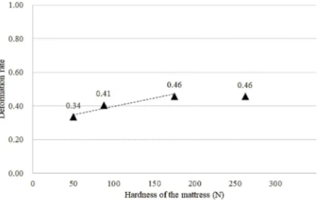
Deformation rate of CS was higher with harder mattresses, yet de- formation rates of CS with 175- and 262.5-N mattresses were the same (Table 3;Fig. 6), suggesting that the skin and soft tissue over the iliac crests were significantly more compressed with a harder mattress, but this degree of compression remained unchanged after a certain degree of hardness. We deduced that there exists a limit to the compression of the skin and soft tissue had a limit regardless of increase in hardness after a certain degree. Shabshin et al. have reported that the com- pression of the soft tissue over the ischial tuberosity significantly de- creased with the use of wheelchair cushions with an elastic modulus of 64 kPa compared to that with the use of wheelchair cushions with an elastic modulus of 85 kPa [28]. These findings are contradictory to our Results that reclining on harder mattress decreased the compression of the skin and soft tissue over the anterior superior iliac spines. There- fore, we concluded that the compression of the soft tissue over the is- chial tuberosity decreased with the use of wheelchair cushions with an elastic modulus of 64 kPa compared to that with wheelchair cushions with an elastic modulus of 85 kPa because the former were “bottoming out” by difference in physique of subjects [BMI of patients in Shabshin et al. and our study was 23.5 (3.2) and 22.1 (3.9) kg/m2, respectively].
Under the experimental conditions of this study using mattresses with four degrees of hardness and two shapes (with and without 20°
inclination), deformation rate of DM was approximately 1.3-times higher while reclining on TC mattresses than that while reclining on RC mattress, suggesting that the skin over the iliac crests, which was af- fected by 20° inclination of the mattress, was 1.3-time more displaced.
We deduced that the mattress with this inclination exerted 1.3-times greater shear force on the skin surface than did the mattress without the Fig. 5.Comparisons of deformation rates between the displacement of marker
on the skin surface (DM) and vein in the internal soft tissue (DV) while reclining on mattresses with two shapes (N= 20).
†P< 0.01, pairedt-test.
Table 4
Effective independent variables for deformation rates of the compression of the skin and soft tissue (CS).
Unstandardized Coefficients Standardized Coefficients t p The 95% confidence interval
B St. Error Beta The lower limit The upper limit
(Constant) 0.386 0.026 14.699 0.000 0.334 0.438
Underweight −0.430 0.031 −0.690 −13.844 0.000 −0.491 −0.369
Overweight 0.116 0.027 0.214 4.295 0.000 0.063 0.170
175 N 0.123 0.033 0.214 3.705 0.000 0.057 0.189
262.5 N 0.122 0.033 0.211 3.660 0.000 0.056 0.187
87.5 N 0.071 0.033 0.123 2.131 0.035 0.005 0.136
A significant regression equation of deformation rate of CS is shown [F (5, 154) = 59.427;P< 0.001], with anR2of 0.659.
inclination, thereby increasing the risk of causing pressure ulcers.
Deformation rate of DM 3.1-times higher than that of DV (RC and TC by approximately 2.4 and 3.7 times, respectively, with values averaged). DM increased and DV decreased by the application of ex- ternal force (Table 3;Fig. 5), suggesting a difference in displacement between the skin surface and internal soft tissue. Therefore, we deduced that the anatomical dynamics of the skin surface tissue and muscle and fat tissue of the internal skin varied. Precisely, DM was longer because the skin surface was pulled, and DV was shorter because the internal soft tissue was compressed.
This study has two limitations. First, the examination was limited because subjects reclined in the prone position on the mattresses in a narrow MRI device with a body coil of 30-cm height and 119-cm cir- cumference. Therefore, obese subjects (BMI ≥30 kg/m2) could not participate in MRI examination. Second, because the mattress thickness was constant at 9 cm, the skin and soft tissue deformation could not be assessed with other thicknesses. However, to elucidate effective mat- tress hardness,in vivoexaminations of subjects with different BMI while using mattresses of different thickness are difficult. In future, therefore, we will consider the simulation of compression and displacement using a finite element approach on the basis of these MRI data.
5. Conclusions
This study evaluated using MRI, the compression and displacement of the skin and soft tissue over the iliac crests with external force while reclining in the prone position on mattresses with four degrees of hardness and two shapes. The compression of the skin and soft tissue depends on mattress hardness; however, a threshold value (175 N) for hardness exists, above which no further changes in the parameters were observed. The displacement of the skin and soft tissue does not depend on mattress hardness but rather on mattress shape. Furthermore, mat- tress inclination increases the displacement of the skin surface. The skin surface and internal soft tissue parameters used to examine the dis- placement of skin and soft tissue are useful. We believe that this study would contribute to the prevention of pressure ulcers during surgery in the prone position, which underlies the highest incidence of pressure ulcers.
Conflicts of interest
We received one gratis 175-N TC among 8 kinds of mattresses from a Cape Co., Ltd., which, notably, is not involved in all processes of our study.
We received research funding from the Support Unit for Conducting Clinically Essential Studies at St. Luke’s International University.
References
[1] Bulfone G, Marzoli I, Quattrin R, Fabbro C, Palese A. A longitudinal study of the incidence of pressure sores and the associated risks and strategies adopted in Italian operating theatres. J Perioperat Pract 2012;22:50–6.https://doi.org/10.1177/
175045891202200202.
[2] Engels D, Austin M, McNichol L, Fencl J, Gupta S, Kazi H. Pressure Ulcers: factors contributing to their development in the OR. AORN J 2016;103:271–81.https://
doi.org/10.1016/j.aorn.2016.01.008.
[3] Karadag M, Gümüskaya N. The incidence of pressure ulcers in surgical patients: a sample hospital in Turkey. J Clin Nurs 2006;15:413–21.https://doi.org/10.1111/j.
1365-2702.2006.01369.x.
[4] Kaizawa M, Momose M, Momose M, Sugiyama A, Ota K. Intraoperative pressure ulcers in prone and lateral position: causes and prevention. Jpn J PU 2000;2:304–9.
[5] Shaw LF, Chang PC, Lee JF, Kung HY, Tung TH. Incidence and predicted risk factors of pressure ulcers in surgical patients: experience at a medical center in Taipei, Taiwan. BioMed Res Int 2014;2014:416896.https://doi.org/10.1155/2014/416896.
Table 5
Effective independent variables for deformation rate of the displacement of marker on the skin surface (DM).
Unstandardized Coefficients Standardized Coefficients t p The 95% confidence interval
B St. Error Beta The lower limit The upper limit
(Constant) −0.579 0.053 −11.000 0.000 −0.683 −0.475
Overweight 0.434 0.059 0.491 7.293 0.000 0.316 0.551
Female 0.171 0.055 0.210 3.123 0.002 0.063 0.279
Trapezoid cube −0.125 0.054 −0.155 −2.303 0.023 −0.233 −0.018
A significant regression equation of deformation rate of DM is shown [F (3, 156) = 21.835;P< 0.001], with anR2of 0.296.
Table 6
Effective independent variables for deformation rate of the displacement of the vein in the internal soft tissue (DV).
Unstandardized Coefficients Standardized Coefficients t p The 95% confidence interval
B St. Error Beta The lower limit The upper limit
(Constant) 0.182 0.009 19.694 0.000 0.164 0.200
Underweight −0.062 0.013 −0.367 −4.752 0.000 −0.088 −0.037
Obese −0.042 0.011 −0.286 −3.698 0.000 −0.065 −0.020
Female −0.029 0.010 −0.209 −2.859 0.005 −0.048 −0.009
A significant regression equation of the deformation rate of DV is shown [F (3, 156) = 11.071;P< 0.001], with anR2of 0.176.
Fig. 6.Correlation between deformation rate of the compression of the skin and soft tissue (CS) and hardness of mattress (N= 20).
Dotted line shows linear forecast of hardness from 50 N to 175 N.
A. Kumagai et al. Journal of Tissue Viability 28 (2019) 14–20
19
[6] DePasse JM, Palumbo MA, Haque M, Eberson CP, Daniels AH. Complications as- sociated with prone positioning in elective spinal surgery. World J Orthoped 2015;6:351–9.https://doi.org/10.5312/wjo.v6.i3.351.
[7] Schonauer C, Bocchetti A, Moraci A, Barbagallo G, Albanese V. Positioning on surgical table. Haemost Spine Surg 2005:50–5.https://doi.org/10.1007/3-540- 27394-8_8.
[8] Kumagai A, Sugama J, Okuwa M, Okuda T, Kakiuti N, Kanno A, et al. Relationship between interface pressure and development of pressure ulcer in prone position surgery using hall frame. Jpn J PU 2011;13:576–82.
[9] Nagai Y, Furuta K, Noda Y, Sekine Y, Kaitani T, Kataoka H, et al. JSPU guidelines for the prevention and management of pressure ulcers. third ed. 2014. p. 12–90.
[10] Pan AWMA. Pacific clinical practice guideline for the prevention and management of pressure injury. 2012http://www.woundsaustralia.com.au/publications/2012_
AWMA_Pan_Pacific_Abridged_Guideline.pdf#search=%27AWMA.+Pan+Pacific +Clinical+Practice+Guideline+for+the+Prevention+and+Management+of +Pressure+Injury.+2012.%27, Accessed date: 30 April 2018.
[11] National NPUAP. Pressure ulcer advisory panel support surface standards initiative - terms and definitions related to support surfaces. 2007. p. S31http://www.npuap.
org/wp-content/uploads/2012/03/NPUAP_S3I_TD.pdf, Accessed date: 30 April 2018.
[12] Soppi E, Lehtio J, Saarinen H. An overview of polyurethane foams in higher spe- cification foam mattresses. Ostomy/Wound Manag 2015;61:38–46.
[13] Kumagai A, Heinai M, Matsui Y, Konya C, Shimada K, Kawakami S, et al.
Effectiveness of highly resilient urethane foam mattresses in pressure ulcer pre- vention for special operative positions. Jpn J PU 2015;17:1–9.
[14] Aronovitch SA, Wilber M, Slezak S, Martin T, Utter D. A comparative study of an alternating air mattress for the prevention of pressure ulcers in surgical patients.
Ostomy/Wound Manag 1999;45. 34-40-44.
[15] Defloor T, De Schuijmer JD. Preventing pressure ulcers: an evaluation of four op- erating-table mattresses. Appl Nurs Res 2000;13:134–41.https://doi.org/10.1053/
apnr.2000.7653.
[16] Keller BPJA, Overbeeke J, van der Werken C. Interface pressure measurement during surgery: a comparison of four operating table surfaces. J Wound Care 2006;15:5–9.https://doi.org/10.12968/jowc.2006.15.1.26858.
[17] Kirkland-Walsh H, Teleten O, Wilson M, Raingruber B. Pressure mapping compar- ison of four OR surfaces. AORN J 2015;102.https://doi.org/10.1016/j.aorn.2015.
05.012. 61.e1-61.e9.
[18] McInnes E, Jammali-Blasi A, Bell-Syer SEM, Dumville JC, Middleton V, Cullum N.
Support surfaces for pressure ulcer prevention. Cochrane Database Syst Rev 2015;9:CD001735.https://doi.org/10.1002/14651858.CD001735.pub5.
[19] Moysidis T, Niebel W, Bartsch K, Maier I, Lehmann N, Nonnemacher M, et al.
Prevention of pressure ulcer: interaction of body characteristics and different
mattresses. Int Wound J 2011;8:578–84.https://doi.org/10.1111/j.1742-481X.
2011.00814.x.
[20] Nixon J, McElvenny D, Mason S, Brown J, Bond S. A sequential randomised con- trolled trial comparing a dry visco-elastic polymer pad and standard operating table mattress in the prevention of post-operative pressure sores. Int J Nurs Stud 1998;35:193–203.https://doi.org/10.1016/S0020-7489(98)00023-6.
[21] Russell JA, Lichtenstein SL. Randomized controlled trial to determine the safety and efficacy of a multi-cell pulsating dynamic mattress system in the prevention of pressure ulcers in patients undergoing cardiovascular surgery. Ostomy/Wound Manag 2000;46:5.
[22] Wu T, Wang ST, Lin PC, Liu CL, Chao YFC. Effects of using a high-density foam pad versus a viscoelastic polymer pad on the incidence of pressure ulcer development during spinal surgery. Biol Res Nurs 2011;13:419–24.https://doi.org/10.1177/
1099800410392772.
[23] Brienza D, Vallely J, Karg P, Akins J, Gefen A. An MRI investigation of the effects of user anatomy and wheelchair cushion type on tissue deformation. J Tissue Viability 2017.https://doi.org/10.1016/j.jtv.2017.04.001.
[24] Call E, Hetzel T, McLean C, Burton JN, Oberg C. Off loading wheelchair cushion provides best case reduction in tissue deformation as indicated by MRI. J Tissue Viability 2017;26:172–9.https://doi.org/10.1016/j.jtv.2017.05.002.
[25] Linder-Ganz E, Shabshin N, Itzchak Y, Gefen A. Assessment of mechanical condi- tions in sub-dermal tissues during sitting: a combined experimental-MRI and finite element approach. J Biomech 2007;40:1443–54.https://doi.org/10.1016/j.
jbiomech.2006.06.020.
[26] Linder-Ganz E, Shabshin N, Itzchak Y, Yizhar Z, Siev-Ner I, Gefen A. Strains and stresses in sub-dermal tissues of the buttocks are greater in paraplegics than in healthy during sitting. J Biomech 2008;41:567–80.https://doi.org/10.1016/j.
jbiomech.2007.10.011.
[27] Makhsous M, Lin F, Cichowski A, Cheng I, Fasanati C, Grant T, et al. Use of MRI images to measure tissue thickness over the ischial tuberosity at different hip flexion. Clin Anat 2011;24:638–45.https://doi.org/10.1002/ca.21119.
[28] Shabshin N, Zoizner G, Herman A, Ougortsin V, Gefen A. Use of weight-bearing MRI for evaluating wheelchair cushions based on internal soft-tissue deformations under ischial tuberosities. J Rehabil Res Dev 2010;47:31–42.https://doi.org/10.1682/
JRRD.2009.07.0105.
[29] Shabshin N, Ougortsin V, Zoizner G, Gefen A. Evaluation of the effect of trunk tilt on compressive soft tissue deformations under the ischial tuberosities using weight- bearing MRI. Clin Biomech 2010;25:402–8.https://doi.org/10.1016/j.clinbiomech.
2010.01.019.
[30] Sonenblum SE, Sprigle SH, Cathcart JMK, Winder RJ. 3D anatomy and deformation of the seated buttocks. J Tissue Viability 2015;24:51–61.https://doi.org/10.1016/
j.jtv.2015.03.003.