Introduction
Since the 1990s, a small thecate dinoflagellate has been known to form vast red tides off the Changjiang River mouth in the East China Sea. In 1995, particularly the red tides caused by this species expanded over more than 100 km. The organism causing this huge red tide off the Chanjiang River mouth in the East China Sea was de- scribed as a new species, Prorocentrum donghaiense Lu, 2001 (Lu & Goebel 2001). However, a species which is morphologically similar to P. donghaiense was earlier de- scribed as Prorocentrum shikokuenseHada, 1975 based on cells collected from Iwamatsu Bay in the Bungo Channel on the western coast of Shikoku, Ehime Prefecture, Japan on 8 June 1973 by Hada (1975). Prorocentrum donghaiense has now been observed in China and Korea, while P. shikokuense is reported from Japan.
Lu et al. (2005) reported variation of cell shape with careful examination of many cells of P. donghaienseusing light and scanning electron microscopy (LM & SEM), and also compared the morphology of P. donghaiensewith that of the original description of P. shikokuense. Based on these
observations, Lu et al. (2005) concluded that it is difficult to determine whether or not the two species are the same. The reason for this difficulty is due to limited information on P.
shikokuense,because of the description and handwritten il- lustrations based on only LM observations. For adequate identification, information on the fine morphology of P.
shikokuense using LM and SEM is required. In addition, molecular data on P. shikokuense are lacking, although ri- bosomal DNA (rDNA) sequences of P. donghaiense were determined.
In this study, in order to compare P. shikokuense with P.
donghaiense based on morphological and molecular data, we collected cells of P. shikokuensefrom the Iwamatsu Bay type locality as well as Uwajima Bay close to the type lo- cality where Hada also found them in 1973, and from some additional locations where the specimens had been identi- fied as Prorocentrum dentatum Stein, 1883 we then ob- served them with a light microscope and a scanning elec- tron microscope and obtained molecular data (small subunit rDNA and internal transcribed spacer 1 (ITS1)-5.8S rDNA- ITS2 sequences).
A comparative study between Prorocentrum shikokuense and P. donghaiense (Prorocentrales, Dinophyceae)
based on morphology and DNA sequences
Y
OSHIHITOT
AKANO& K
AZUMIM
ATSUOKA*
Institute for East China Sea Research, Nagasaki University, 1–14 Bunkyo-machi, Nagasaki 852–8521, Japan Received 16 March 2010; Accepted 8 March 2011
Abstract: The taxonomic position of an armored dinoflagellate Prorocentrum donghaienseLu, 2001, which is one of the causative species of large red tides occurring off Changjiang River mouth was examined and compared to a simi- lar species P. shikokuense. In both species, the following morphological features were shared; a weakly concave cell with a tiny apical spine around the periflagellar area, one side of the cell being slightly extended on the anterior end, a rounded posterior end, a rounded nucleus located in the posterior region and similar size of cells. DNA sequences of small subunit rDNA and internal transcribed spacer 1 (ITS1)-5.8S rDNA-ITS2 regions of these two species were largely the same. Morphological and molecular data suggest that P. donghaienseand P. shikokuenseare synonymous and P.
shikokuensehas priority over P. donghaiense.
Key words: dinoflagellate, HAB, Prorocentrum dentatum, Prorocentrum donghaiense, Prorocentrum shikokuense
Corresponding author: Kazumi Matsuoka; E-mail, kazu-mtk@nagasaki- u.ac.jp
Plankton & Benthos Research
© The Plankton Society of Japan
Materials and Methods Sampling and culture
Plankton samples containing Prorocentrum shikokuense cells were collected from the surface waters of Iwamatsu Bay in July 2010. Cells of P. shikokuense were collected with a 10mm-mesh plankton net from Uwajima Bay in June 2005. The plankton samples from Iwamatsu Bay were fixed with formalin at a final concentration of 2%. Glutaralde- hyde was added to part of the field samples collected from Uwajima Bay (final concentration 2%) and they were kept as stock samples. For enrichment cultures, samples were added to a plastic cup containing Daigo’s IMK Medium for Marine Microalgae (Daigo, Tokyo, Japan) and placed in a culture cabinet at 15°C with a photon flux density of about 50mmol photons · m2· s1 under a 16 h : 8 h light : dark regime. Clonal cultures were established by capillary- pipette isolation and maintained under the conditions de- scribed above. Other clonal cultures of the targeted species
“Prorocentrum dentatum” (Culture No. ND117 and ND118) were established using plankton cells collected from Ago Bay, Mie Prefecture in July 2005. Several addi- tional samples for morphological observation of the species called Prorocentrum sp. aff. dentatum by Matsuoka et al.
(2006) were examined from the following locations; off Mie Fishing port in Nagasaki Prefecture, around the Danjo Islands, and from Imari Bay in Nagasaki Prefecture (Table 1; Fig. 1).
Light microscopy
Cells were observed using an Olympus BX51 micro- scope equipped with Nomarski interference optics (Olym- pus Co. Ltd., Tokyo, Japan), and micrographs were taken with an Olympus DP50 digital camera attached to the mi- croscope.
Scanning electron microscopy
The selected cells were allowed to adhere to a 0.1% poly- L-lysine coated glass plate. Thereafter, to remove mucilage on the cell surface, 2 N sodium hydroxide solution was used. The cells on the glass plate were rinsed a few times in distilled water, dehydrated in an ethanol series, and critical point dried using a critical point dryer HCP-2 (Hitachi Koki, Tokyo, Japan). Dried cells were coated with plat- inum-palladium in a JFC-1600 ion-sputter (JEOL, Tokyo, Japan). The materials were examined with a JEOL JSM- 6390 scanning electron microscope.
Molecular analysis
Cells of Prorocentrum shikokuense collected from Iwa- matsu Bay, Uwajima Bay, and “P. dentatum” cultures (ND117 and ND118) from Ago Bay were centrifuged, ho- mogenized using a plastic stick, and then used as DNA templates. We used external primers SR1, LSUR2 and
for the first round of PCR (Takano & Horiguchi 2006). In the second round of PCR, 0.5mL of the PCR products was used as a DNA template with combinations of internal primers and TaKaRa Ex TaqTM (TAKARA BIO INC., Japan). Details of the PCR and sequencing protocol are provided by Takano & Horiguchi (2004, 2006).
We used two sets of alignments i.e. SSU rDNA and ITS regions, respectively. The SSU rDNA sequences were aligned manually based on the published secondary struc- ture of the SSU rRNA molecule, available at the rRNA Web server (http://www.psb.ugent.be/rRNA/ssu/index.html) and alignments were refined manually. The ITS1-5.8S rDNA- ITS2 sequences were aligned manually with only a few gaps. Neighbor joining (NJ), maximum parsimony (MP) and maximum likelihood (ML) methods were implemented by PAUP version 4.0b10 (Swofford 2002) for the phyloge- netic analyses. The program Modeltest version 3.7 (Posada
& Crandall 1998) was used to explore the model of se- quence evolution that best fits the data set by the Akaike In- formation Criterion (AIC) (Akaike 1974). The model se- lected for the SSU alignment set was the GTRIG; the general time-reversible (GTR) model plus the proportion of invariable sites (I) and the variable sites (G; gamma distrib- ution with shape parameter) with the following parameters:
assumed nucleotide frequencies A0.2565, C0.2008, G0.2652, and T0.2775; substitution-rate matrix with AC1.1158, AG3.5526, AT1.4707, CG0.7050, CT8.5060, and GT1; I0.4701; G0.6959, and the number of rate categories=4, and that for the ITS regions alignment set was the K81uf (Kimura three-parameters with unequal base frequencies, Kimura 1981) with the fol- lowing parameters: assumed nucleotide frequencies A0.1977, C0.2445, G0.2623, and T0.2955; substi- tution-rate matrix with AC1, AG2.9693, AT0.4181, CG0. 4181, CT2.9693, and GT1. ML was performed using the heuristic search option with a branch-swapping algorithm (tree bisection-reconnection; TBR) and the start- ing tree obtained by stepwise addition with the random ad- dition of sequences (10 replicates). The distance matrix was calculated using the Kimura two-parameters distances (Kimura 1980), and the distance tree was constructed using the NJ method (Saitou & Nei 1987). MP was performed using the heuristic search option with random addition of sequences (1000 replicates) and the branch-swapping algo- rithm (TBR). All characters were weighted equally and gaps were treated as missing. Bootstrap analyses with 100 and 1,000 replicates for ML (using the heuristic search op- tion with a branch swapping algorithm, nearest neighbor in- terchange (NNI) and a starting tree obtained by neighbor joining) of the SSU and the ITS regions, respectively, and 1,000 replicates of both sets for NJ and MP (with the ran- dom addition of sequences, 10 replicates, and TBR) were applied to examine the robustness and statistical reliability of the topologies (Felsenstein 1985).
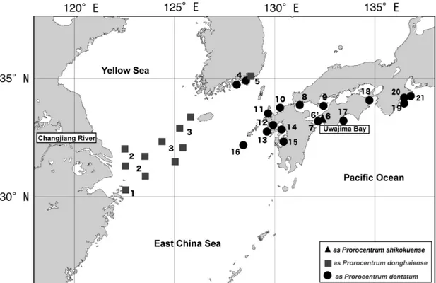
Table1.Occurrence records of Prorocentrum shikokuense(“P. shokokuensis”by Hada, 1975, P. donghaienseLu, 2001 and P. obtsusidens by Adachi 1972) around the East China Sea. Number LocationSpecies nameAge of appearanceReference Supplment 1Near Gouqi Islad in the Chanjeang as P. donghaienseLu D.-D. 2001Original description River Estuary 2Off Chanjiang Riveras P. donghaienseLu D.-D, et al. 200529–32°N122–123.5°E China?as P. dentatumHan X.-T. et al. 2005 (unpublished)AY551273 (GenBank) 3Chanjeang River Estuary - West of as P. donghaienseYoon Y.-H. et al. 2003 Jeju Iskand, Korea 4Masan Bay of Koreaas P. donghaienseJeong et al. 2004 (unpublished)AJ841810 (GenBank) 5Masan Bay of Koreaas P. dentatumJune 1985, July 1986, July 1990Park, S.-J. 1991 5Hengan Bay of Koreaas P. dentatumJune 1985, July 1990Park, S.-J. 1991 5Bukshin Bay of Koreaas P. dentatumJuly 1990Park, S.-J. 1991 5Gohyun Bay of Koreaas P. dentatumJuly 1990Park, S.-J. 1991 6Iwamatsu Bay of the Bungo Channelas P. shikokuensisJuly 1973Hada 1975Original description 6Iwamatsu Bay of the Bungo ChannelP. shikokuenseJuly 2010This studyDNA analysis; Onservation 7Bungo Channelas P. dentatum2008Natl. Res. Inst. Fish. & Environ. Inland Sea 2008 8Suo Nada, Yamaguchi Prefas P. dentatum2003, 2004, 2005, 2007Natl. Res. Inst. Fish. & Environ. Inland Sea 2007 9Seto Inland Seaas P. dentatum2003Natl. Res. Inst. Fish. & Environ. Inland Sea 2003 10Fukuoka Bayas P. dentatum2005Natl. Res. Inst. Fish. & Environ. Inland Sea 2005 11Imari BayJuly 2006This study 12Omura Bay, Nagasaki Pref.as P. dentatum2005Natl. Res. Inst. Fish. & Environ. Inland Sea 2005 13Off Mie Fishing Port, Nagasaki Pref.May 2006This study 14Ariake Bay, Kumamoto Pref.as P. dentatum2005Natl. Res. Inst. Fish. & Environ. Inland Sea 2005 15Yatsushiro Sea, Kumamoto Pref.as P. dentatum2005Natl. Res. Inst. Fish. & Environ. Inland Sea 2005 16Danjo Island WestApril of 2006This study 17Nomi Bay, Kochi Pref.as P. dentatum2008Natl. Res. Inst. Fish. & Environ. Inland Sea 2008 18Tachibana Bay of the Kii Channelas P. shikokuensisMay 1974Hada 1975 19Kumano Nada, Mie Pref.as P. dentatum2006Natl. Res. Inst. Fish. & Environ. Inland Sea 2006 20Owase Bay, Mie Pref.as P. dentatum2003, 2005Natl. Res. Inst. Fish. & Environ. Inland Sea 2005 21Ago Bay, Mie Pref.as P. dentatum2002, August 2005Hata 2005 (personal com.)ND 117, ND 118; DNA analysis; Observation 21Ago Bay, Mie Pref.as P. obtusidensAdachi 1972 South Pacific, South Americaas P. dentatumAY803742 CCMP1517 Canary Island, the Atlanticas P. donghaiense2009Ellegaard, M. (coll.), Hansen, G. (Iso.)SCCAP K-1260
Results
Morphology of Prorocentrum shikokuense
Light microscopy. Cells are variable in shape, elongated and asymmetric, narrowing toward the posterior ends, with the shape being somewhat similar to that of a sunflower seed (Fig. 2a–l). In blooming seasons, chains consisting of
more than four cells are frequently observed. One side of the anterior end is often extends more than the other (Fig.
2a–c; a black arrow). The periflagellar area of the cell is slightly concave with a tiny apical spine that is rarely visi- ble with a light microscope (Fig. 2d; a black arrow). The posterior end of most cells is generally rounded, but that of some cells is attenuated (Fig. 2g–l, k). Cells from Iwamatsu Fig. 1. Geographical distribution of Prorocentrum shikokuenseincluding those formally referred to P. donghaiense, P. denta- tumand P.sp. aff. dentatumin and around the East China Sea. Locations with numbers are shown in Table 1.
Fig. 2. Light micrographs of fixed cells of Prorocentrum shikokuense collected from Iwamatsu Bay (a–k) and Uwajima Bay (l) showing individual variations in cell shape and size. a–c. A black arrow indicates shoulder slightly developed on the anterior end.
d. A black arrow indicates shallow indentation around the flagellar pore. l. “n” indicates a spherical nucleus. Scale bars10mm.
Bay, type locality, and Uwajima Bay near the type locality are from 20.2 to 31.7mm long (mean25.9mm, n40) and from 8.0 to 14.3mm wide (mean10.5, n40). The range of long/wide ratio is from 1.79 to 3.88. The rounded nu- cleus is located in the posterior region of the cell (Fig. 2l).
Scanning electron microscopy. The thecal plates have many knob-like spines spread densely over the surface (Fig. 3a–f) and trichocyst pores (approx. 240 nm in diameter; Fig. 3g),
that are mainly distributed around the peripheral margin of the cells (Fig. 3a, b), particularly around the antapex (Fig. 3f). The periflagellar area of the right thecal plate is V-shaped and con- cave with an ear-shaped collar structure that varies in shape and size (Fig. 3d), and one of the left thecal plats is slightly concave (Fig. 3a, b). Inside the cells, there are small hollows, probably corresponding to the trichocyst pores outside. The megacytic zones are well developed in natural cells (Fig. 3a, e) Fig. 3. Scanning electron micrographs of fixed cells of Prorocentrum shikokuense collected from Iwamatsu Bay. a; Right valve view showing megacytic zones (arrow) (Scale bar5mm). B; Left valve view showing gently swelling shoulder (Scale bar5mm).
c; Typically slender cell (Scale bar5mm). d; ‘ear-shaped’ apical collar structure around the periflagellar area and trichocyst pores (Scale bar1mm. e; High magnification view of the cell showing tiny knobs distributed perpendicularly to the antero-posterior axis (Scale bar1mm). f; High magnification view of the posterior region showing trichocyst pores mainly distributed around the cell margin (Scale bar0.5mm). g; Small valve pores and knob-like spines densely distributed over the surface (Scale bar1mm).
h; Inner surface of the valve showing small hollows probably corresponding to trichocyst pores (Scale bar1mm).
and dense rows of tiny knobs are arranged on the surface of the cell margin (Fig. 3e). Small hollows were observed on the inner surface of the valve (Fig. 3h).
Molecular data on Prorocentrum shikokuense
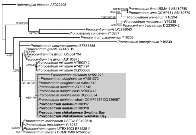
We determined the sequences of SSU rDNA (1731 bp) and the ITS regions (534 bp) of P. shikokuense collected from Iwamatsu Bay and Uwajima Bay, and SSU rDNA of so-called “P. dentatum” (ND117 and ND118). The se- quences of SSU rDNA of P. shikokuense are identical to those of so-called “P. dentatum” (ND117 and ND118), P.
dentatum CCMP1517 (AY803742 and DQ336057) and P.
donghaiense (AY803743 and DQ336054) (Fig. 4). The se- quences of the ITS regions of P. shikokuense from Iwa- matsu Bay and Uwajima Bay are completely identical.
Prorocentrum shikokuenseshares the same sequences with P. donghaiense presented in Figure 11 by Lu et al. (2005) and P. dentatum CCMP1517. Prorocentrum shikokuense has 1 bp degenerate site; Y (C and T). At the same site P.
donghaiense of Lu et al. (2005) has C and P. dentatum CCMP1517 has T. Therefore, P. shikokuensehas two base pair substitutions from P. donghaiense (AY465116) (Fig. 5).
Fig. 4. Maximum likelihood tree constructed from SSU rDNA sequences, showing phylogenetic affinities of Prorocentrum shikokuensecollected from Iwamatsu Bay and Uwajima Bay and “P. dentatum”(ND117 and ND118) collected from Ago Bay in- dicated with bold face, to sequences deposited as P. donghaiense and“P. dentatum”. For details of the evolutionary model and parameters used, see Materials and Methods. Bootstrap percentages (50%) for NJ/MP/ML methods are presented at each node.
Fig. 5. Maximum likelihood tree constructed from ITS regions sequences, showing phylogenetic affinities of Prorocentrum shikokuensecollected from Iwamatsu Bay and Uwajima Bay, indi- cated with bold face, to sequences deposited as P. donghaiense and“P. dentatum”. For details of the evolutionary model and pa- rameters used, see Materials and Methods. Bootstrap percentages (50%) for NJ/MP/ML methods are presented at each node.
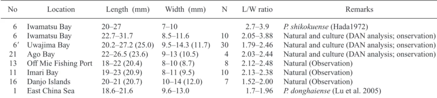
Morphometric measurement of cells reported as Proro- centrum dentatum
Cells collected from other areas are from 18.0 to 27.2mm long and from 7 to 14.3mm wide. The range of long/wide ratio is from 1.52 to 3.9 (Table 2).
Discussion
The morphology of our specimens collected from Iwa- matsu Bay, the type locality for Prorocentrum shikokuense and Uwajima Bay near the type locality is identical with re- spect to cell shape and size to the original description of P.
shikokuense(Hada 1975). Using a scanning electron micro- scope, we confirmed the characteristic distribution of tri- chocyst pores, development of a tiny anterior horn, and the presence of well developed megacytic zones. We were also able to newly observe several important morphological fea- tures of P. shikokuense, especially an ear-shaped collar structure at the apical end and valve pores. When the ear- shaped collar structure is large, it appears as a spine under LM as Hada (1975) described and illustrated, but that of most cells was not visible under LM. This structure is iden- tical to that of P. donghaienseshown in Figure 6 by Lu et al. (2005). In comparison, SEM images of P. donghaiense given by Lu et al. (2005) have rounder posterior ends, though this feature is rather variable as shown in Figs 2 and 3. Also the long/wide ratio of the original P. shikokuense was slightly larger than that of P. donghaiense, making the former more elongate. Although the over all cell shapes of P. shikokuense and P. donghaiense are slightly different, they are not clearly distinguishable because of overlapping characters. Therefore, P. shikokuense and P. donghaiense can be considered to be the same species.
Previously, cells were reported in the East China Sea and Japanese–Korean coastal waters as “P. dentatum”by Yoo &
Lee (1986), Park (1991), Horiguchi (1990) and the National Institute of Fisheries & Environment of the Inland Sea (2002–2008) and as Prorocentrum sp. aff. dentatum by Matsuoka et al. (2006). They also can be considered to be the same species as P. shikokuensebased on the cell shape and size. The cell shape and the size of P. shikokuense are variable to some degree. Additionally, with regard to a comparison between P. shikokuense(as P. donghaiense) and
the true P. dentatum, we agree with Lu et al. (2005) that P.
shikokuense is a different species from P. dentatum (Stein 1883) in terms of cell shape and size.
In addition to morphological aspects, our molecular data, including specimens collected from the type locality, show that cells that have been identified as P. donghaiense and “P.
dentatum” in the East China Sea and Japanese–Korean coastal waters are the same species as P. shikokuense, be- cause their sequences, including those of P. donghaiense determined by Lu et al. (2005), are identical to those of P.
shikokuense. The sequences of the ITS regions of P. dong- haiense deposited under AY465116 have two base pairs substituted from others, but we have limited information on other data on P. dentatumaccumulated in Genbank.
Prorocentrum shikokuense was originally described by Hada (1975, as Shikokuensis) under ICZN in 1975, with an English description and illustrations but without designation of a type. Lu et al. (2005) pointed out that P. shikokuense was an invalid name under both the International Code of Botanical Nomenclature (ICBN) and the International Code of Zoological Nomenclature (ICZN), due to lacking a Latin diagnosis and designation of a type, respectively. However, the ICZN accepts dinoflagellate species regardless of trophic nature (whether autotroph, heterotroph or mixotroph) and designation of a type was not a mandatory requirement but a recommendation before the fourth edition of the ICZN (Ride et al. 1999). The ICBN in Article 45.4 recognizes that a taxon validly described even under other nomenclatures has priority for its name and authorship. Therefore, P.
shikokuense has validity under the ICZN and has priority over P. donghaiensediscribed by Lu & Goebel (2001).
All of the previous occurrences of P. shikokuense are listed in Table 1 and arranged in Figure 1, which shows the wide distribution in and around the East China Sea and where it has formed massive blooms in the Changjiang River mouth.
Taxonomic appendix
Prorocentrum shikokuenseHada, 1975
Synonym: Prorocentrum donghaiense. Lu, 2001. Lu &
Goebel (2001) p. 339, fig. 2.
Lectotype designated herein. figs. 1, 2 of Hada (1975) as Prorocentrum shikokuensis.
Table 2. Cell size and ratios of length/width of Prorocentrum shikokuense collected from several different sites, including its type location.
No Location Length (mm) Width (mm) N L/W ratio Remarks
6 Iwamatsu Bay 20–27 7–10 2.7–3.9 P. shikokuense(Hada1972)
6 Iwamatsu Bay 22.7–31.7 8.5–11.6 10 2.05–3.88 Natural and culture (DAN analysis; onservation) 6 Uwajima Bay 20.2–27.2 (25.0) 9.5–14.3 (11.7) 30 1.79–2.46 Natural and culture (DAN analysis; onservation) 21 Ago Bay 22–26.5 (23.6) 9–13 (10.5) 4 2.03–2.44 Natural and culture (DAN analysis; onservation) 13 Off Mie Fishing Port 18–22 (20.4) 8–10 (8.7) 8 2.12–2.48 Natural (Observation)
11 Imari Bay 19–23 (20.9) 8–11 (9.5) 10 2.13–2.38 Natural (Observation) 16 Danjo Islands 20–21 (20.7) 10–14 (12.0) 7 1.52–2.00 Natural (Observation)
1 East China Sea 18.6–21.6 9.6–13.0 1.7–1.96 P. donghaiense(Lu et al. 2005) N: Number of cells observed
Remarks:
Because of an orthographic reason, the spelling of the species name, Prorocentrum shikokuensis, should be changed from sihokuensisto shikokuense.
Acknowledgements
This work was financially supported by the Ministry of Education, Culture, Sports Science and Technology, and the Japan Science and Technology Agency under the project “A Japanese, Korean and Chinese joint study for harmful algal blooms in the East China Sea”. We appreciate Dr. Y.
Koizumi who collected plankton samples from Iwamatsu Bay, Prof. T. Horiguchi who provided the plankton sample from Uwajima Bay, Dr. N. Hata who sent plankton samples from Ago Bay, and Dr. H. Takayama who advised us to pre- pare SEM samples of P. shikokuense. Also we would like to thank Dr. Gregory N. Nishihara for correcting our English.
References
Adachi R (1972) A taxonomical study of the red-tide organisms. J Fac Fish Pref Univ Mie 9: 9–145.
Akaike H (1974) A new look at the statistical model identification.
IEEE Transact Automat Cont AC-19: 716–723.
Felsenstein J (1985) Confidence limits on phylogenies: an ap- proach using the bootstrap. Evolution 39: 783–791.
Hada Y (1975) On two new species of the genus Prorocentrum Ehrenberg belonging to Dinoflagellida. Hiroshima Shudo Daigaku Ronshu 16: 31–38.
Horiguchi T (1990) Prorocentrum dentatumStein. In: Red Tide Organisms in Japan—An Illustrated Taxonomic Guide— (eds Fukuyo Y, Takano H, Chihara M, Matsuoka K). Uchida Rokakuho, Tokyo, pp. 26–27.
Kimura M (1980) A simple method for estimating evolutionary rates pf base substitutions through comparative studies of nu- cleotide sequences. J Mol Evol 16: 111–120.
Kimura M (1981) Estimation of evolutionary distances between homologous nucleotide sequences. Proc Natl Acad Sci USA 78:
454–458.
Lu D, Goebel J (2001) Five red tide species in genus Prorocentrum including the description of Prorocentrum donghaiense Lu sp.
nov. from the East China Sea. Chin J Ocean Limn 19: 337–344.
Lu D, Goebel J, Qi Y, Zou J, Han X, Gao Y, Li Y (2005) Morpho- logical and genetic study of Prorocentrum donghaiense Lu from the East China Sea, and comparison with some related Prorocentrumspecies. Harm Alg 4: 493–505.
Matsuoka K, Iwataki M, Yamatogi T (2006) Harmful phytoplank- ton occurring around Nagasaki. Nagasaki Prefectural Organiza- tion for Industry and Japan Science and Technology Agency, Nagasaki, 29 pp.
National Institute of Fisheries & Environment of Inland Sea (2002) Memorandum of the Sub-meeting of Red-tides and
National Institute of Fisheries & Environment of Inland Sea (2003) Memorandum of the Sub-meeting of Red-tides and Shellfish Poisoning, 23 pp., Hiroshima, Japan. (in Japanese) National Institute of Fisheries & Environment of Inland Sea
(2004) Memorandum of the Sub-meeting of Red-tides and Shellfish Poisoning, 28 pp., Hiroshima, Japan. (in Japanese) National Institute of Fisheries & Environment of Inland Sea
(2005) Memorandum of the Sub-meeting of Red-tides and Shellfish Poisoning, 32 pp., Hiroshima, Japan. (in Japanese) National Institute of Fisheries & Environment of Inland Sea
(2006) Memorandum of the Sub-meeting of Red-tides and Shellfish Poisoning, 29 pp., Hiroshima, Japan. (in Japanese) National Institute of Fisheries & Environment of Inland Sea
(2007) Memorandum of the Sub-meeting of Red-tides and Shellfish Poisoning, 28 pp., Hiroshima, Japan. (in Japanese) National Institute of Fisheries & Environment of Inland Sea
(2008) Memorandum of the Sub-meeting of Red-tides and Shellfish Poisoning, 27 pp., Hiroshima, Japan (in Japanense).
Park J-S (1991) Red tide occurrence and countermeasures in Korea. In: Recent approaches on red tide (eds Park J-S, Kim H- G), Proceedings of “1990 Korean-French Seminar on Red tides”, National Fisheries Research and Development Agency, Korea, pp. 1–24.
Posada D, Crandall KA (1998) MODELTEST: testing the model of DNA substitution. Bioinformatics 14: 817–818.
Ride WDL, Cogger HG, Dupuis C, Kraus O, Minelli A, Thomp- son FC, Tubbs PK (1999) International Code of Zoological Nomenclature: adopted by the International Union of Biological Sciences. Internat Trust Zool Nomen, London.
Saitou N, Nei M (1987) The neighbor-joining method: A new method for reconstructing phylogenetic trees. Mol Biol Evol 4:
406–425.
Stein F (1883) Der Organismus der Infusionsthiere nach eigenen Forschungen in systematischer Reihenfolge bearbeitet. III. Die Naturgeschichte der arthrodelen Flagellaten. Wilhelm En- glmann, Leipzig, 30 pp.
Swofford DL (2002) PAUP*: Phylogenetic Analysis Using Parsi- mony (*and Other Methods), version 4.0b10. Sinauer, Sunder- land, MA.
Takano Y, Horiguchi T (2004) Surface ultrastructures and molecu- lar phylogenetics of four unarmored heterotrophic dinoflagel- lates, including the type species of the genus Gyrodinium (Dinophyceae). Phycol Res 52: 107–116.
Takano Y, Horiguchi T (2006) Acquiring scanning electron micro- scopical, light microscopical and multiple gene sequence data from a single dinoflagellate cell. J Phycol 42: 251–256.
Yoo K, Lee J-B (1986) Taxonomical studies on dinoflagellates in Masan Bay. 1. Genus Prorocentrum Ehrenberg. J Ocean Soc Korea 21: 46–55.
Yoon Y-H, Park J-S, Soh H-Y, Hwang D-J 2003 Spatial distribu- tion of phytoplankton community and red tide of dinoflagellate, Prorocentrum donghaiencein the East China Sea during early summer. Korean J Environ Biol 21: 132–141 (in Hangul with English abstruct).