and sharing with colleagues.
Other uses, including reproduction and distribution, or selling or licensing copies, or posting to personal, institutional or third party
websites are prohibited.
In most cases authors are permitted to post their version of the article (e.g. in Word or Tex form) to their personal website or institutional repository. Authors requiring further information
regarding Elsevier’s archiving and manuscript policies are encouraged to visit:
http://www.elsevier.com/copyright
Prostate brachytherapy
MRI-based preplanning in low-dose-rate prostate brachytherapy
Osamu Tanakaa,b,*, Shinya Hayashia,b, Masayuki Matsuoa,b, Masahiro Nakanoc, Yasuaki Kubotac, Sunaho Maedaa,b, Kazuhiro Ohtakaraa,b,
Takashi Deguchic, Hiroaki Hoshia
aDepartment of Radiology,bDivision of Radiation Oncology,cDepartment of Urology, Gifu University School of Medicine, Japan
Abstract
Purpose: To compare the dosimetric results between MRI-based and TRUS-based preplanning in permanent prostate brachytherapy, and to estimate the accuracy of MRI-based preplanning by comparing with CT/MRI fusion-based postimplant dosimetry.
Methods and materials: Twenty-one patients were entered in this prospective study with written informed consent.
MRI-based and TRUS-based preplanning were performed. The seed and needle locations were identical according to MRI- based and TRUS-based preplanning. MRI-based and TRUS-based preplanning were compared using DVH-related parameters. Following brachytherapy, the accuracy of the MRI-based preplanning was evaluated by comparing it with CT/MRI fusion-based postimplant dosimetry.
Results: Mean MRI-based prostate volume was slightly underestimated (0.73 cc in mean volume) in comparison to TRUS-based volume. There were no significant differences in the mean DVH-related parameters except with rectal V100(cc) between TRUS-based and MRI-based preplanning. Mean rectal V100(cc) was 0.74 cc in TRUS-based and 0.29 cc in MRI-based preplanning, respectively, and the values demonstrated a statistical difference.
There was no statistical difference in mean rectalV150(cc), and rectal V100(cc) between MRI-based preplanning and CT/
MRI fusion-based postimplant dosimetry.
Conclusion: Prostate volume estimation and DVH-related parameters in MRI-based preplanning were almost identical to TRUS-based preplanning. From the results of CT/MRI fusion-based postimplant dosimetry, MRI-based preplanning was therefore found to be a reliable and useful modality, as well as being helpful for TRUS-based preplanning. MRI-based preplanning can more accurately predict postimplant rectal dose than TRUS-based preplanning.
c 2007 Elsevier Ireland Ltd. All rights reserved. Radiotherapy and Oncology 88 (2008) 115–120.
Keywords: Prostate brachytherapy; MRI; CT; TRUS; Preplanning
Transperineal prostate brachytherapy has become an increasingly popular treatment over the past 15 years [11,12]. Image-based preplanning is an important part of the modern practice of permanent prostate brachytherapy.
Transrectal ultrasound (TRUS) images enable multiplanar visualization of the prostate capsule, bladder, rectum, and, with the introduction of a catheter or contrast agent, the urethra[1,2,7,13,15,22–24]. Based on TRUS-based pre- planning, the exact number of seeds required for the im- plant (plus a few extra) is ordered for each individual patient [3]. However, TRUS-based preplanning is inconve- nient and uncomfortable due to the insertion of a probe into the rectum.
The discrepancies between Computed tomography (CT)- based, Magnetic resonance imaging (MRI)-based and TRUS- based prostate volume have been well discussed [1,2,5–
7,9,14–16,20]. Narayana et al., using two different ap- proaches for preplanning (CT-based vs. TRUS-based), found
a significant difference in the CT volumes, which were markedly greater (47%) than the TRUS volumes [13]. On the other hand, several authors have reported the TRUS- based prostate volume to be similar to the MRI-based pros- tate volume[1,7,15,24]. The cost for performing MRI is still high, but recently, it has been used to detect the location of prostate cancer and extracapsular extension to establish a pretreatment diagnosis[4]. We hypothesized that MRI could be replaced as an imaging modality for preplanning of per- manent prostate implants. If similar results as accurate as those obtained through TRUS-based preplanning could be obtained by MRI-based preplanning, then it would be com- fortable and convenient for patients. Therefore, the accu- racy of MRI-based preplanning was estimated through comparisons with TRUS-based preplanning. In addition, we also estimated the accuracy of MRI-based preplanning by comparing it with CT/MRI fusion-based postimplant dosimetry.
www.thegreenjournal.com
0167-8140/$ - see front matter c 2007 Elsevier Ireland Ltd. All rights reserved. doi:10.1016/j.radonc.2007.09.012
Methods and materials
Between April 2006 and December 2006, 21 patients with local prostate cancer were entered in this prospective study. All patients gave their written informed consent be- fore entry into the study. The patient’s ages ranged from 62 to 74 years and nine patients underwent neoadjuvant hor- mone therapy. All patients were treated with loose 125I radioactive seeds with an activity of 0.432U (0.34 mCi) per source (Oncoseed; Nihon Mediphysics Co., Tokyo, Japan) using a Mick applicator (Mick Radio-Nuclear Instruments, Inc., Bronx, New York, USA). Thirteen patients with low-risk prostate cancer (prostate-specific antigen [PSA] <10 ng/mL, Gleason score <7, Union Internationale Contre le Cancer 1997 clinical stage T1–T2b) were treated with an implant of 145 Gy alone. Eight patients with intermediate-risk pros- tate cancer (PSA 10–20 ng/mL and Gleason score 6, or Glea- son score 7 and PSA <10 ng/mL) were treated with an implant boost of 104 and 40 Gy of external beam radiother- apy 4 weeks after implantation. All patients underwent CT- based, MRI-based and TRUS-based prostate volume studies 4 weeks before implantation. Simultaneously, MRI-based and TRUS-based preplanning was performed. The mean number of seeds and needles was 53.6 and 15.3, respectively.
CT, MRI, and TRUS preplanning image acquisition and organ contouring
Preplanning CT, MRI, and TRUS were obtained within 2 h (Fig. 1). Initially, CT was obtained using a CT scanner with 16 detector arrays (LightSpeed Ultra 16; GE Healthcare, WI) in supine position. Axial CT images of 5-mm thickness with a 5-mm interval were obtained. A field of view of 15 cm, a 512-square matrix, and a standard reconstruction algorithm were used. No i.v. contrast material was used. The patients were not catheterized for a CT scan and MRI. The CT images were transported to the Variseed system (Variseed 7.1; Var- ian Medical Systems, Inc., Palo Alto, CA) to calculate the CT-based prostate volume by a radiation oncologist (Fig. 1a). CT-based preplanning was not performed because prostate volume contoured by CT was very different from that done by TRUS, as previously reported[2,6].
Next, MRI was also performed using an MR imager (Intera Achieva 1.5T. Pulsar, Philips Medical Systems, Eindhoven, the Netherlands). MRI was performed within 1 h after CT in supine position. MRI was performed using a five-channel sense cardiac coil (5-mm section thickness with no intersec- tion gap, and a 16-cm field of view). The MR imaging se- quence was a T2-weighted fast spin-echo (repetition time [TR]/echo time [TE] in millisecond: 3000/80). The MR images were transported to the Variseed system to calcu- late the MRI-based prostate volume and to perform the MRI-based preplanning. The radiation oncologist contoured the urethra under the guidance of the MRI, but in some cases, the urethra was contoured as a surrogate when visu- alization was difficult based on the MRI findings. The surro- gate urethra was drawn as a 5-mm diameter circle. It was contoured through the geometric center at the apex to the base which was slightly posterior to the center at the base of the prostate on each MR image based on subjective judgments. This method was derived because Lee et al.
demonstrated the location of the actual urethra at the base
of the prostate to be more anterior than the geometric cen- ter of the prostate [10]. The rectum was contoured as a whole outer wall.
Fig. 1. Comparison between the three modalities at the same slice level in preplanning imaging. (a) Computed tomography (CT)-based prostate volume study. (b) Magnetic resonance imaging (MRI)-based preplanning. (c) Transrectal ultrasound (TRUS)-based preplanning.
Red lines represent the prostate. Green circles represent the urethra. Blue lines posterior to the prostate represent rectum.
Green dots represent the seeds and yellow circles represent needles. Blue, yellow and white lines represent 100%, 150% and 200% of the prescription dose, 145 Gy.
116 MRI-based preplan in prostate brachytheraph
Preplanning (seed and needle location)
Preplanning was performed by the radiation oncologist using a Variseed system, following a modified peripheral seed placement approach (Fig. 1b). The planning target vol- ume (PTV) was formed by adding margins to the prostate contours in accordance with RTOG protocol P-0019 guide- lines, namely 3 mm anteriorly and laterally, and 5 mm cra- nially and caudally. No margin was added posteriorly at the rectal interface. The seed locations were chosen manu- ally within a volume extending not more than 3 mm outside the PTV, to meet the following dose-volume constraints:
prostate V100> 96%, prostate D90> 120%, urethra D90< 150%, and rectumV100< 1 cc (Vndenotes the percent- age (or cc) of a structure volume receivingn% of the pre- scribed dose and Dn denotes the percentage of the prescribed dose coveringn% of the structure).
TRUS-based preplanning in the lithotomy position was performed by a urologist using ALOKA transrectal ultrasound machine with a rectal biplanar transducer (UST-672/7.5).
CIVCO Assist Stabilizer and EXII Stepper were used in all pa- tients. A urethral catheter was used for TRUS-based pre- planning. The urethra was contoured on the catheter and only the anterior one-third of the rectal wall was contoured, because the TRUS field is restricted to this area. After organ contouring on TRUS, the MRI-based preplanning (seed and needle locations) was manually fused to the TRUS-based contouring image on the Variseed system (Fig. 1c).
We compared the accuracy of MRI-based preplanning with TRUS-based preplanning. This analysis included a com- parison of the prostate volume, prostate V100(%), prostate D90(%), urethral D30(%), urethral D5(%), urethral V150(cc), rectalV150(cc), and rectalV100(cc).
Brachytherapy procedure
The required number of seeds (plus a few extra) was or- dered based on MRI-base preplanning except in one patient.
The required number of seeds was identical to the MRI-based preplanning except in this patient. The prostate volume of this patient was 11.5 cc on preplanning MR imaging and 14.9 cc on preplanning TRUS imaging, therefore we ordered the seeds based on TRUS-based preplanning. Following organ contouring on TRUS in the operating room, MRI-based pre- planning (seed and needle locations) was manually fused to the TRUS-based intraoperative image on the Variseed system.
In 2 of 21 cases, MRI-based preplannings were partly modified using TRUS-based intraoperative image before implantation (however, the number of seeds was identical to the number determined using MRI-based preplanning, and therefore the location of the seeds was moved.), because a prostate volume or shape varied from MRI-based preplanning.
CT/MRI fusion-based postimplant dosimetry
Postimplant CT and MRI were obtained on the day after the procedure. The CT and MRI axial images (3-mm section thickness with no intersection gap, 15-cm displayed field of view for CT, 16-cm field of view for MR imaging) were ob- tained within 30 min of each other. The MR imaging se- quence was T2-weighted fast spin-echo (TR/TE: 3000/80).
The details of the CT/MRI fusion-based postimplant dosime- try have been previously reported[21]. The accuracy of the
MRI-based preplanning was evaluated by comparing it with CT/MRI fusion-based postimplant dosimetry.
Statistical methods
The comparison of TRUS and MRI prostate volume was estimated by measuring the agreement methods. The group comparisons for the volumes and dosimetric parameters were performed using a paired t test and a p value of
<0.05 was considered statistically significant.
Results
Comparison of the prostate volume between TRUS- based, MRI-based, and CT-based preplanning
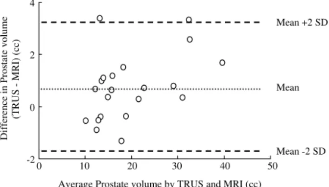
The mean prostate volume was 20.00 ± 8.71 cc, 19.26 ± 8.15 cc and 23.42 ± 8.43 cc in TRUS-based, MRI- based and CT-based preplanning, respectively. The prostate volume contoured by MRI was slightly underestimated in comparison to that done by TRUS (however, the difference in the mean prostate volume was less than 1 cc), the pros- tate volume as contoured by MRI thus closely agreed with that done by TRUS (Fig. 2). The prostate volume contoured by CT was overestimated by about 1.17 times (3.4 cc) in comparison to that done by TRUS.
Comparison of the DVHs between TRUS-based and MRI-based preplanning
The mean DVH-related parameters estimated by TRUS- based and MRI-based preplanning are shown in Table 1.
Although we found some discrepancies in the DVH related parameters between TRUS-based and MRI-based preplan- ning due to the prostate deformations caused by examina- tions with or without the rectal probe, there were no significant differences in the mean DVH related parameters except between rectal V100.
ProstateV100were over 96% both on MRI-based and TRUS- based preplanning in most cases, but there was one case with 99.0% on MRI-based preplanning and 84.6% on TRUS- based preplanning (Fig. 3a). Regarding prostate D90, this case shows 130.6% on MRI-based preplanning and 86.62%
on TRUS-based preplanning (Fig. 3b) The prostate volume of this case was 11.5 cc on MR imaging and 14.9 cc on TRUS imaging, about 1.3 times.
-2 0 2 4
0 10 20 30 40 50
Average Prostate volume by TRUS and MRI (cc) Difference in Prostate volume (TRUS - MRI) (cc)
Mean
Mean -2 SD Mean +2 SD
Fig. 2. Prostate volume measured with TRUS and MRI. Differences against mean for prostate volume.
Urethral D30, D5 and V150 showed agreement between MRI-based and TRUS-based preplanning. Rectal V100 and V150contoured by TRUS were overestimated in comparison to that done by MRI.
Comparison of MRI-based preplanning and CT/MRI fusion-based postimplant dosimetry
A mean prostate volume and DVH-related parameters are shown in Table 1. The postimplant prostate volumes in- creased by prostatic edema in comparison to preplanning.
Postimplant prostateV100andD90 were decreased in com- parison to the MRI-based preplanning. However, there was no statistical difference in the rectal V150(cc), and rectal V100(cc) values between MRI-based preplanning and CT/MRI fusion-based postimplant dosimetry. The rectal V100(cc) value between MRI-based preplanning and CT/MRI fusion-based postimplant dosimetry showed a close correla- tion (Fig. 4). The time course of postimplant dosimetry in comparison to the preplanning findings was similar to that reported in previous studies.
Discussion
Prostate volume and dose
TRUS is commonly used before implantation to deter- mine the prostate volume and boundaries. These determi- nations have been shown to correlate closely with the
volume of the prostate during pathologic evaluations [3,12]. Sosna et al. in a series of 20 patients found a Table 1
Comparisons between TRUS-based preplanning, MRI-based pre- planning and CT/MRI fusion-based postimplant dosimetry in the prostate volume and dose volume histograms
80 85 90 95 100
80 85 90 95 100
MRI-based Prostate V100 (%)
TRUS-based Prostate V100 (%)
80 100 120 140 160
80 100 120 140 160
MRI-based Prostate D90 (%)
TRUS-based Prostate D90 (%)
a
b
Fig. 3. (a) Comparison of the prostateV100 value between TRUS- based preplanning and MRI-based preplanning. R2= 0.1704. (b) Comparison of the prostate D90 value between TRUS-based pre- planning and MRI-based preplanning.R2= 0.5322.
0 0.5 1 1.5
0 0.5 1 1.5
CT/MRI fusion-based postimplant Rectal V100 (cc)
MRI-based preplanning Rectal V100 (cc)
Fig. 4. Comparison of the rectalV100between MRI-based preplan- ning and CT/MRI fusion-based postimplant dosimetry.R2= 0.5115.
118 MRI-based preplan in prostate brachytheraph
strong correlation between volumes calculated from MRI with pathologic specimens [19]. Hricak et al. measured prostate volume using US and MRI in 15 patients prior to undergoing a radical prostatectomy [7]. The accuracy of US and MRI was evaluated by recording the difference be- tween prostate size using imaging and that of the actual surgical specimen. Both US and MRI predicted the prostate size with a high degree of accuracy. Hoffelt et al. re- ported a comparison of CT and TRUS-based prostate vol- ume in 36 patients with untreated prostate cancer [6].
A strong correlation was found between the CT and TRUS measurements of the prostate volume, however, CT over- estimated the prostate volume by approximately 50%.
Roach et al. compared CT with MRI-measured prostate volume. The mean prostate volume in the 10 patients studied was 32% larger using CT than with MRI [18]. Kag- awa et al. also reported a 26% mean overestimation when delineating prostate volume with CT images in comparison to MRI images [9]. Based on the above studies, the pros- tate can be identified with a high degree of accuracy with MRI or US imaging but, at least in the hands of some investigators, CT overestimates the size of the prostate considerably and is associated with a high interobserver variation.
Our current results correlated with previous findings in the literature. The mean MRI-based prostate volume was similar to the mean TRUS-based prostate volume (the differ- ence in the mean prostate volume was less than 1 cc), and the mean CT-based prostate volume was 17% larger than mean TRUS-based prostate volume. A strong correlation was found between the MRI and TRUS measurements of the prostate volume. Although MRI was performed in supine position and TRUS was performed in lithotomy position, there was no statistical difference in the prostate volume or the prostate DVH-related parameters regarding preplanning.
The prostate V100 and D90 values in MRI-based pre- planning were correlated to those of TRUS-based pre- planning except in one patient. The prostate volume of this patient was 11.5 cc on MRI and 14.9 cc on TRUS imaging. The number of seeds was identical between the TRUS-based and MRI-based preplanning, therefore the seeds required according to TRUS-based preplanning were insufficient in comparison to those required based on MRI-based preplanning. The fact that an underesti- mation of the prostate volume strongly influences the DVH-related parameters in the postimplant setting has been thoroughly described by Polo et al. [14]. In our series, the discrepancy between the TRUS and MRI pros- tate volume for this patient was caused by the miscon- touring of either the apex or the base of the prostate because the prostate size was too small. The MRI-based prostate volume was similar to TRUS-based one in most patients, we believe that the MRI-based volume estima- tion is useful, and comfortable in comparison to TRUS- based.
The CT/MRI fusion-based postimplant prostate volumes increased due to prostatic edema in comparison to preplan- ning by approximately 40% (7.7 cc). DVH-related parameters decreased in postimplant dosimetry in comparison to the preplanning parameters.
Urethral location and dose
Lee et al. reported that using a surrogate urethra at the geometric center of the prostate may significantly overesti- mate the urethral dose in a postimplant setting [10]. We used the T2-weighted spin echo MRI which helps to deter- mine the urethra in the prostate. A surrogate urethra was used when the urethra was unable to be visualized on MRI, a 5-mm diameter circle was drawn at the geometric center at apex to posterior slightly than center at the base of the prostate (not at the geometric center through the prostate) on each MR image. Fig. 1shows that urethral discrepancy was found in A–P direction between MRI-based and TRUS- based preplanning, but we found no significant difference in urethral dose between them. This was due to better ure- thral visualization on MRI in some cases and contouring the modified surrogate urethra as shown above. Using a urethral catheter is the best way to visualize the urethra, but using T2-weighted imaging and a new surrogate model is there- fore sometimes reliable in MRI-based preplanning. There was no statistical difference in urethral V150(cc) between the preplanning and postimplant dosimetry findings. As a re- sult, we believe that MRI-based preplanning is thus a reli- able modality for predicting the appropriate urethral dose in postimplant dosimetry.
Rectal location and dose
We found a significant difference in the MRI-based and TRUS-based preplanning findings regarding the rectal dose.
On TRUS-based preplanning, the shape of the rectum changes with the insertion of the probe, namely it tends to take on an unnaturally straight course[8]. On MRI-based preplanning and CT/MRI fusion-based postimplant dosime- try, the shape of the rectum maintains its natural form.
Therefore, the rectal dose tended to be overestimated in TRUS-based preplanning in comparison to MRI-based pre- planning, due to the increased surface of the rectal wall adjacent to the prostate in the TRUS-based preplanning findings (Fig. 1). Although using an endorectal coil MRI is similar to using the TRUS image, it is uncomfortable and the shape of the rectum is unnatural such as in TRUS posi- tion. In fact, we found no significant difference in the rectal dose between the MRI-based preplanning and CT/MRI fu- sion-based postimplant dosimetry. Based on these findings, MRI-based preplanning are thus considered to be superior to TRUS-based preplanning in predicting rectal dose.
During the implant procedure, we did not experience any pubic arch interference in the examined patients. One rea- son for this is considered to be due to the fact that our pa- tients mostly have relatively small prostate volumes. In addition, MRI-based preplanning helps to predict pubic arch interference such as TRUS-based preplanning. Recently, intraoperative planning has been used at many centers to eliminate the need for a preplan volume study[25]. How- ever, the use of an operating room has now become a very expensive cost factor, and the cost of performing intraoper- ative planning in an operating room is far greater than that done in an outpatient examination room. Reynier et al. re- ported that using MRI linked with TRUS improves TRUS im- age segmentation, and this may significantly modify the prostate volume definition and therefore strongly influence
the treatment planning[17]. At a center which uses intraop- erative planning, MRI-based preplanning may thus shorten the intraoperative planning time by combining MRI-based preplanning with TRUS-based intraoperative planning.
Although prostate volume estimation in very small pros- tates may show a significant difference between the findings determined by TRUS-based and MRI-based imaging, usual or larger prostate volumes tend to be the same for both TRUS- based and MRI-based imaging. We believe that MRI-based preplanning is therefore equally useful to that of TRUS- based preplanning, while it can more accurately predict the rectal dose than TRUS-based preplanning.
Conclusions
To our knowledge, our series is the first to report the findings of dosimetric comparisons between TRUS-based and MRI-based preplanning. Prostate volume estimation and DVH-related parameters, with the exception of the rec- tal dose, on MRI-based preplanning is almost identical to TRUS-based preplanning. From the results of the CT/MRI fu- sion-based postimplant dosimetry, MRI-based preplanning is therefore considered to be a reliable and useful modality, in addition to TRUS-based preplanning. Besides, we found no significant difference in the rectal dose between the MRI- based preplanning and CT/MRI fusion-based postimplant dosimetry. MRI-based preplanning can therefore more accu- rately predict the postimplant rectal dose than TRUS-based preplanning.
* Corresponding author. Osamu Tanaka, Department of Radiol- ogy, Gifu University School of Medicine, 1-1, Yanagido, Gifu City, 501-1194, Japan. E-mail address:[email protected]
Received 2 February 2007; received in revised form 30 April 2007;
accepted 20 September 2007; Available online 22 October 2007
References
[1] al-Rimawi M, Griffiths DJ, Boake RC, et al. Transrectal ultrasound versus magnetic resonance imaging in the estima- tion of prostatic volume. Br J Urol 1994;74:596–600.
[2] Badiozamani KR, Wallner K, Cavanagh W, et al. Comparability of CT-based and TRUS-based prostate volumes. Int J Radiat Oncol Biol Phys 1999;43:375–8.
[3] Blasko JC. Low-dose-rate brachytherapy for prostate cancer:
preplanning vs. intraoperative planning – preplanning is best.
Brachytherapy 2006;5:139–42.
[4] Claus FG, Hricak H, Hattery RR. Pretreatment evaluation of prostate cancer: role of MR imaging and 1H MR spectroscopy.
Radiographics 2004;24:167–80.
[5] Dubois DF, Prestidge BR, Hotchkiss LA, et al. Intraobserver and interobserver variability of MR imaging- and CT-derived prostate volumes after transperineal interstitial permanent prostate brachytherapy. Radiology 1998;207:785–9.
[6] Hoffelt SC, Marshall LM, Garzotto M, et al. A comparison of CT scan to transrectal ultrasound-measured prostate volume in untreated prostate cancer. Int J Radiat Oncol Biol Phys 2003;57:29–32.
[7] Hricak H, Jeffrey RB, Dooms GC, et al. Evaluation of prostate size: a comparison of ultrasound and magnetic resonance imaging. Urol Radiol 1987;9:1–8.
[8] Ishiyama H, Kitano M, Satoh T, et al. Difference in rectal dosimetry between pre-plan and post-implant analysis in transperineal interstitial brachytherapy for prostate cancer.
Radiother Oncol 2006;78:194–8.
[9] Kagawa K, Lee WR, Schultheiss TE, et al. Initial clinical assessment of CT-MRI image fusion software in localization of the prostate for 3D conformal radiation therapy. Int J Radiat Oncol Biol Phys 1997;38:319–25.
[10] Lee HK, D’Souza WD, Yamal JJ, et al. Dosimetric conse- quences of using a surrogate urethra to estimate urethral dose after brachytherapy for prostate cancer. Int J Radiat Oncol Biol Phys 2003;57:355–61.
[11] Lee WR, deGuzman AF, Bare RL, et al. Postimplant analysis of transperineal interstitial permanent prostate brachytherapy:
evidence for a learning curve in the first year at a single institution. Int J Radiat Oncol Biol Phys 2000;46:83–8.
[12] Nag S, Beyer D, Friedland J, et al. American Brachytherapy Society (ABS) recommendations for transperineal permanent brachytherapy of prostate cancer. Int J Radiat Oncol Biol Phys 1999;44:789–99.
[13] Narayana V, Roberson PL, Pu AT, et al. Impact of differences in ultrasound and computed tomography volumes on treat- ment planning of permanent prostate implants. Int J Radiat Oncol Biol Phys 1997;37:1181–5.
[14] Polo A, Cattani F, Vavassori A, et al. MR and CT image fusion for postimplant analysis in permanent prostate seed implants.
Int J Radiat Oncol Biol Phys 2004;60:1572–9.
[15] Rahmouni A, Yang A, Tempany CM, et al. Accuracy of in-vivo assessment of prostatic volume by MRI and transrectal ultra- sonography. J Comput Assist Tomogr 1992;16:935–40.
[16] Rasch C, Barillot I, Remeijer P, et al. Definition of the prostate in CT and MRI: a multi-observer study. Int J Radiat Oncol Biol Phys 1999;43:57–66.
[17] Reynier C, Troccaz J, Fourneret P, et al. MRI/TRUS data fusion for prostate brachytherapy. Preliminary results. Med Phys 2004;31:1568–75.
[18] Roach III M, Faillace-Akazawa P, Malfatti C, et al. Prostate volumes defined by magnetic resonance imaging and com- puterized tomographic scans for three-dimensional confor- mal radiotherapy. Int J Radiat Oncol Biol Phys 1996;35:1011–8.
[19] Sosna J, Rofsky NM, Gaston SM, et al. Determinations of prostate volume at 3-Tesla using an external phased array coil:
comparison to pathologic specimens. Acad Radiol 2003;10:846–53.
[20] Tanaka O, Hayashi S, Sakurai K, et al. Importance of the CT/
MRI fusion method as a learning tool for CT-based post implant dosimetry. Radiother Oncol 2006;81:303–8.
[21] Tanaka O, Hayashi S, Matsuo M, et al. Comparison of MRI- based and CT/MRI fusion-based postimplant dosimetric anal- ysis of prostate brachytherapy. Int J Radiat Oncol Biol Phys 2006;66:597–602.
[22] Terris MK, Stamey TA. Determination of prostate volume by transrectal ultrasound. J Urol 1991;145:984–7.
[23] Terris MK, McNeal JE, Stamey TA. Estimation of prostate cancer volume by transrectal ultrasound imaging. J Urol 1992;147:855–7.
[24] Tewari A, Indudhara R, Shinohara K, et al. Comparison of transrectal ultrasound prostatic volume estimation with mag- netic resonance imaging volume estimation and surgical specimen weight in patients with benign prostatic hyperplasia.
J Clin Ultrasound 1996;24:169–74.
[25] Zelefsky MJ. Low-dose-rate brachytherapy for prostate can- cer: preplanning vs. intraoperative planning –Intraoperative planning is best. Brachytherapy 2006;5:143–4.
120 MRI-based preplan in prostate brachytheraph