Takao A RAI1, 2, Yuzuru H ASEGAWA2, Toshihide T ANAKA2, and Yuichi M URAYAMA3
, Toshihide T ANAKA2, and Yuichi M URAYAMA3
【症例報告】
慈恵医大誌 2016;131:83-8.
1東京慈恵会医科大学附属第三病院脳神経外科
2東京慈恵会医科大学附属柏病院脳神経外科
3東京慈恵会医科大学脳神経外科学講座
(受付 平成 28 年 3 月 30 日)
Craniopharyngioma is a histologically benign tumor, and the ideal first choice of treatment is total removal via surgery. For complete resection of a tumor, craniotomy or transsphenoidal surgery or both are required. On the other hand, because of advances in endoscopic techniques, cyst fenestration and the resection of some tumors is possible with neuroendoscopic surgery. By combining the conveniences of all available techniques, we used a 2-stage method of surgical management, in which endoscopic cyst fenestration is followed by craniotomy for tumor resection, to treat 2 patients with cystic craniopharyngioma and obtained favorable results. This method has 3 advantages. First, urgent decompression of the optic nerve can be performed. Second, craniotomy can be less complex because the tumor is better visualized after cyst shrinkage. Third, time is available for the patientʼs general condition to improve and for endocrine evaluation before craniotomy. This method of surgical management can be an effective strategy for treating some patients with cystic craniopharyngioma.
(Tokyo Jikeikai Medical Journal 2016;131:83-8)
1
Department of Neurosurgery, The Jikei University Daisan Hospital
2
Department of Neurosurgery, The Jikei University Kashiwa Hospital
3
Department of Neurosurgery, The Jikei University School of Medicine
ENDOSCOPIC CYST FENESTRATION AND EXCISION BY CRANIOTOMY AS A TWO-STAGE SURGICAL MANAGEMENT IN CYSTIC
CRANIOPHARYNGIOMA: TECHNICAL CASE REPORTS
荒 井 隆 雄1, 2 長谷川 譲2 田 中 俊 英2 村 山 雄 一3
開頭腫瘍摘出術前の神経内視鏡的嚢胞開窓術が有効であった 嚢胞性頭蓋咽頭腫の 2 例
Key words; craniotomy, cyst fenestration, cystic craniopharyngioma, neuroendoscpic surgery, surgical management
Ⅰ.緒 言
頭蓋咽頭腫は組織学的良性腫瘍であることか ら,治療の第一選択は手術による全摘出が理想で ある.しかし多くの場合患者の年齢・術前の神経 症状や腫瘍の発生部位などを考慮した上で,各々 の症例に応じて手術・放射線治療やホルモン補充 療法を組み合わせた集学的治療を検討しているの が実状である.今回我々は,嚢胞性頭蓋咽頭腫の 2 例に対して開頭腫瘍摘出術を前提とした神経内 視鏡的嚢胞開窓術を行い良好な結果を得たので,
本法を用いた治療戦略の有用性と注意点について
報告する.
Ⅱ.症 例
症 例 1:51 歳 女 性.201X年 X 月 上 旬 に 視 野 の異常を自覚した.その後,左視力が急速に低下
したため X月 21 日に東京慈恵会医科大学附属柏
病院脳神経外科を紹介受診した.来院時,意識清 明で視力は右 0.4 で左光覚弁だった.視野検査で は右側視野に耳側半盲が認められた.PRL 値 71.5
ng/ml 以外には異常所見は認められず,ホルモン
負 荷 試 験 で は 異 常 所 見 は 認 め ら れ な か っ た.
Computed tomography(CT)ではトルコ鞍上部か ら第三脳室へと伸展する嚢胞性病変が認められ た. 石灰化は認められず, 水頭症は呈していなかっ た(Fig. 1A, B) .T
1-weighted MR images with gadolinium(MRI T1WI Gd)ではトルコ鞍上部か ら第三脳室と左側頭葉内側に伸展する多房性嚢胞 性病変が認められた(Fig. 1C, D) .以上より頭蓋 咽頭腫の診断のもと二期的手術を計画した.はじ めに視神経の緊急減圧を目的とした神経内視鏡的 嚢胞開窓術および腫瘍生検を行い,その後に開頭 腫瘍摘出術を行う方針とした.X 月 30 日に, もっ とも大きな 2 つの嚢胞を開窓する方針で内視鏡手
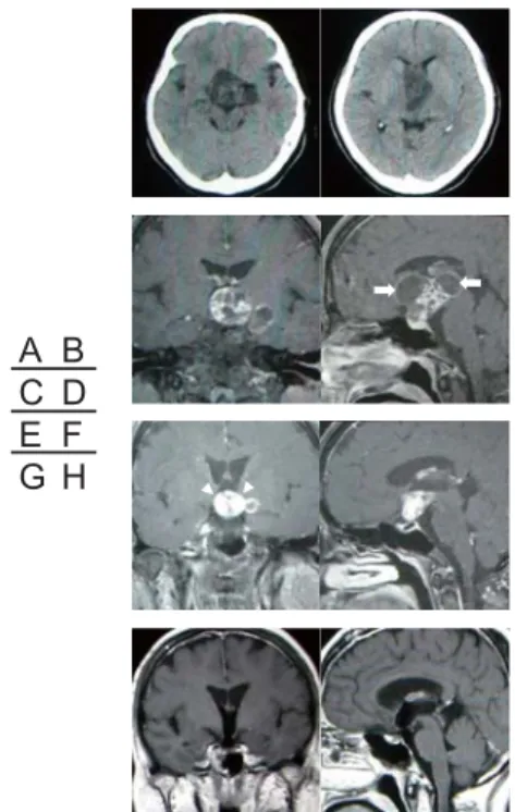
Fig. 1. Preoperative computed tomography scans showing a cystic mass extending from the suprasellar region to the third ventricle. (A, B) Preoperative coronal (C) and sagittal (D) contrast-enhanced T1-weighted magnetic resonance images showing a multicystic lesion extending from the suprasellar region to the third ventricle and left medial temporal lobe. The arrows show the two largest cysts which was fenestrated by endoscopic surgery. Coronal (E) and sagittal (F) contrast-enhanced T1-weighted magnetic resonance images on day 6 after endoscopic surgery revealed that not only the two fenestrated cysts but the cyst in the left temporal lobe have all been reduced. The arrow heads show the adhesion site between the hypothalamus and the tumor.
Coronal (G) and sagittal (H) contrast-enhanced T1- weighted magnetic resonance images 17 months after craniotomy revealed no residual tumor or recurrence.
$ &
( *
% ' ) +
$%
& '
Fig. 2. Intraoperative photographs demonstrating the surgical procedure of endoscopic cyst fenestration. A: A 4Fr Fogertyʼs balloon catheter (arrow head) inserted into the cyst and the cyst wall was fenestrated. The arrow shows the choroid plexus in the right lateral ventricle. B: Inside the cavities, each cyst was surrounded by thin membranes and the cysts were communicating with each other.
The arrow head shows a cholesterol crystals floating around inside the cysts. C: The second cyst was tapped and fenestrated by the balloon catheter (arrow head). D:
The aqueduct (arrow head) was patent and no abnormal
fi ndings were observed on the ventricular wall.
術を行った(Fig. 1D) .内視鏡は軟性鏡(町田製 作所 東京)を使用した.右前角穿刺で側脳室内 を観察すると,拡大したモンロー孔に腫瘍の嚢胞 壁が認められた.嚢胞壁を穿刺するとモーターオ イル様の内容物が吸引された.穿刺部位に 4Fr.の フォガティーバルーンカテーテルを挿入して開窓 した(Fig. 2A) .嚢胞内部を観察すると,すだれ 状の薄い嚢胞被膜が多数認められた.また嚢胞内 部にはコレステリン結晶と思われる脂肪塊が浮遊 していた(Fig. 2B) .嚢胞の外を回って第三脳室 内へ進むと第二の嚢胞が観察された.この嚢胞の 圧は既に減じており,第一の嚢胞と内部で交通し ていたことが判明した.同様の手技でこの嚢胞を 穿刺・開窓した(Fig. 2C) .縮小した第二の嚢胞
の奥に中脳水道が開存しているのが確認できた
(Fig. 2D) .人工髄液(ARTCEREB ®)で嚢胞内 と脳室内を十分洗浄して手術を終了した.病理診 断は頭蓋咽頭腫であった.術後は髄膜炎などの合 併症なく経過した. 術後6日目の MRI T1WI Gd では,
開窓した 2 つの嚢胞のみならず左側頭葉の嚢胞を 含むすべての嚢胞が縮小し,視床下部や中脳への 圧迫が解除された(Fig. 1E, F) .これにともない 腫瘍と第三脳室壁の癒着部位が明らかとなった
(Fig. 1E) .術後の視力は,右 1.0 左 0.4 と視力は回 復し,視野は改善し両耳側半盲を呈していた.術 後12日にbasial interhemispheric translaminaterminalis approachによる開頭術で腫瘍を全摘出した.開頭 術後の視力は右 1.0,左 0.4 と悪化は認められず,
視野検査では両耳側半盲は改善していた.術後 17 ヵ月の MRI T1WI Gd では残存腫瘍および再発は 認められない(Fig. 1G, H) .Levothyroxine sodium hydrate , hydrocortisone によるホルモン補充療法 を行いながら社会復帰している.
症例 2:12 歳 女児.10 歳の時から低身長で小 児科に通院していた.11 歳の時から時々頭痛を 自覚していた.201X 年 X月より頭痛と嘔気が出 現した.X 月 19 日に複視が出現,X 月 22 日に意
Fig. 3. Computed tomography scans on admission showing a cystic lesion with calcifi cation extending from the sella turcica to the suprasellar region. (A, B) Preoperative coronal (C) and sagittal (D) contrast-enhanced T1 - weighted magnetic resonance images revealed a monolocular cystic tumor, and the foramen of Monro was blocked. Coronal (E) and sagittal (F) contrast-enhanced T1-weighted magnetic resonance images on day 3 after endoscopic surgery revealed that since the cyst had been reduced. The arrow head is showing the adhesion site of the hypothalamus and tumor. Coronal (G) and sagittal (H) contrast-enhanced T1-weighted magnetic resonance images 5 months after craniotomy revealed no residual tumor or recurrence.
$ &
( *
% ' ) +
$%
& '
Fig. 4. Intraoperative photographs demonstrating the surgical procedure of endoscopic cyst fenestration. A: The fenestrated cyst wall and the fl oor of the third ventricle were observed from the enlarged foramen of Monro. The arrow is showing the fl oor of the third ventricle that was elevated by the tumor. B: Calcification was observed inside the cyst wall, and a number of biopsies were performed from the same site. C: When the endoscope moved forward into the third ventricle, the adhesion site between the third ventricle wall and tumor was observed.
The arrow heads are showing the adhesion site between
the hypothalamus and the tumor. D: The aqueduct (arrow
head) was patent and no abnormal fi ndings were observed
on the ventricular wall.
識障害が出現して東京慈恵会医科大学附属柏病院 に救急搬送された.来院時の身長は 128 cm(−
4SD) ,体重は 28 ㎏(− 2SD)であった.来院時 意識レベルは E3V4M6/G.C.S.で,左外転神経麻痺 が認められた.視力は右 1.0 左 1.2 で,視野障害と うっ血乳頭は認められなかった.GH 0.5 ng/ml,
IGF- 1 118 ng/mlと低値であり,PRL が 54.1 ng/ml と軽度上昇していた.ホルモン負荷試験では GH
と ACTHの分泌不全が認められた.頭部 CTでは
トルコ鞍から鞍上部にかけて石灰化を伴う嚢胞性 病変が認められ,水頭症を呈していた(Fig. 3A, B) .MRI T1WI Gd では単房性の嚢胞性腫瘍が鞍 上部から第三脳室に伸展し,モンロー孔と中脳水 道を閉塞していた(Fig. 3C, D) .以上より頭蓋咽 頭腫の診断のもと,はじめに非交通性水頭症の解 除を目的とした神経内視鏡的嚢胞開窓術と腫瘍生 検を行い,後日二期的に開頭腫瘍摘出術を行う方 針とした. 来院 6 日目に神経内視鏡手術を行った.
内視鏡は軟性鏡(町田製作所 東京)を使用した.
右前角穿刺を行うと,拡大したモンロー孔から腫 瘍が観察された.腫瘍を穿刺すると内容液が吸引 され嚢胞の圧が減じた(Fig. 4A) .穿刺部位に 4Fr. のフォガティーバルーンカテーテルを挿入し 嚢胞壁を開窓した.嚢胞内に進入すると石灰化を 伴う嚢胞壁が観察され,同部位を数か所生検した
(Fig. 4B) . つぎに第三脳室内へ内視鏡を進めると,
減圧された腫瘍と第三脳室壁の癒着部位が確認さ れた(Fig. 4C) .縮小した嚢胞の外側を周ると開 存した中脳水道が観察された(Fig. 4D) .最後に 人工髄液(ARTCEREB ®)で嚢胞内と脳室内を 十分洗浄し手術を終了した.病理診断は頭蓋咽頭 腫であった.術後は合併症なく経過した.術後 3
日目の MRI T1WI Gdでは,嚢胞が縮小したこと
により腫瘍と第三脳室壁の癒着部位が明瞭となっ た(Fig. 3E, F) .術後16日目にbasial interhemispheric translaminaterminalis approach による開頭術で腫瘍 を全摘出した.術後の視力は,右 1.2,左 1.5 で視 野の悪化も認められなかった.術後 5 ヵ月目の
MRI T1WI Gd では,残存および再発腫瘍は認め
られなかった(Fig. 3G, H) .Levothyroxine sodium hydrate,hydrocortisone,DDAVP,somatropin によ るホルモン補充療法を行いながら復学した.
Ⅲ.考 察
頭蓋咽頭腫は組織学的良性腫瘍であることか ら,治療の第一選択は手術による全摘出が理想で ある.しかし現実には,患者の年齢(若年者・高 齢者) ,症状(視神経障害・水頭症・下垂体機能 障害など) ,腫瘍の局在(トルコ鞍内・鞍上部・
脳室内など)や腫瘍の性状(充実性・嚢胞性・石 灰化など)などにより,個々の症例で手術治療・
放射線治療とホルモン補充療法を組み合わせる オーダーメイド治療が行われているのが実状であ る.
根治的な腫瘍摘出術に関しては,開頭手術と顕 微鏡的経蝶形骨洞手術(顕微鏡的transsphenoidal surgery; TSS)あるいは内視鏡的経蝶形骨洞手術
(内視鏡的TSS)がある
1)-15). 開頭手術では一般 的に interhemispheric approach (IH), pterional approach, orbitozygomatic approach, subtemporal approach, transpetrosal approahやこれらの combined approachが選択され,本例で用いたIHでは全摘 出率は 71.0%-80.4%との報告がある
7)9)13)14). 顕 微 鏡 的TSSに 関 し て は, 全 摘 出 率 が 27.3% 〜 90.0%で術後髄液漏が 0%- 17.5% と報告によりば らつきがあることから,本アプローチを行う場合 には腫瘍の局在や性状に応じた手術適応が重要と なる
1)3)5)12)16). すなわち,石灰化を伴う大きな 腫瘍や内頚動脈を越えて大きく外側に伸展する腫 瘍は,本アプローチでは摘出困難であり適応外と なる.さらには術者が視交叉・下垂体柄・後交通 動脈や脳底動脈およびその穿通枝,視床下部など の重要構造物を温存する技術と,経鼻的に頭蓋内 に到達することにより生じる術後髄液漏の修復技 術を有していることが必要条件となる
15).
頭蓋咽頭腫に対する神経内視鏡手術は,神経内 視鏡の進歩と術者の技量の上達にともない,その 役割が術中観察から嚢胞開窓術さらには腫瘍摘出 術へと発展してきた
2)4)6)8)10)11)16)-24). 術中観察
に関しては,開頭手術や顕微鏡的TSSにおいて神
経内視鏡を補助的に使用することにより腫瘍摘出
率の向上に有用である
19)21). また嚢胞性頭蓋咽
頭腫に対しては,神経内視鏡観察下にOmmaya カ
テーテル留置術が行われ,安全かつ確実に手術を
行うことが可能となった.
16)19)23)24)さらに嚢胞
性頭蓋咽頭腫に対しては,嚢胞を開窓して腫瘍を 縮小させた後にガンマナイフを施行する報告もあ り,摘出術のリスクが高い高齢者や全身状態不良 の患者に対しては有効な治療戦略である
18)22). 一方,近年頭蓋咽頭腫に対しても経鼻的アプロー チによる神経内視鏡的腫瘍摘出術が試みられるよ う に な っ て き た
2)4)6)8)10)11). そ の 全 摘 出 率 は 70%〜91%,術後髄液漏は 15%〜58%と報告され ている.その手術適応には十分な検討が必要であ るが,神経内視鏡技術と術者の技量の今後のさら なる進歩により,手術適応の拡大と治療成績の向 上が期待される.
今回我々は,嚢胞性頭蓋咽頭腫の 2 例に対して 開頭腫瘍摘出術を前提に神経内視鏡的嚢胞開窓術 を行う二期的治療を行い良好な結果を得た.本法 の利点は以下の 3 点と考える.第一に,水頭症の 治療と視神経の減圧を緊急で行うことができるこ とである.それに加えて開頭術に先駆けてあらか じめ視神経の除圧を図ることは,開頭摘出操作が 視神経に与えるダメージを軽減できる可能性があ る.今回の症例 1 のように,急速に進行する視力 視野障害に対して嚢胞の減圧を行うことで,視力 視野ともに回復させることができた.また症例 2 では術後も視力視野に悪化を見ず良好に温存され ていた.第二に,症例 1,2 とも嚢胞開窓術で嚢 胞を縮小させることにより,腫瘍と視床下部との 癒着部位を開頭術を行う前に評価することができ た.このことは鞍上部から第三脳室へ進展する腫 瘍を安全に摘出するうえで有用な術前情報となっ た.さらに症例1においては,IH では到達困難 な左側頭葉内側へ伸展する嚢胞が嚢胞開窓術によ り縮小したことも摘出の難易度の軽減につながっ た.なお頭蓋咽頭腫に対する嚢胞開窓術は単一の 嚢胞が良い適応との報告があるが,本例が示すよ うに画像上は多房性であっても実際には嚢胞の内 部が交通していることもあり開窓術の効果が期待 できる症例があることが示唆された.
22)第三に,
可及的に嚢胞を縮小させることにより,開頭術前 に患者の全身状態の改善や内分泌的評価を行う時 間を確保できることである.症例 2 のように水頭 症や下垂体機能不全により全身状態の悪い患者に おいて,開頭術に向けて全身状態の改善や内分泌 的評価を行ったうえで開頭術に臨めたことは本治
療の利点であった.以上の利点がある一方で,本 法を行う上で以下の 3 点に注意が必要である.第 一に,嚢胞開窓術を行う際には,後に行う開頭手 術を考慮して皮膚切開と穿頭部位を決める必要が ある.第二に ,嚢胞液の流出による c h e m i c a l
meningiitis とそれに伴う腫瘍と周囲組織との癒着
の懸念である.これを回避するためには開窓直前 に嚢胞液を十分吸引して排除すること,そして開 窓後には人工髄液(ARTCEREB ®)を用いて嚢 胞内および脳室内を十分に洗浄することが重要で ある.今回の 2 症例では,これらの操作により術 後の chemical meningiitsは発症せず,また開頭術 の術中所見では嚢胞開窓術に起因する腫瘍と周囲 組織との癒着は認められなかった.第三に,高度 の石灰化を伴う症例では嚢胞壁が固いために,腫 瘍縮小による視神経の減圧や視床下部との癒着評 価が可能となるかどうかは不明である.この点に 関しては,今後の症例の蓄積が必要と考える.
もちろん卓越した術者であれば,今回の 2 症例 に対して腫瘍を一期的に摘出することも可能であ ろう.しかし,脳神経外科医にとって最も高度な 手術技術と緻密な周術期管理を必要とする頭蓋咽 頭腫をより安全に治療するための戦略の一つとし て,開頭術前神経内視鏡的嚢胞開窓術が有用であ ると考え報告した.
著者の利益相反 (confl ict of interest:COI) 開示:
本論文の研究内容に関連して特に申告なし
文 献
1)
A b e T, L ü d e c k e D K . T r a n s n a s a l s u r g e r y f o r infradiaphragmatic craniopharyngiomas in pediatric patients. Neurosurgery. 1999; 44: 957 -
64.2)
Cavallo LM, Prevedello DM, Solari D, Gardner PA, Esposito F, Snyderman CH, et al. Extended endoscopic endonasal transsphenoidal approach for residual or recurrent craniopharyngiomas. J Neurosurg. 2009;
111:578-89.
3)
Chakrabarti I, Amar AP, Couldwell W, Weiss MH. Long- term neurological, visual, and endocrine outcomes following transnasal resection of craniopharyngioma. J Neurosurg. 2005; 102: 650-7.
4)
de Divitiis E, Cappabianca P, Cavallo LM, Esposito F, de
D i v i t i i s O , M e s s i n a A . E x t e n d e d e n d o s c o p i c
t r a n s s p h e n o i d a l a p p r o a c h f o r e x t r a s e l l a r craniopharyngiomas. Neurosurgery. 2007; 61: 219-27.
5)
Fahlbusch R, Honegger J, Paulus W, Huk W, Buchfelder M.
Surgical treatment of craniopharyngiomas: experience with
168 patients. J Neurosurg. 1999; 90: 237-50.6)
Frank G, Pasquini E, Doglietto F, Mazzatenta D, Sciarretta V, F a r n e t i G , e t a l . T h e e n d o s c o p i c e x t e n d e d transsphenoidal approach for craniopharyngiomas.
Neurosurgery. 2006; 59: 75-83.
7)
Fujitsu K, Sekino T, Sakata K, Kawasaki T. Basal interfalcine approach through a frontal sinusotomy with vein and nerve preservation. Technical note. J Neurosurg.
1994; 80: 575
-
9.8)
Gardner PA, Kassam AB, Snyderman CH, Carrau RL, Mintz AH, Grahovac S, et al. Outcomes following endoscopic, expanded endonasal resection of suprasellar craniopharyngiomas: a case series. J Neurosurg. 2008;
109: 6-16.
9)
Hori T, Kawamata T, Amano K, Aihara Y, Ono M, Miki N.
Anterior interhemispheric approach for 100 tumors in and around the anterior third ventricle. Neurosurgery. 2010;
66: 65-74.
10)
Kassam AB, Prevedello DM, Carrau RL, Snyderman CH, Thomas A, Gardner P, et al. Endoscopic endonasal skull base surgery: analysis of complications in the authors' initial 800 patients. J Neurosurg. 2011; 114: 1544 -
68.11)
Kitano M, Taneda M. Extended transsphenoidal surgery for suprasellar craniopharyngiomas: infrachiasmatic r a d i c a l r e s e c t i o n c o m b i n e d w i t h o r w i t h o u t a suprachiasmatic trans-lamina terminalis approach. Surg Neurol 2009; 71: 290-8.
12)
Maira G, Anile C, Albanese A, Cabezas D, Pardi F, Vignati A. The role of transsphenoidal surgery in the treatment of craniopharyngiomas. J Neurosurg. 2004; 100: 445-51.
13)
Shibuya M, Takayasu M, Suzuki Y, Saito K, Sugita K.
B i f r o n t a l b a s a l i n t e r h e m i s p h e r i c a p p r o a c h t o craniopharyngioma resection with or without division of the anterior communicating artery. J Neurosurg.1996; 84:
951
-
6.14)
Shirane R, Ching-Chan S, Kusaka Y, Jokura H, Yoshimoto T . S u r g i c a l o u t c o m e s i n
3 1p a t i e n t s w i t h craniopharyngiomas extending outside the suprasellar
cistern: an evaluation of the frontobasal interhemispheric approach. J Neurosurg. 2002; 96: 704-12.
15)
Yamada S, Fukuhara N, Oyama K, Takeshita A, Takeuchi Y, Ito J, et al. Surgical outcome in 90 patients with craniopharyngioma: an evaluation of transsphenoidal surgery. World Neurosurg. 2010; 74: 320-30.
16)倉本晃一
, 内門久明, 田島裕, 徳富孝志 , 重森稔. 神経
内視鏡にて嚢胞内リザーバー留置を行った高齢者再 発頭蓋咽頭腫の 1 例
. Neurol Surg. 2005; 33: 1207-12.
17)
Abdullah J, Caemaert J. Endoscopic management of craniopharyngiomas: a review of 3 cases. Minim Invasive Neurosurg. 1995; 38: 79 -
84.18)
Barajas MA, Ramírez - Guzmán G, Rodríguez - Vázquez C, Toledo-Buenrostro V, Velásquez-Santana H, del Robles RV, et al. Multimodal management of craniopharyngiomas:
neuroendoscopy, microsurgery, and radiosurgery. J Neurosurg. 2002; 97: 607-09.
19)
Joki T, Oi S, Babapour B, Kaito N, Ohashi K, Ebara M, et al. Neuroendoscopic placement of Ommaya reservoir into a cystic craniopharyngioma. Childs Nerv Syst. 2002;
18:629-33.
20)
Kadri H, Mawla AA. Endoscopy-assisted microsurgical total resection of craniopharyngioma in childhood. Minim Invasive Neurosurg. 2006; 49: 369-72.
21)
Kim EH, Ahn JY, Kim SH. Technique and outcome of endoscopy-assisted microscopic extended transsphenoidal surgery for suprasellar craniopharyngiomas. J Neurosurg.
2011; 114: 1338-49.
22)
Nakahara Y, Koga H, Maeda K, Takagi M, Tabuchi K.
Nakahara Y, et al. Neuroendoscopic transventricular surgery for suprasellar cystic mass lesions such as cystic craniopharyngioma and Rathke cleft cyst. Neurol Med Chir (Tokyo). 2004; 44: 408-13.
23)
Nakamizo A, Inamura T, Nishio S, Inoha S, Ishibashi H, F u k u i M . N e u r o e n d o s c o p i c t r e a t m e n t o f c y s t i c craniopharyngioma in the third ventricle. Minim Invasive Neurosurg. 2001; 44: 85-7.
24)