Role of Protease Activated Receptors in the Intracellular Calcium Dynamics of Neurons and Satellite Cells in the Rat Superior Cervical Ganglia
Hitoshi Miura1, Tomoyuki Saino2, Masahito Sato1, Yoh-ichi Satoh2*
1Division of Dental Anesthesiology, Department of Oral and Maxillofacial Surgery, School of Dentistry, Iwate Medical University, 1-3-27 Chuo-dori, Morioka, Iwate 020-8505, Japan, 2Department of Anatomy (Cell Biology), Iwate Medical University, 2-1-1 Nishitokuta, Yahaba-cho, Shiwa-gun, Iwate 028-3694, Japan
Summary
Protease-activated receptors (PARs) mediate cellular responses to various proteases in numerous cell types, including nerve cells. The issue of whether stimulation of PARs induces responses in neurons and satellite cells of sympathetic superior cervical ganglia (SCG) of rats was examined with special reference to PAR mRNA levels and to intracellular Ca2+ ([Ca2+]i) changes, since [Ca2+]i is a key factor in intracellular signaling. SCG whose essential struc- tural integrity was maintained intact were used.
RT-PCR showed that SCG expressed mRNAs encoding PAR1, 2 and 3, and PAR2 expression was the highest. Confocal microscopic analysis indicated that thrombin and trypsin induced an increase in [Ca2+]i in some neurons and many satellite cells.
These proteases initially elicited a [Ca2+]i increase in satellite cells and a subsequent [Ca2+]i change in neurons. Synchronized [Ca2+]i changes in satellite cells were often observed. Neither the removal of extracellular Ca2+ nor Ca2+ channel blockers affected the trypsin-induced or PAR2-activating peptide (PAR2-AP)-induced [Ca2+]i changes in satellite cells, thus suggesting that these changes were caused by Ca2+ mobilization from an internal store, but not by Ca2+ influx. However, neither the phospholipase C inhibitor, U73122, nor the IP3 receptor antagonist, heparin, could inhibit the [Ca2+]i changes of satellite cells, whereas these reagents considerably inhibited the [Ca2+]i changes of neurons. These findings demonstrate the presence of PARs in sympathetic nervous tissue, and establish that proteases induce [Ca2+]i changes in both neurons and satellite cells via Ca2+
mobilization.
Keywords
protease activated receptor, intracellular calcium ion dynamics, superior cervical ganglia
Introduction
Protease activated receptors (PARs) represent a novel class of seven transmembrane domain G-protein coupled receptors (GPCRs) that are activated by proteolytic cleavage (Déry et al., 1998). Four members of this class of receptors have been identified; PAR1, 2, 3 and 4 (Vu et al., 1991; Nystedt et al., 1994; Ishihara et al., 1997; Kahn et al., 1998; Xu et al., 1998;
Hollenberg, 1999). The mechanism of PAR activation involves the proteolytic unmasking of an N-terminal sequence that acts as a tethered ligand (Déry et al., 1998). Thrombin functions as an agonist for PAR1, 3 and 4 (Vu et al., 1991; Ishihara et al., 1997; Kahn et al., 1998; Xu et al., 1998) and trypsin is a major agonist for PAR2 (Nystedt et al., 1994). In addition, mast cell tryptase as well as coagulation factors VIIa and Xa are generally thought to serve as significant agonists of PAR2 (Nystedt et al., 1994; Camerer et al., 2000;
Kawabata et al., 2000). Therefore, the PAR activation of cells by various proteases can play an essential role in inflammation and pain as well as in other physiological and pathological responses (Macfarlane et al., 2001; Ossovskaya and Bunnett, 2004).
PAR2 activation sensitizes dorsal root ganglion (DRG) neurons, and this activation may play a role in inflammatory pain (Hoogerwerf et al., 2001; Dai et al., 2004, 2007; Grant et al., 2007). In addition to somatic sensory neurons, myenteric neurons may be excited by proteases (Corvera et al., 1999; Gao et al., 2002), and vagal sensory neurons display a PAR2-activated channel current (Gu and Lee, 2009). Despite the various potential interactions between the inflammation, pain and function of sympathetic nervous system, there have been no studies regarding the response of the sympathetic nervous system to proteases.
Generally, peripheral nervous tissues consist of neuronsandsupportingsatellitecells.Mostreportsof
*To whom all correspondence should be addressed.
E-mail: [email protected]
peripheral nervous tissue have studied responses of
“neurons” rather than those of satellite cells. However, a previous study described changes in intracellular calcium ion concentration ([Ca2+]i) that are thought to be essential events in various cell functions (Berridge, 2009) in both neurons and satellite cells in the sympathetic superior cervical ganglia (SCG) of the rat.
There are ATP-induced [Ca2+]i changes in satellite cells as well as in neurons, but different purinoceptors are expressed in both types of cells (Kumagai and Saino, 2001). Extracellular ATP may cause the pain and the inflammation under pathological conditions (Burnstock, 2007). The ATP-induced responses of SCG satellite cells indicate that satellite cells can actively participate in nervous function; they are never simple “silent” supporting cells.
The first aim of this study was to determine whether neurons and satellite cells of the sympathetic SCG express PARs. The reverse transcription polymerase chain reaction (RT-PCR) was used to examine PAR expression in intact rat SCG, which are composed of various types of cells. The signaling mechanism of PAR-induced [Ca2+]i changes in SCG were also examined. Intact SCG were used for these studies, because it is possible that the signaling mechanism of primary cells may be altered upon culture. A previous study showed that inflammatory mediators function by activating specific receptors, and that many of these receptors are G-protein-coupled receptors (GPCRs).
They used Gq or a G11 type of Gα for signaling, and activated phospholipase C (PLC) (Linley et al., 2008), which cleaves a membrane-bound phosphatidylinositol biphosphate to generate inositol triphosphate (IP3) and diacylglycerol. IP3 causes Ca2+ mobilization from internal stores (Berridge, 2009). PARs are one of GPCRs, thus PARs should activate [Ca2+]i responses, and such activation should be suppressed by inhibition of PLC or of the IP3 receptor. However, preliminary experiments on exocrine glandular cells showed that a treatment with a PLC inhibitor, U73122, did not suppress PAR2-mediated [Ca2+]i responses. The second aim of the present study was therefore to determine the IP3-dependency of PAR2-mediated [Ca2+]i changes in SCG cells.
Materials and Methods Preparation of SCG
Experiments were conducted in a manner consistent with the guidelines of the ethics committee for animal treatment of Iwate Medical University. Adult male rats (Wistar-Kyoto; age, 8–12 weeks, body weight, 250–400 g) were perfused with Ringer’s solution via the left
cardiac ventricle following carbon dioxide inhalation.
The SCGs were then taken out and associated connective tissues were carefully removed in HEPES- buffered Ringer’s solution (HR), containing 118 mM NaCl, 4.7 mM KCl, 2.5 mM CaCl2, 1.1 mM MgCl2, 1.0 mM NaH2PO4, 5.5 mM glucose, 0.2% bovine serum albumin (Sigma, St. Louis, Mo., USA), MEM amino acid solution (Gibco, Grand Island, N.Y., USA), 2.0 mM L-glutamine, and 10 mM HEPES, pH 7.4, adjusted with NaOH. Ca2+-deficient solutions were prepared by replacing CaCl2 with EGTA (1.0 mM; Sigma).
Ca2+-sensitive dye loading and stimulation Spatiotemporal changes in [Ca2+]i in SCG were deter- mined by ratiometry using a fluorescent dye, Indo-1, whose emission spectrum shifts upon binding to Ca2+. Dye loading was facilitated by using acetoxy-methyl esters of this dye (Indo-1/AM; Dojindo, Kumamoto, Japan). The specimens were transferred to HR contain- ing 300 U/ml purified collagenase (Elastin Products, Owensville, Mo., USA), 0.02% cremophor EL (Nacalai Tesque, Kyoto, Japan), and 10 μM Indo-1/AM, follow- ed by incubation for 1 h at 37°C. The specimens were placed on coverslips coated with Cell-Tak (a non-toxic adhesive reagent; Collaborative Biomedical, Bedford, Mass., USA) in Sykus-Moor chambers, and then con- tinuously perfused with HR which contained selected stimulants.
Acquisition of digital images of [Ca2+]i dynamics A real-time confocal microscope (RCM/Ab, revised type of RCM-8000; Nikon, Tokyo, Japan) was used.
The specimens were illuminated with an excitation UV beam (351 nm) produced by an argon ion laser and were placed on the stage of an inverted microscope (TE-300; Nikon). The fluorescence emission was guided through a water-immersion objective (Nikon C Apo 40×, N.A. 1.15) to a pinhole diaphragm. A dichroic mirror split the emission at 440 nm and directed it into two separate photomultipliers.
The acquisition time per image frame was 1/15 s. Eight frames were integrated to obtain a confocal image with maximal spatial resolution. Images were immediately stored on a high-speed hard disk, and the ratio of the fluorescence intensity measured at a wavelength shorter than 440 nm to that measured at a wavelength longer than it was computed. This ratio (<440 nm/>440 nm) was used as a measure of relative [Ca2+]i values. Ratio images contained 512 × 480 pixels with a density resolution of approximately 0.3 μm, and were displayed in pseudocolor with 256 colors (i.e., 8 bits/pixel). A pink color represents a higher ratio (high
[Ca2+]i) and blue and green shades represent lower ratios (low [Ca2+]i; Figs. 2–6).
Perfusion
SCG [Ca2+]i dynamics were observed in a Sykus-Moor perfusion chamber (capacity was 1 ml) as quickly as possible after the dye loading. Specimens were perfused at 1 ml/min at room temperature (ca. 23°C).
SCGs were perfused with the standard HR for a few minutes, then intact neurons and satellite cells were selected and examined under the microscope. SCGs that showed sufficient fluorescence signals were selected to reduce the possibility of differential load- ing of Indo-1/AM into neurons and satellite cells.
These SCGs were perfused by replacing the standard HR. The specimens were briefly washed in HR, then continuously perfused with HR containing agonists and/or antagonists: thrombin derived from bovine plasma (1 U/ml; Sigma), trypsin derived from porcine pancreas (1,000 U/ml), PAR2-activating peptide (PAR2-AP), a synthetic agonist for PAR2 (SLIGRL- NH2; 50 and 100 µM; Bachem AG, Bubendorf, Switzer- land), heparin (so called, an antagonist of the IP3 receptor; Sigma, 5 mg/ml), U73122 (2.5 µM; a specific inhibitor of phospholipase C; Sigma), diltiazem (50 µM; an L-type Ca2+ channel blocker; Sigma), Gd3+
(100 µM; a nonspecific Ca2+ channel blocker; Sigma), and thapsigargin (2 μM, a microsomal Ca2+-ATPase inhibitor; Alomone Laboratories, Jerusalem, Israel).
Doses of proteases, PAR2-AP and others used in the present study were determined after a preliminary examination. The inhibitory effect of extracellular
heparin is suspect, because such large molecules may be difficult to penetrate the cell membrane. However, extracellular heparin previously considerably sup- pressed ATP-induced Ca2+ mobilization in rat lacrimal glandular cells (Kamada et al., 2012), therefore it was used, although the results should be carefully inter- preted.
RNA extraction and RT-PCR
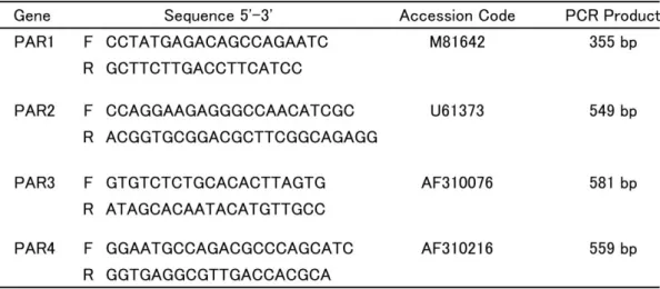
Total RNA was extracted from the SCG using the RNeasy Micro Kit (Qiagen, Hilden, Germany) according to the manufacturer’s instructions. The RNA concentra- tion was determined by spectroscopy at 260 nm. Isolated RNA was analyzed by RT-PCR that was performed with a thermal cycler (PC-701, ASTEC, Fukuoka, Japan) using the ReverTra Ace-α-® kit (TOYOBO, Osaka, Japan). The primer sequences are shown in Table 1 and were described by Rohatgi et al. (2003) except for the PAR-2 primers. The primers targeted to PAR-2 were 5’-CCAGGAAGAGGGCCAACATCGC-3’and 5’-ACGGTGCGGACGCTTCGGCAGAGG-3’, which yielded amplified fragments of 549 bp.
The thermal cycling protocol was; 42°C for 10 min followed by 94°C for 2 min, then 35 cycles of 94°C for 45 s, 54°C for 30 s and 72°C for 2 min, followed by 72°C for 10 min. The PCR products were separated by electrophoresis in 2% agarose gels and stained with ethidium bromide. Images of the gels were captured using a camera (Polaroid MP4 Land Camera, Minnea- polis, MN, USA). Each RT-PCR was repeated in three independent experiments.
Table 1. Primers used for the PCR analysis of PAR receptor expression. The primers listed are as described by Rohatgi et al. (2003) except for the PAR2 primers. The run conditions are provided in the Materials and Methods. F, forward primer; R, reverse primer. All primers were assessed for their sequence homology with other genes using BLAST searches (http://blast.ncbi.nlm.nih.gov/Blast.cgi).
Results
mRNA expression of PARs in SCG
RT-PCR analysis showed expression of PAR1, 2 and 3 mRNA in the SCG, but not PAR4 mRNA (Fig. 1).
PAR2 showed the highest level of mRNA expression.
Effect of proteases on [Ca2+]i dynamics
Relatively large round neurons (ca. 20–30 µm in diameter) and satellite cells, which are small and thin (ca. 5–10 µm in diameter at perinuclear regions) cells that surround neurons, were observed in the digital image (Fig. 2; thick and thin arrows indicate neurons and satellite cells, respectively). Nuclei of neurons were often clearly observed, because of the high fluorescence. These SCG specimens were perfused with HR for 5–10 minutes before stimulation with selected reagents, and spontaneous [Ca2+]i changes were rarely observed. Generally, damaged cells showed high [Ca2+]i levels. Therefore, SCG cells, which showed such high [Ca2+]i under resting conditions were excluded from subsequent analyses (Fig. 2, shown by asterisk).
Thrombin activates PAR1, 3 and 4 (Vu et al., 1991;
Ishihara et al., 1997; Kahn et al., 1998; Xu et al., 1998). This reagent (1 U/ml; number of independent chambers analyzed = 10) caused spike-like fluctuations in [Ca2+]i in many satellite cells and a few neurons.
The dynamics were biphasic; the initial acute increase was followed by a gradual decline (Fig. 3). The fluctua- tions were sometimes repeated (‘oscillatory’ fluctuat- ions).
Extracellular trypsin (1,000 U/ml, n = 6), which activates PAR2, caused a spike-like transient increase in [Ca2+]i in satellite cells, and subsequently [Ca2+]i changes in neurons were also detected (Fig. 4). HR containing the PAR2-AP agonist (50 µM, n = 8; 100 µM, n = 8; Effects on [Ca2+]i dynamics were almost same) was applied to confirm that the trypsin-induced changes in [Ca2+]i were related to PAR2-activation.
PAR2-AP induced responses similar to those induced by trypsin (Fig. 5). The patterns of the [Ca2+]i changes over time that were induced by trypsin and by PAR2-AP were similar to those induced by thrombin.
The biphasic pattern was also observed. Further study focused on the detailed mechanism of PAR2-mediated responses, since PAR2 mRNA expression was higher than that of the other PARs, and since somatic neurons express PAR2.
The characteristic [Ca2+]i changes during PAR2 stimulation were synchronized among neighboring satellite cells (Figs. 2 and 6). The perikaryon of a neuron were surrounded by a few satellite cells, and these satellite cells showed simultaneous [Ca2+]i changes (Satellite A and C in Fig. 6). In addition, many satellite cells in a region responded simul- taneously (Satellite B in Fig. 6). The synchronization indicated intercellular communications among the satellite cells. The gap junction protein connexin is expressed in satellite cells of dorsal root ganglia (Procacci et al., 2008). However, no intercellular propagation of the [Ca2+]i changes between neurons and satellite cells was observed.
Ca2+ influx or Ca2+ mobilization
The possibility of Ca2+ influx from the extracellular environment during protease stimulation has to be excluded when considering protease induced changes in intracellular [Ca2+]i. Neither trypsin- (1,000 U/ml; n
= 3) nor PAR2-AP (50 µM; n = 10, 100 µM; n = 14)- induced [Ca2+]i changes in satellite cells of SCG were inhibited in the absence of extracellular Ca2+
([Ca2+]o-free; Fig. 7). The effect of Gd3+ (100 µM), a nonspecific cation channel blocker, was the same as that observed in the absence of Ca2+ (n = 6; data not shown). Furthermore, diltiazem (50 µM), an L-type Ca2+ channel blocker, did not inhibit PAR2-AP-induc- ed [Ca2+]i changes in satellite cells (n = 5; data not shown). The responses of the neurons were consider- ably inhibited, but not completely. The responses of both neurons and satellite cells were almost inhibited Fig. 1. RT-PCR. Amplified PAR mRNA fragments obtained using
RT-PCR were analyzed by ethidium-bromide agarose gel electro- phoresis. GAPDH was used as a control marker.
100bp PAR1 PAR2 PAR3 PAR4 GAPDH100bp
Markers Markers
by pretreatment of thapsigargin (resulting in consum- ing internal Ca2+ store; 1 µM, n = 6; data not shown).
These results indicate that the satellite cell responses induced by PAR2 activation were exclusively due to a Ca2+ influx-independent mechanism, whereas Ca2+
influx (probably store-operated Ca2+ entry) was important in neurons.
The role of PLC and IP3 in PAR2-mediated [Ca2+]i responses of SCG was investigated. The effect of U73122, an inhibitor of phospholipase C (PLC), was observed. U73122 (2.5 µM) did not inhibit trypsin (n
= 8)- or PAR2-AP (n = 16)-induced [Ca2+]i changes in
satellite cells (Fig. 8). Therefore, PAR2-mediated Ca2+-mobilization is PLC-independent in rat SCG.
Moreover, the addition of a high dose of heparin (5 mg/ml), which may be an IP3 receptor blocker, did not block [Ca2+]i responses of satellite cells during trypsin (n = 7) or PAR2-AP (n = 6) stimulation (Fig. 9). In contrast to satellite cells, the responses of neurons were considerably inhibited by U73122 and heparin.
However, the inhibition was not complete; the initial increase was still observed in the presence of U73122 (Fig. 8).
Fig. 2. Digital images of SCG. Digital images of superior cervical ganglia loaded with Indo-1. A: fluorescent image of shorter than 440 nm. B: fluorescent image of longer than 440 nm. C and D: Pseudocolor images showing ratio of <440 nm/>440 nm indicating [Ca2+]i at resting condition (C) and during trypsin (1000 U/ml) stimulation (D). Color scale bar: Fluorescence ratio is representing [Ca2+]i. Thick and thin arrows indicate: neurons and satellite cells, respectively. Cells showing higher [Ca2+]i at resting condition (asterisk) are excluded from subsequent analyses. Dark round profiles are poorly loaded neurons.
Fig. 3. Effect of thrombin. Digital pseudo color image showing ratio of <440 nm/>440 nm indicating [Ca2+]i and time courses of [Ca2+]i dynamics following continuous perfusion with thrombin in specific regions (about 1 µm2) of SCG. The blue and red lines in the graph, and the small blue and red circle in the image represent neuron and satellite cells, respectively. Note the biphasic [Ca2+]i
dynamics; an initial acute increase (thin arrows) was followed by a gradual decline (small thick arrows).
(s)
Fig. 4. Effect of trypsin. [Ca2+]i changes in SCG following continuous perfusion with trypsin was analyzed as described for Fig. 3.
Biphasic pattern is shown.
(s)
Fig. 5. Effect of PAR2-AP. [Ca2+]i changes in SCG following continuous perfusion with PAR2-AP was analyzed as described for Fig.
3. Biphasic pattern is shown.
(s)
Fig. 6. Synchronizing of [Ca2+]i changes of satellite cells. Digital image and time course of satellite cells of SCG shown in Fig. 2.
During trypsin stimulation, oscillatory fluctuations were detected. Satellite cells surrounded neurons (Satellite A1, 2 and C1-4) and in a region (Satellite B1-7) show synchronizing [Ca2+]i changes. Biphasic pattern is shown.
(s)
Fig. 7. Effect of extracellular Ca2+-free ([Ca2+]o -free) conditions. Time courses of [Ca2+]i changes induced by trypsin (A) and PAR2-AP (B) in specific regions (about 1 µm2). Solid and dotted lines indicate time course of neurons and satellite cells, respectively.
Fig. 8. Effect of U73122. Time courses of [Ca2+]i changes induced by trypsin (A) and PAR2-AP (B) in specific regions (about 1 µm2). Solid and dotted lines indicate time course of neurons and satellite cells, respectively. Note the biphasic pattern (thin and thick arrows) of satellite cells, but neuron shows only acute initial phase (thin arrows).
Discussion
Various proteases play important roles under patho- logical conditions such as inflammation and injury, and sympathetic nerves may be involved in tissue responses. However, no investigation of the potential role of PARs in sympathetic nervous tissue has been reported. The present study is the first to demonstrate PAR expression in sympathetic SCG, to show that proteases induce [Ca2+]i changes in both neurons and satellite cells, and to implicate proteases in sympathet- ic nerve activation.
PAR2 activation induces a [Ca2+]i decrease in the smooth muscle cells of brain blood vessels, indicating the possibility of induction of blood vessel dilation (Misaki et al., 2006). A negative-feedback mechanism is required to prevent potential acceleration of inflammatory edematous processes by an increase in blood supply in vivo. Protease-induced [Ca2+]i increases might enhance noradrenaline release from sympathetic nerve endings, thereby contracting peripheral vessels, thus the resulting decrease in tissue blood supply might inhibit acceleration of inflammatory processes.
A previous study reported the expression of P2Y receptors on neurons and of P2X receptors on rat SCG satellite cells (Kumagai and Saino 2001), which suggested that these receptors may be of functional significance for neuron-satellite cell signaling pathways.
However, their functional role remains a topic of considerable speculation. Many hypotheses concerning the role of satellite cells have been proposed since their discovery (Pannese, 1981). Proteases elicited a [Ca2+]i increase in many satellite cells, and a sub- sequent [Ca2+]i change in some neurons in the current study. It is unclear whether a part of neurons possess PAR2; because neurons were covered tightly by satellite cell envelopes, it was difficult to determine whether all neurons were directly exposed to HR containing the stimulants. PARs are GPCRs, and in general, changes in [Ca2+]i upon GPCR activation are thought to be due to IP3-mediated Ca2+ mobilization from an internal store such as the sarco/endoplasmic reticulum (Berridge 2009). A considerable inhibition of trypsin-induced and PAR2-AP-induced responses by PLC in neurons indicates that the PAR2-mediated [Ca2+]i changes are mainly IP3-dependent.
Fig. 9. Effect of heparin. Time courses of [Ca2+]i changes induced by trypsin (A) and PAR2-AP (B) in specific regions (about 1 µm2). Solid and dotted lines indicate time course of neurons and satellite cells, respectively.
On the other hand, PAR2-mediated [Ca2+]i mobiliza- tion of satellite cells may be IP3-independent, because such mobilization is not exclusive to IP3. Therefore, there are a variety of Ca2+-mobilizing second messenger systems in addition to IP3, such as Ca2+ itself, which act through a process of Ca2+-induced Ca2+ release, nicotinic acid-adenine dinucleotide phosphate signaling or cyclic ADP-ribose signaling. Furthermore, polycystin- 2 is a channel that plays a role in Ca2+-mobilization from the endoplasmic reticulum (Anyatonwu and Ehrlich, 2004). PAR2-mediated [Ca2+]i changes were not inhibited by xestospongins (which antagonize the calcium-releasing action of IP3) and U73122 in preliminary experiments using exocrine cells which is consistent with the lack of involvement of IP3 in PAR2-mediated [Ca2+]i changes in satellite cells of SCG. U73122 could not completely inhibit the initial increase in neurons, although the peak level was low and the gradual decline phase disappeared, suggesting that neurons possessed IP3-independent as well as IP3-dependent Ca2+ mobilization systems. In summary, PAR2-mediated responses in various cells appear to be IP3-independent, although the exact mechanism by which Ca2+ is mobilized remains to be elucidated.
In conclusion, sympathetic nerves can be activated by proteases released in response to inflammation and other pathological conditions. It is conceivable that [Ca2+]i changes in satellite cells stimulated by proteases are exclusively caused by Ca2+ mobilization from internal stores and it is IP3-independent, whereas Ca2+ influx and IP3-dependent Ca2+ mobilization play an important role in neuron responses to proteases.
Satellite cell activation by proteases may affect neuronal activity, although the responses of neurons and satellite cells were independent in the present study. Furthermore, the current data indicate that the PAR2-mediated intracellular signaling mechanism is worthy of extensive further study.
Acknowledgments
We thank Prof. S. Joh (Department of Oral and Maxillofacial Surgery) for his continued encouragement during this work.
This work was supported by research grants from the Ministry of Education, Culture and Science of Japan (Y.S.;
23590241) and from the Promotion and Mutual Aid Corporation for Private Schools of Japan. A portion of this work was performed at the Advanced Medical Science Center of Iwate Medical University, with its financial support.
Received December 20, 2011; revised version received January 7, 2012; accepted January 10, 2012
References
Anyatonwu, G. I. and Ehrlich, B. E. (2004). Calcium signaling and polycystin-2. Biochem. Biophys. Res.
Commun., 322: 1364-1373.
Berridge, M. J. (2009). Inositol trisphosphate and calcium signalling mechanisms. Biochim. Biophys.
Acta, 1793: 933-940.
Burnstock, G. (2007). Physiology and pathophysiology of purinergic neurotransmission. Physiol. Rev., 87:
659-797.
Camerer, E., Huang, W. and Coughlin, S. R. (2000).
Tissue factor- and factor X-dependent activation of protease-activated receptor 2 by factor VIIa. Proc.
Natl. Acad. Sci. U.S.A., 97: 5255-5260.
Corvera, C. U., Déry, O., McConalogue, K., Gamp, P., Thoma, M., Al-Ani, B., Caughey, G. H., Hollenberg, M. D. and Bunnett, N. W. (1999). Thrombin and mast cell tryptase regulate guinea-pig myenteric neurons through proteinase-activated receptors-1 and -2. J. Physiol., 517: 741-756.
Dai, Y., Moriyama, T., Higashi, T., Togashi, K., Kobayashi, K., Yamanaka, H., Tominaga, M. and Noguchi, K. (2004). Proteinase-activated receptor 2-mediated potentiation of transient receptor potential vanilloid subfamily 1 activity reveals a mechanism for proteinase-induced inflammatory pain. J. Neurosci., 24: 4293-4299.
Dai, Y., Wang, S., Tominaga, M., Yamamoto, S., Fukuoka, T., Higashi, T., Kobayashi, K., Obata, K., Yamanaka, H. and Noguchi, K. (2007). Sensitization of TRPA1 by PAR2 contributes to the sensation of inflammatory pain. J. Clin. Invest., 117: 1979-1987.
Déry, O., Corvera, C. U., Steinhoff. M. and Bunnett, N.
W. (1998). Proteinase-activated receptors: novel mechanisms of signaling by serine proteases. Am. J.
Physiol., 274: C1429-C1452.
Gao, C., Liu, S., Hu, H. Z., Gao, N., Kim, G. Y., Xia, Y.
and Wood, J. D. (2002). Serine proteases excite myenteric neurons through protease-activated recep- tors in guinea pig small intestine. Gastroenterology, 123: 1554-1564.
Grant, A. D., Cottrell, G. S., Amadesi, S., Trevisani, M., Nicoletti, P., Materazzi, S., Altier, C., Cenac, N., Zamponi, G. W., Bautista-Cruz, F., Lopez, C. B., Joseph, E. K., Levine, J. D., Liedtke, W., Vanner, S., Vergnolle, N., Geppetti, P. and Bunnett, N. W.
(2007). Protease-activated receptor 2 sensitizes the transient receptor potential vanilloid 4 ion channel to cause mechanical hyperalgesia in mice. J.
Physiol., 578: 715-33.
Gu, Q. and Lee, L. Y. (2009). Effect of protease- activated receptor 2 activation on single TRPV1 channel activities in rat vagal pulmonary sensory
neurons. Exp. Physiol., 94: 928-936.
Hollenberg, M. D. (1999). Protease-activated receptors:
PAR4 and counting: how long is the course? Trends Pharmacol. Sci., 20: 271-273.
Hoogerwerf, W. A., Zou, L., Shenoy, M., Sun, D., Micci, M. A., Lee-Hellmich, H., Xiao, S. Y., Winston, J. H. and Pasricha, P. J. (2001). The proteinase-activated receptor 2 is involved in nociception. J. Neurosci., 21: 9036-9042.
Ishihara, H., Connolly, A. J., Zeng, D., Kahn, M. L., Zheng, Y. W., Timmons, C., Tram, T. and Coughlin, S. R. (1997). Protease-activated receptor 3 is a second thrombin receptor in humans. Nature, 386:
502-506.
Kamada, Y., Saino, T., Oikawa, M., Kuroaska, D. and Satoh, Y. (2012). P2Y purinoceptors induce intra- cellular calcium dynamics of acinar cells in rat lacrimal glands. Histochem. Cell Biol., 137:97-106.
Epub 2011 Nov 8.
Kahn, M. L., Zheng, Y. W., Huang, W., Bigornia, V., Zeng, D., Moff, S., Farese, R. V. Jr, Tam, C. and Coughlin, S. R. (1998). A dual thrombin receptor system for platelet activation. Nature, 394: 690-694.
Kawabata, A., Kuroda, R., Kuroki, N., Nishikawa, H., Kawai, K. and Araki, H. (2000). Characterization of the protease-activated receptor-1-mediated contrac- tion and relaxation in the rat duodenal smooth muscle. Life Sci., 67: 2521-2530.
Kumagai, M. and Saino, T. (2001). Effects of ATP on intracellular calcium dynamics of neurons and satellite cells in rat superior cervical ganglia.
Histochem. Cell Biol., 115: 285-292.
Linley, J. E., Rose, K., Patil, M., Robertson, B., Akopian, A. N. and Gamper N. (2008). Inhibition of M current in sensory neurons by exogenous pro- teases: a signaling pathway mediating inflammatory
nociception. J. Neurosci., 28: 11240-11249.
Macfarlane, S. R., Seatter, M. J., Kanke, T., Hunter, G.
D. and Plevin, R. (2001). Proteinase-activated receptors. Pharmacol. Rev., 53: 245-282.
Misaki, T., Satoh, Y., Saino, T. and Ogawa, A (2006).
The role of protease activated receptors on intracellular calcium ion dynamics of vascular smooth muscles with special reference to cerebral arterioles. Arch. Histol. Cytol., 69: 49-60.
Nystedt. S., Emilsson, K., Wahlestedt, C. and Sundelin, J. (1994). Molecular cloning of a potential proteinase activated receptor. Proc. Natl. Acad. Sci.
U.S.A., 91: 9208-9212.
Ossovskaya, V. S. and Bunnett, N. W. (2004). Protease- activated receptors: contribution to physiology and disease. Physiol. Rev., 84: 579-621.
Pannese E. (1981). The satellite cells of the sensory ganglia. Adv. Anat. Embryol. Cell Biol., 65: 1-111.
Procacci, P., Magnaghi, V. and Pannese, E. (2008).
Perineuronal satellite cells in mouse spinal ganglia express the gap junction protein connexin43 throughout life with decline in old age. Brain Res.
Bull., 75: 562-569.
Rohatgi, T., Sedehizade. F., Sabel, B. A. and Reiser, G.
(2003). Protease-activated receptor subtype expres- sion in developing eye and adult retina of the rat after optic nerve crush. J. Neurosci. Res., 73:
246-254.
Vu, T. K., Wheaton, V. I., Hung, D. T., Charo, I. and Coughlin, S. R. (1991). Domains specifying thrombin- receptor interaction. Nature, 6345: 674-677.
Xu, W. F., Andersen, H., Whitmore, T. E., Presnell, S.
R., Yee, D. P., Ching, A., Gilbert, T., Davie, E. W.
and Foster, D. C. (1998). Cloning and character- ization of human protease-activated receptor 4. Proc.
Natl. Acad. Sci. U.S.A., 95: 6642-6646.

![Fig. 4. Effect of trypsin. [Ca 2+ ] i changes in SCG following continuous perfusion with trypsin was analyzed as described for Fig](https://thumb-ap.123doks.com/thumbv2/123deta/6655755.2193361/6.892.106.778.637.1040/effect-trypsin-changes-following-continuous-perfusion-analyzed-described.webp)
![Fig. 5. Effect of PAR2-AP. [Ca 2+ ] i changes in SCG following continuous perfusion with PAR2-AP was analyzed as described for Fig](https://thumb-ap.123doks.com/thumbv2/123deta/6655755.2193361/7.892.120.799.109.518/fig-effect-changes-following-continuous-perfusion-analyzed-described.webp)
![Fig. 7. Effect of extracellular Ca 2+ -free ([Ca 2+ ] o -free) conditions. Time courses of [Ca 2+ ] i changes induced by trypsin (A) and PAR2-AP (B) in specific regions (about 1 µm 2 )](https://thumb-ap.123doks.com/thumbv2/123deta/6655755.2193361/8.892.114.772.109.522/effect-extracellular-conditions-courses-changes-induced-trypsin-specific.webp)
![Fig. 9. Effect of heparin. Time courses of [Ca 2+ ] i changes induced by trypsin (A) and PAR2-AP (B) in specific regions (about 1 µm 2 )](https://thumb-ap.123doks.com/thumbv2/123deta/6655755.2193361/9.892.134.788.110.531/effect-heparin-courses-changes-induced-trypsin-specific-regions.webp)
