Tyrphostin AG 1478 Accelerates Hydrogen Peroxide-Induced Apoptosis in A431 Cells
Toshiaki TSUKADA 1), Hiroaki IDA 2), Atsushi KAWAKAMI2), Katsumi EGUCHI2), Raymond C. HARRIS 3)
1) Department of arthritis and Connective Tissue Disease, Nagasaki Rosai Hospital 2) First Department of Internal Medicine, Nagasaki University School of Medicine
3) Kidney and Urologic Diseases Center and Division of Nephrology, Department of Medicine, Vanderbilt University School of Medicine and Department of Veteran's Affairs
Oxidative stress is a potent inducer of apoptosis and acti- vates protein tyrosine kinases and cytokine receptors, such as the epidermal growth factor receptor (EGFR). Previous studies suggest that cytokine receptors are potential effectors for anti-apoptotic signals, but it has not previ- ously been determined whether cytokine receptors regulate down-stream protein kinases. To investigate the role of EGFR on oxidative stress-induced apoptosis and its down- stream protein kinases, we blocked EGFR activation with Tyrphostin AG1478, a highly selective EGFR inhibitor. We determined that Tyrphostin AG1478 accelerated hydrogen peroxide-induced apoptosis in A431 cells, with activation of caspases 3 and 9, and decreased mitochondrial membrane potential. Hydrogen peroxide induced-activation of EGFR, Akt/PKB, MAPK, and Bad (both Ser-112 and Ser-136 resi- dues) were inhibited by Tyrphostin AG1478. These results suggest that early upstream signaling events, such as EGFR activation, exert anti-apoptotic effects by regulating MAPK, Akt/PKB, and phosphorylation of Bad.
ACTA MEDICA NAGASAKIENSIA 48: 117-123, 2003
Key Words: EGFR, H202, Tyrphostin AG1478, apoptosis
Introduction
Oxidative stresses are known to induce apoptosis.
On the other hand, it is also recognized that oxidative stresses activate a number of protein kinases, includ- ing P13 - kinase, Akt / PKB1-3', MAPK4-6', p38 MAPK7-8', and JNK9' 10' and cytokine receptors, such as EGFR11-14' Results of recent studies indicate that activation of MAPK and P13 - kinase - Akt / PKB participate in cell
Address Correspondence: Toshiaki Tsukada, M.D.
Department of arthritis and connective tissue disease, Nagasaki Rosai Hospital. 2-12-5 Setogoshi, Sasebo 857-0134, Japan.
TEL: +81-956-49-2191 FAX: +81-956-49-2358 E-mail: [email protected]
death induced by oxidative stress3' 4'. Under certain circumstances, apoptosis may be controlled by at least two distinctive anti-apoptotic signals, the P13 - kinase - Akt / PKB, and MAPK pathways. It is also known that non - small cell lung cancer (NSCLC), that overexpress EGFR are resistant to anti-cancer drugs"-"'. The re- sults demonstrate that blocking EGFR activation by Tyrphostin AG1478, augmented hydrogen peroxide - induced apoptosis, which was accompanied by activa- tion of both caspase 9, 3, and decreased mitochondrial membrane potential and marked reduced activation of MAPK, Akt / PKB, and a downstream target, Bad (both Ser-112 and Ser-136). These results suggest that acti- vation of cytokine receptors, such as EGFR exert po- tential anti - apoptotic effects by regulating MAPK and via two distinctive pathways that phosphorylate both Ser - 112 and 136 residues of Bad.
Materials and Methods Reagents.
Hydrogen peroxide (H202) was purchased from Wako (Tokyo, Japan). Fetal bovine serum (FBS) was obtained from GIBCO (Grand Island, NY). Propidium iodide (PI) was purchased from Sigma Chemical Co.
(St. Louis, MO). Monoclonal anti-phospho-Erk antibody, polyclonal anti-Erk 1/2 antibody, polyclonal anti- phospho-Akt/PKB antibody, polyclonal anti-Akt/PKB antibody and polyclonal anti-phospho-Bad (Ser-136, Ser-112) antibody were purchased from New England Biolabs (Beverly, MA). Monoclonal anti-caspase 9 anti- body was obtained from MBL (Nagoya, Japan).
Polyclonal anti-caspase 3 antibody was obtained from PharMingen. (San Diego, CA). Monoclonal anti-EGFR antibody and anti-Bad antibody were purchased from Transduction Lab (Lexington, KY). Monoclonal anti- phospho-EGFR antibody was obtained from Upstate Biotechnology, Inc. (Lake Placid, NY).
Cell culture.
A431 cells were obtained from ATCC and cultured in plastic dishes with DMEM supplemented with 10
FBS at 37°C in 5% CO2 /95% air gas mixture. A431 / wt PDGFR - beta cells"' were cultured as previously described.
TBS and incubated with 1 : 1000 dilution of donkey anti-mouse IgG antibody or 1 : 5000 goat-anti-rabbit IgG antibody coupled with horseradish peroxidase. The enhanced chemiluminescence (ECL) system (Amersham Pharmacia Biotech, MA) was used for detection. Filters were subsequently exposed to film for 15 s and the latter was processed.
Apoptosis Assays.
Apoptosis was analyzed in two ways :1) staining cells with the potential sensitive fluorescent dye DiOC6(3)
(3,3'-dihexyloxacarbocyanine iodide) from Lambda Co.
(Graz, Austria) to evaluate mitochondrial Aim as pre- viously reported","'. Briefly, cells were stained for 15 min at 37°C with 40 nM of DiOC6(3). After washing once with PBS, the decrement of mitochondrial mem- brane potential was measured by flow cytometry. 2) For hypodiploid cell assessment, cells (5X 10⁵) were washed twice in PBS and fixed overnight in ethanol (70% in water, at 4°C). Cells were then resuspended in 0.5 ml of PBS containing 50 fig/ml propidium iodide (PI) (Sigma) and incubated for 30 min at 4°C The DNA content of 104 cells was monitored by flow cytometry21.
Western blot analysis.
A431 cells (9 x 10⁵) were grown to subconfluence on 6 well culture dishes and starved by serum - free me- dium containing 1 % BSA for 24 h. The cells were treated with dimethyl sulfoxide (0.15% : vol/vol) or indicated concentrations of Tyrphostin AG1478 (0.05, 0.15, 0.5, 1.5, 5, 15 u M ; 0.15% : vol/vol) prior to stimulation with hydrogen peroxide (0.5 mM) for the indicated times (0, 5, 10, 15, 30, 60, 90, 120, 180 min).
A431 cells were washed with cold PBS and lysed by the addition of lysis buffer (1% NP-40, 50 mM Tris, [ pH 7.5 ], 150 mM NaCI, 1 mM EDTA, 1.0 mM PMSF, 5 ug/ml aprotinin, 5 ug/ml leupeptin, 5 uM phenyl- methyl sulfoxide) for 20 min at 4°C, and insoluble ma- terials were removed by centrifugation at 13,000 x g for 20 min at 4°C. The supernatant was collected and the protein concentration was determined by the Bio- Rad protein assay kit. An identical amount of protein (20 ug) for each lysate was subjected to 6-15% SDS - PAGE . For investigation of Bad phosphorylation, pro- teins were separated on low bis-acrylamide gels.
Proteins were transferred to a PVDF membrane, and the filters were blocked for 1.5 h using 5% bovine serum albumin in TBS (50 mM Tris, 0.15 M NaCI, pH 7.5) containing 0.1 % Tween 20, washed with TBS and incubated at room temperature for 2 h in an indicated dilution of antibody. Filters were later washed with
Immunoprecipitation
Cell lysates (200 pg/protein) were immunoprecipitated with anti-Bad antibody (4 pg) for 6 h and then 20 ul of protein G (50% ; vol/vol) was added and shaken for 1 hr. After washing 3 times with lysis buffer, 20 ul of 1 x reducing buffer was added to protein G and boiled and samples were collected.Statistics. All values were expressed as mean ± S.E.M. of n experiments.
Data were analyzed using the paired t-test. A p value
< 0.05 denoted statistical significance.
RESULTS
Tyrphostin AG1478 accelerates hydrogen peroxide in- duced apoptosis accompanied by caspase 9 and 3 activa- tion and decreased mitochondrial membrane potential in A431 cells.
Previous study demonstrated the potency of EGFR as anti-apoptotic effect for oxidative stresses by phosphorylation of downstream protein kinases, such as Akt / PKB3' or MAPK4' and prevent apoptosis. To determine whether blockade of EGFR activation leads to acceleration of hydrogen peroxide-induced apoptosis, we therefore investigated whether blockade of EGFR activation leads to acceleration of hydrogen peroxide- induced apoptosis. Using flow cytometry with propidium iodide staining, we determined the optimal concentration of hydrogen peroxide and exposure time for induction of apoptosis. The increased induction of apoptosis was observed in a time and dose dependent manner (data not shown). To determine the effect of Tyrphostin AG 1478 on apoptosis induced by hydro- gen peroxide, we investigated the apoptosis ratio in 0.5 mM of hydrogen peroxide insults for 30 h with or without the inhibitor. As shown in Figure 1 A, at con- centration of Tyrphostin AG 1478 >_ 5 u M, signifi- cantly increased apoptosis was observed compared with control (incubated with DMSO and H202 : 0.5 MM).
Recent reports suggest that hydrogen peroxide in- duced - apoptosis is accompanied by caspase 3 activa- tion secondary to mitochondrial injury and release of cytochrome C to the cytoso122'. We utilized western
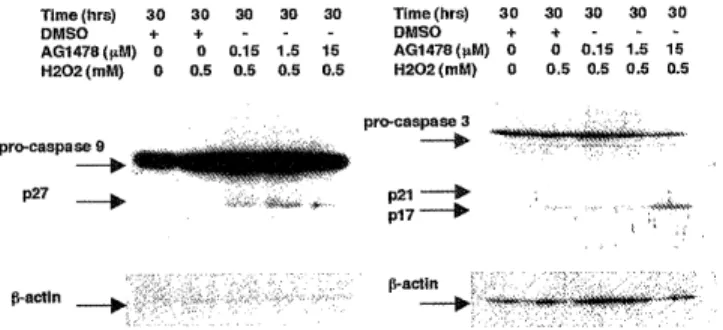
blotting to examine activation of caspase 9 and 3 in response to hydrogen peroxide. As shown in Figure 113, hydrogen peroxide - induced activation of caspase 9 and 3 was increased with Tyrphostin AG 1478 in a dose dependent manner. Recently several reports have suggested that mitochondrial membrane potential is correlated with mitochondrial injury","'. Using flow cytometry with DiOC6(3), we investigated the mitochondrial membrane potential in response to hydrogen peroxide in the absence or presence of Tyrphostin AG 1478. As shown in Figure IC, Tyrphostin AG 1478 significantly decreased the mitochondrial membrane potentials in a dose dependent manner.
P < 0.05
P < 0.05 20
P < 0.05 _r 'e 0 15
y O Q co 10 a
5
o n
0 H202 (0.5mM) (-) (-) (-) (-) (-) (-) (+) (+) (+) (+) (+) (+) DMSO (+) (-) (-) (-) (-) (-) (+) (+) (+) (+) (+) (+) AG1478 (µM) 0 0.15 0.5 1.5 5 15 0 0.15 0.5 1.5 5 15
Figure IA. Effect of Tyrphostin AG1478 on hydrogen perox- ide - induced apoptosis. Cells were treated with DMSO or with T yrphostin AG 14 7 8 at the indicated concentrations for 30 min prior to stimulating with hydrogen peroxide (0.5 mM) for 30 h under serum - free conditions (1% BSA). Cells were collected and analyzed by flow cytometry with propidium io- dide staining and the apoptosis ratio was investigated. All experiments were repeated at least three times with similar results.
AG1478 : 150 nM AG1478 : 15 µM
DMSO/PBS /PBS /PBS
3.1% P 3.9% 18.8%
m.i FL3 LOG 3888 m.l FLI LOG 1888 m,l FLI LOG 18 8
DMSO / H202 0.5 mM AG1478 /H202 150 nM AG1478 15 µM
0.5 mM /H202 0 .5 mM
11.0% 17.6% 24.0%
m.1 18 8
FLI LOG m_1 FLI LOG 1088 FLI LOG 1888
Time (hrs) 30 30 30 30 30 Time (hrs) 30 30 30 30 30
DMSO + + - - - DMSO + + - - -
AG1478(µM) 0 0 0.15 1.5 15 AG1478(µM) 0 0 0.15 1.5 15 H202 (mM) 0 0.5 0.5 0.5 0.5 H202 (mM) 0 0.5 0.5 0.5 0.5
Figure 1B. Effect of Tyrphostin AG1478 on the activation of caspase 9 and 3 by hydrogen peroxide. Quiescent cells were treated with DMSO or Tyrphostin AG1478 as indicated for 30 min prior to inducing apoptosis with hydrogen peroxide (0.5 mM) for 30 h under serum - free condition. Cell lysates (20 ug of protein) were used in Western blots with anti - caspase 9, and 3 antibodies and anti - l9 actin antibody. All experiments were repeated at least three times with similar results.
Figure 1C. Effect of Tyrphostin AG1478 on trans - mem- brane potential by hydrogen peroxide. Cells were treated with DMSO or Tyrphostin AG1478 at the indicated concen- trations for 30 min prior to stimulating with hydrogen per- oxide (0.5 mM) for 30 h under serum - free conditions (1%
BSA). Cells were collected and analyzed by flow cytometry with DiOCb(3) staining, and mitochondrial membrane poten- tial was investigated. Numbers indicate the percentage of gated cells displaying decreased mitochondrial membrane po- tential. All experiments were repeated at least three times with similar results.
Hydrogen peroxide activates EGFR, Akt / PKB, MAPK, and Bad in A431 cells.
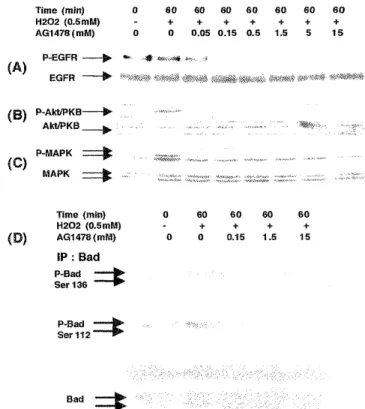
It has been reported that cytokine receptors, such as EGFR11-14) and PDGFR23' are activated by oxidative stress, as well as protein kinases activation, such as MAPK, P13 - kinase, and Akt / PKB. We investigated the activation and phosphorylation of EGFR, P13 - kinase, Akt / PKB, MAPK, and Bad (Ser - 112, Ser - 136) in response to hydrogen peroxide in A431 cells. As re- ported previously, activation of EGFR was observed in response to hydrogen peroxide and was sustained for more than 180 min (Figure 2A). As shown in Figure 2 B, hydrogen peroxide addition activated Akt / PKB and activation of Akt / PKB reached a plateau level by 60 min and was sustained for more than 180 min. Next, we investigated the time course of MAPK activation. As shown in Figure 2 C, hydrogen perox- ide led to MAPK activation, and this activation was also sustained for more than 180 min. Finally, we in- vestigated the time course of phosphorylation of Bad, Both the Ser - 112 and Ser - 136 residue of Bad was phosphorylated and this phosphorylation was sus- tained for more than 180 min (Figure 2D).
Time (min) 0 5 10 15 30 60 90 120 180 H202 (0.5 mM) - + + + + + + + +
P-EGFR ----i•
(~} EGFR
p-Akt/PKB~
B ) AkVPKB
(~) P-MAPK
MAPK
Time (min) 0 30 60 120 180
H202 (0.5mM) + + + + +
(E)IP:Bad
P-Bad Ser 136
P-Bad Ser 112
Bad
Figure 2. Effect of hydrogen peroxide on the activation of EGFR, P13 - kinase, Akt / PKB, MAPK, and phosphorylation of Bad (ser-136, ser-112) Quiescent cells were stimulated with hydrogen peroxide (0.5 mM) for various times as indicated.
Cell lysates (20 ug of protein) were prepared and analyzed by immunoblotting. (A) phosphorylated EGFR and EGFR;
(B) phospho - Akt / PKB and Akt / PKB; (C) phospho - MAPK and MAPK; (D) Lysates (200 ug of protein) were prepared and immunoprecipitated with anti - Bad monoclonal antibody, and the anti-Bad immunoprecipitates were analyzed by Western blots with anti-phosphorylated Bad (Ser-136), anti-phosphory- lated Bad (Ser-112) and anti - Bad antibody. All experiments were repeated at least three times with similar results.
Time (min) 0 60 60 60 60 60 60 60
H202 (0.5mM) - + + + + + + +
AG1478(mM) 0 0 0.05 0.15 0.5 1.5 5 i5
P-EGFR --p EGFR -~'
(B) P-Akt/PKB---® Ak t/PKB _~
P-MAPK ----ice (~) MAPK
(D)
Time (min) 0 60 60 60 60
H202 (0.5mM) - + + + +
AG1478 (mM) 0 0 0.15 1.5 15
IP : Bad P-Bad Ser 136
P-Bad Ser 112
Bad
Figure 3. Effect of Tyrphostin AG1478 on hydrogen perox- ide - induced activation of EGFR, P13 - kinase, Akt / PKB, MAPK, and phosphorylation of Bad (ser-136, ser-112).
Quiescent cells were treated with DMSO or Tyrphostin AG1478 at various concentrations as indicated for 30 min prior to stimulation with hydrogen peroxide (0.5 mM) for 60 min. Cell lysates (20 /i g) were prepared and analyzed by immunoblotting. (A) phosphorylated EGFR, and EGFR; (B) phospho - Akt / PKB, and Akt / PKB; (C) phospho - MAPK, and MAPK; (D) Lysates (200 fig of protein) were prepared and immunoprecipitated with anti - Bad monoclonal anti- body, and the anti - Bad immunoprecipitates were also ana- lyzed by Western blots with anti-phosphorylated Bad (Ser- 136), anti - phosphorylated Bad (Ser-112) and anti - Bad antibody. All experiments were repeated at least three times with similar results.
Tyrphostin AG1478 inhibited hydrogen peroxide induced phosphorylation of EGFR, Akt / PKB, MAPK, and Bad.
Using western blot analysis, we investigated the ef- fect of Tyrphostin AG 1478 on hydrogen peroxide- induced EGFR stimulation. Tyrphostin AG1478 inhib- ited EGFR activation by hydrogen peroxide in a dose dependent manner (Figure 3A). At the time of 60 min, a similar inhibitory effect of Tyrphostin AG1478 on the activation of Akt / PKB (Figure 3B) and MAPK (Figure 3C). Finally we investigated the effect of Tyrphostin AG 1478 on the phosphorylation of Bad at the time of 60 min. Phosphorylation of both serine resi- dues (Ser-136, and Ser-112) of Bad in response to hy- drogen peroxide was significantly prevented (Figure 3D).
Confirmation of a specificity of Tyrphostin AG1478 for EGFR.
Tyrphostin AG1478 is a member of the class of quinazoline inhibitors of the EGFR tyrosine kinases.
It blocks EGFR phosphorylation and Src kinase activ- ity in vivo but does not inhibit the PDGFR24-Z". To de- termine whether the observed inhibitory effects of Tyrphostin AG 1478 on the activation of downstream protein kinases were mediated through inhibition of EGFR, we investigated the effect of Tyrphostin AG1478 on the activation of MAPK and Akt / PKB in response to PDGF - BB. As reported previously, A431 cells lack endogenous PDGFR18'. Using the A431 / wt PDGFR beta cells in which wild type PDGFR beta
was introduced"', we stimulated A431 / wt PDGFR beta cells with PDGF BB with or without indicated concentrations of Tyrphostin AG 1478 (150 nM, 1.5 p M, 15 ;cM ), for 15 min. As shown in Figure 4A, ac- tivation of MAPK by PDGF - BB was slightly pre- vented at 15 M of Tyrphostin AG 1478, but even at 15 _M of Tyrphostin AG 1478, Akt / PKB activation was unaffected (Figure 4B).
DMSO (%) 0.15 0.15 0 0 0
AG1478([M) 0 0 0.15 1.5 15
PDGF-BB (ng/ml) 0 30 30 30 30
Time (min) 0 15 15 15 15
p-MAPK
(A) MAPK
p-AktIPKB ---I>
(B) Akt/PKB
---
Figure 4. Effect of Tyrphostin AG1478 on PDGF - BB stimu- lated MAPK activation in A431 /wt PDGF beta cells.
Quiescent A431/wt PDGF beta cells were treated for 30 min with DMSO or Tyrphostin AG1478 at various concentration as indicated prior to stimulation with PDGF BB (30 ng/ml) for 15 min. Cell lysates (20 p g of protein) were prepared and analyzed by immunoblotting. (A) phosphorylated MAPK, MAPK. (B) phosphorylated Akt/PKB, Akt/PKB. All experi- ments were repeated at least three times with similar results.
ABBREVIATION EGF : epidermal growth factor, EGFR : epi- dermal growth factor receptor, PDGF: platelet-derived growth factor, PDGFR : platelet-derived growth factor receptor, MAPK : mitogen-activated protein kinase, JNK : C-jun N terminal kinase, P38MAPK : P38 mitogen-Activated protein kinase, PI3K : phosphoinositide 3-kinase, Akt/PKB : Akt/protein kinase B, PKC : protein kinase C, PKB : protein kinase B, MEK : mitogen-activated protein kinase kinase
DISCUSSION
Oxidative stress elicits rapid, highly regulated adap- tive responses involving cordinate control of multiple signal transduction pathways. Previous studies sug- gested that early signal transduction events after oxi- dative stress appear to occur at or near the cell membrane"'. However, there are few studies that have directly addressed the consequences of the activation of cytokine receptors and their cordination of down- stream kinases in response to oxidative insults. The aim of the present study was to examine possible anti - apoptotic effects of cytokine receptors . We focused upon EGFR activation by hydrogen peroxide and used A431 cells, which have abundant EGFRs.
From our results, the following conclusions were ob- tained : 1) In A431 cells, Tyrphostin AG 1478 acceler- ated hydrogen peroxide - induced apoptosis, which was accompanied by activation of caspases 3 and 9 and decreased mitochondrial membrane potential ; 2) hydrogen peroxide induced sustained activation of EGFR, Akt / PKB, MAPK, and Bad (both Ser-112 and Ser-136 residue), and activation of EGFR, Akt / PKB, MAPK, and Bad with hydrogen peroxide were inhib- ited by Tyrphostin AG1478 in a dose dependent man- ner, 3) The effect of Tyrphostin AG1478 on the EGFR activation was specific, since there were no effects upon PDGFR-mediated activation of MAPK, Akt / PKB.
Yang E. et al demonstrated that Bad ( Ser - 136 ) is one of the targets of Akt / PKB activation; in addi- tion, Bad (Ser - 112) is a target of P90 RSK which is thought to be located downstream of PKC29-34' and MAPK35-36' Therefore the results of the present studies suggest that one mechanism of accelerated hydrogen peroxide-induced apoptosis may be the result of accel- erating caspase activation by mitochondrial injury due to prevention of Bad phosphorylation.
It is known that administration of a MEK inhibitor, PD98059 or a P13 - kinase inhibitor, Wortmannin will accelerate oxidative stress- induced apoptosis3' h' These previous studies suggest that upstream kinases, such as MEK or P13 - kinase partially regulates anti- apoptotic downstream protein kinases, such as MAPK or Akt / PKB in response to oxidative stress. On the other hands, phosphorylation of these protein kinases and cytokine receptors has been suggested to be due to inactivation of phosphatases by oxidative stress37.
It was unclear whether EGFR cordinate these down- stream kinses in the previous studies with Tyrphostinn AG 1478. However, it is still unclear whether the cytokine receptors systemically regulate anti - apoptotic protein kinases, such as MAPK or P13 - kinase. In re- gard to the signaling system, there are two different scenarios concerning the activation of protein kinases in response to oxidative stresses. The first scenario is that once cytokine receptors are activated in response to oxidative stress and regulate their downstream pro- tein kinases systemically, which protect the cells from apoptosis. The second scenario is that in spite of par- tial regulation of protein kinases by upstream kinases, each protein kinase and each receptor is activated at random due to inactivation of specific phosphatases.
As expected, Tyrphostin AG 1478 inhibited the activa- tion of MAPK, P13 - kinase, and Akt / PKB, as well as EGFR activation. Tyrphostin AG1478 also inhibited the phosphorylation of both serine residues of Bad in re- sponse to hydrogen peroxide. Therefore, our results
Toshiaki Tsukada et al : Anti-apoptotic Effect in EGFR Signaling
support the first scenario and suggest that EGFR regulates downstream protein kinases systemically and prevents apoptosis by phosphorylation of Bad.
Tyrphostin AG1478 has been reported to be highly selective as an EGFR blocker2^-z". In the present study, we examined whether Tyrphostin AG1478 would inhibit the activation of protein kinases in re- sponse to a different, structurally dissimilar receptor tyrosine kinase. We utilized A431 / wt PDGFR beta cells, in which wild type PDGFR beta was introduced"'.
Activation of Akt / PKB by PDGF - BB was not af- fected by Tyrphostin AG 1478, and activation of MAPK, by PDGF - BB was only slightly affected by Tyrphostin AG1478. From these results, we conclude that Tyrphostin AG1478 does not have any non - spe- cific inhibitory effect for Akt / PKB activation but cannot rule out that it may have some minor, non - specific inhibitory effect for MAPK activation.
Therefore, in conclusion, we demonstrate that EGFR is involved in, and regulates cell survival signaling pathways, such as MAPK and Akt / PKB and its downstream target, Bad in response to oxidative in- sults. These results suggest potential anti - apoptotic effects of cytokine receptors by regulating down- stream kinases in response to hydrogen peroxide.
REFFERENCES
1) Qin S, Chock B. : Implication of phosphatidylinositol 3-kinase membrane recruitment in hydrogen peroxide-induced activation of
P13K and Akt. Biochemistry 42 :2995-3003, 2003
2) Jeon BW, Kim KT, Chang SI, Kim HY. : Phosphoinositide 3-OH kinase/protein kinase B inhibits apoptotic cell death induced by
reactive oxigen spicies in Saccharomyces cerevisiae. J Biochem
131: 693-699, 2003
3) Wan YS, Wang ZO, Shao Y, Voorhees JJ, Fisher GJ. : Ultraviolet irradiation activates PI 3-kinase/Akt surrvival pathway via EGF
receptors in human skin in vivo. Int J Oncol 18 : 461-466, 2001 4) Wang X, Martindale JL, and Liu Y, Holbrook NJ.: The cellular re-
sponse to oxidative stress: influences of mitogen-activated protein
kinase signalling pathways on cell survival. Biochem J 333 : 291-
300, 1998
5) Crossthwaite AJ, Hassan S, Williams RJ. : Hydrogen peroxide- mediated phosphorylation of ERK1/2, Akt/PKB and JNK in corti-
cal neurones: dependemce on Ca(2+) and P13-kinase. J Neurochem
80 : 24-35, 2002
6) Sano M, Fukuda K, Sato T, et al. : ERK and p38 MAPK, but not NF-kappaB are critically involved in reactive oxigen species-
mediated induction of IL-6 by angiotensin II in cardiac fibroblasts.
Circ Res 89 : 661-669, 2001
7) Tamagno E, Robino G, Obbili A, et al.: H202 and 4-hydroxynoneal
mediate amyloid beta-induced neuronal apoptosis by activating JNKs and p38MAPK. Exp Neurol 180 : 144-155, 2003
8) Wang JY, Shum AY, Ho YJ, Wang JY. : Oxidative neurotoxicity
in rat cerebral cortex neeurons: syergistic effects of H202 and NO on apoptosis involving activation off p38 mitogen-activated pro-
tein kinase and caspase-3. J Neurosci Res 72 : 508-519, 2003 9) Inoshita S, Takeda K, Terada Y, et al. : Phosphorylation and inac-
tivation of myeloid cell leukemia 1 by JNK in response to oxida-
tive stress. J Biol Chem 277 : 4370-4374, 2002
10) Chen YR, Shrivastava A, Tan TH : Down-regulation of the c-Jun
N-terminal kinase (JNK) M3/6 and activation of JNK by hydrogen peroxide and pyrrolidine dithiocarbamate. Oncogene 20 : 367-374,
2001
11) Meves A, Stock SN, Beyerle A, Pittelkow MR, Peus D.: H(2)O(2) mediates oxidative stress-induced epidermal growth factor recep-
tor phosphorylation. Toxicol Lett 122 : 205-214, 2001
12) Chen K, Vita JA, Berk BC, Keaney JF Jr. : c-Jun N-terminal kinase activation by hydrogen peroxide in endothelial cells involves
SRC-dependent epidermal growth factor receptor transactivation.
J Biol Chem 276 : 16045-16050, 2001
13) Takayama K, Dabbagh K, Jeong Shim J, et al. : Oxidative stress causes mucin synthesis via transactivation of epidermal growth
factor receptor: role of neutrophils. J Immunol 164 : 1546-1552,
2000
14) Peus D, Meves A, Beyerle A, et al. : H202 is required for UVB- induced EGF receptor and downstream signaling pathway activa-
tion. Free Radic Biol Med 11-12 : 1197-1202, 1999
15) Rosell R, Fossella F, Milas L : Molecular markers and targeted therapy with novel agents: prospects in the treatment of non-
small cell lung cancer. Lung Cancer 38 : 43-49, 2002
16) Grunwald V, Hidalgo M : Development of the epidermal growth factor receptor inhibitor OSI-774. Semin Oncol 30 : 23-31, 2003 17) Schiller JH : New directions for ZD 1839 in the treatment of solid
tumors. Semin Oncol 30 : 49-55, 2003
18) Assefa Z, Valius M, Vantus T, Agostinis P, Merlevede W, Vandenheede JR.: JNK/SAPK activation by platelet-derived growth
factor in A431 cells requires both the phospholipase C-gamma
and the phosphatidylinositol 3-kinase signaling pathways of the
receptor. Biochem Biophys Res Commun 261 : 641-645, 1999 19) Banki K., Hutter E., Gonchoroff NJ, Perl A. : Elevation of
mitochondrial transmembrane potential and reactive oxigen inter-
mediate levels are early events and occur independently from ac-
tivation of caspases in Fas signaling. J Immunol 162 : 1466-1479,
1999
20) Hirsch T, Dallaporta B, Zamzami N, et al. : Proteasome activation occurs at an early, premitochondrial step of thymocyte apoptosis.
J Immunol 161: 35-40, 1998
21) Darzynkiewicz Z, Juan G, Li X, et al. : Cytometry in cell neecrobiology: analysis of apoptosis and accidental cell death (ne-
crosis). Cytometry 27 : 1-20, 1997
22) Yuan J, Murrell GA, Trickett A, Wang 1\4X. : Involvement of cytochrome c release and caspase-3 activation in the oxidative
stress induced apoptosis in human tendon fibroblasts. Biochim
Biophys Acta 1641 : 35-41, 2003
23) Iantomasi T, Favilli F, Catarzi S, Vincenzini MT. : GSH role on platelet-derived growth factor receptor tyrosine phosphorylation
induced by H202. Biochem Biophys Res Commun 280: 1279-1285, 2001
24) Arteaga CL, Ramsey TT, Shawver LK, Guyer CA. : Unliganded epidermal growth factor receptor dimerization induced by direct
interaction of quinazolines with the ATP binding site. J Biol Chem
272 : 22347-22354, 1997
25) Levitzki A, Gazit A. : Tyrosine kinase inhibition: an approach to drug development. Science 267 : 1782-1788, 1995
26) Ossherov N, Levitzki A. : Epidermal-growth-factor-dependent acti- vation of src-family kinases. Eur J Biochem 225 : 1047-1053, 1994 27) Fry DW, Kraker AJ, McMichael A, et al. : A specific inhibitor of epidermal growth factor receptor tyrosine kinase. Science 265
1093-1095, 1994
28) Sachsenmaier C, Radler-Pohl A, Zinck R, et al. : Involvement of growth factor receptors in the mammalian UVC response. Cell 78 : 963-972, 1994
29) Pastorino JG., Tafani M, Farber JL. : Tumor necrosis factor in- duces phosphorylation and translocation of Bad thhrough a
phosphatidylinositide-3-OH kinase-dependent pathway. J Biol Chem
274 : 19411-19416, 1999
30) Bai H, Pollmann MJ, Inishi Y, Gibbons GH. : Regulation of vascu- lar smooth muscle cell apoptosis. Modulation of bad by a
phosphatidylinositol 3-kinase-dependent pathway. Circulation Res
85 : 229-237, 1999
31) Zundel W, Galcia A. : Inhibition of the anti-apoptotic PI(3)K/Akt/
Bad pathway by stress. Genes Develop : 12, 1941-1946, 1998 32) Perez OD, Kinoshita S, hitoshi Y, et al. : Activation of the
PKB/AKT pathway by ICAM-2. Immunity 16 : 51-65, 2002 33) Scheid M, Duronio V. : Dissociation of cytokine-induced
phosphorylation of Bad and activation of PKB/akt: involvement
of MEK upstream of Bad phosphorylation. Proc Natl Acad Sci USA 89 : 9121-9125, 1998
34) Yang E, Zha J, Jockel J, et al. : Bad, a heterodimeric partner for Bcl-xL and Bcl-2, displaces Bax and promote cell death. Cell 80 285-291, 1995
35) Tan Y, Ruan H, Demeter MR, Comb MJ. : p90RSK blocks Bad- mediated cell death via a protein kinase C-dependent pathway. J
Biol Chem : 274, 34859-34867, 1999
36) Jin K, Mao X, Zhu Y, Greenberg DA. : MEK and ERK protect hypoxic cortical neurons via phosphorylation of Bad. J Neurochem
80 : 119-125, 2002
37) Kmata H, Shibukawa Y, Oka SI, Hirata H. : Epidermal growth fac- tor receptor is modulated by redox through multiple mechanisms.
Effects of reductants and H202. Eur J Biochem 267 : 1933-1944, 2000