September 2018
AUNG KO KO OO
Graduate School of Natural Science and Technology (Doctoral Course)
OKAYAMA UNIVERSITY
A dissertation submitted by AUNG KO KO OO in partial fulfilment of the requirements for the Doctor of Philosophy in Engineering in the Graduate
School of Natural Science and Technology, Okayama University, Japan.
September 2018
List of Figures iii
List of Tables iv
PREFACE v
CHAPTER (1) GENERAL INTRODUCTION 1
1.1. CANCER STEM CELLS 3
1.2. iPSC-CSC 7
1.3. CSC AND EPIGENETICS 11
REFERENCES 16
CHAPTER (2) 23
“iPSC DERIVED CSC MODEL WITH LUNG METASTASIS DEVELOPED IN THE MICROENVIRONMENT OF LUNG CARCINOMA”
ABSTRACT 25
2.1. INTRODUCTION 26
2.2. RESULTS 28
2.2.1. Conversion of miPSCs into CSC-like cells with 28 tumorigenic and metastatic potential
2.2.2. Characterization of miPS-LLCcm, Ptdc and LMN cells 34 2.2.3. In vivo tumorigenic differentiation of miPS-LLCcm 35
2.3. MATERIALS AND METHODS 37
2.3.1. Cell Culture 37
2.3.2. Conversion of miPSCs into the CSC-like cells 37
2.3.3. Sphere Formation Assay 38
2.3.4. Animal experiments 38
2.3.5. Preparation of primary cell culture 38 2.3.6. RNA extraction, cDNA synthesis and qPCR analysis 39 2.3.7. Histological analysis and immunohistochemistry (IHC) 40
2.3.8. Statistical analysis 41
2.4. DISCUSSION 41
2.5. CONCLUSIONS 44
CHAPTER (3) 48
“Up-Regulation of PI 3-Kinases and the Activation of PI3K-Akt Signaling Pathway in Cancer Stem-Like Cells Through DNA Hypomethylation Mediated by the Cancer Microenvironment”
ABSTRACT 50
3.1. INTRODUCTION 51
3.2. RESULTS 52
3.2.1. Global DNA methylation analysis in Cancer Stem-like 52 Cells
3.2.2. Analysis of the differentially methylated regions (DMRs) 54 in the cancer stem cell model converted from iPSCs
3.2.3. Overexpression of PI3K-Gamma Candidates in the 58 Model were Relating to Oncogenic Potential
3.2.4. PI3K-Akt Activation Drives iPSC-CSCs Model 59
3.3. MATERIALS AND METHODS 60
3.3.1. RRBS DNA methylation analysis 60
3.3.2. KEGG Pathway Enrichment 62
3.3.3. Western Blotting 62
3.3.4. RNA extraction, cDNA synthesis and qPCR mRNA expression 63 analysis
3.3.4. Statistical Analysis 64
3.4. DISCUSSION 64
3.5. CONCLUSION 66
REFERENCES 67
APPENDIX (Figure) 72
APPENDIX (Table) 78
ACKNOWLEDGEMENT 83
LIST OF PUBLICATION 85
Figure 1.1 The hypothesis of miPSCs conversion into iPS-CSCs 9 Figure 1.2 Key developmental events with global epigenetic 13
modifications and gene-expression patterns mammalian cells.
Figure 2.1. Conversion of miPSCs into miPS-LLCcm 29 Figure 2.2. Tumorigenic and metastatic potential of miPS-LLCcm 30
Figure 2.3. Primary cultured cells 31
Figure 2.4. Comparison of the expression levels of stemness markers 32 by rt-qPCR
Figure 2.5. Histological analyses of allografts of miPS-LLCcm cells 33 Figure 2.6. The localization and expression of CSC markers 34 Figure 2.7. The localization and expression of EMT markers 35
in miPS-LLCcm derived tumors.
Figure 3.1. DMR in each chromosome and methylation pattern 55 changes (Hyper and Hypomethylated DMRs)
Figure 3.2. Relative DNA methylation levels of DMRs 56 Figure 3.3. KEGG pathways nominated with the number 57
of hypomethylated genes
Figure 3.4. Expression of hypomethylated genes significant in 59 KEGG-pathway related to cancer
Figure 3.5. Evaluation of the candidate signaling pathway 60 nominated by the KEGG analysis.
Figure S1. Cumulative distribution of effective sequencing 74 depth of cytosine
Figure S2. The proportion of mCG, mCHG, and mCHH 75 Figure S3. Methylation level distribution in CGI of four 76
different stages of CSC cells
Figure S4. Trace data of codons in Pik3ca and Pten cDNA from 77 miPSCs, miPS-LLCcm cells and Ptdc cells and LMN cells
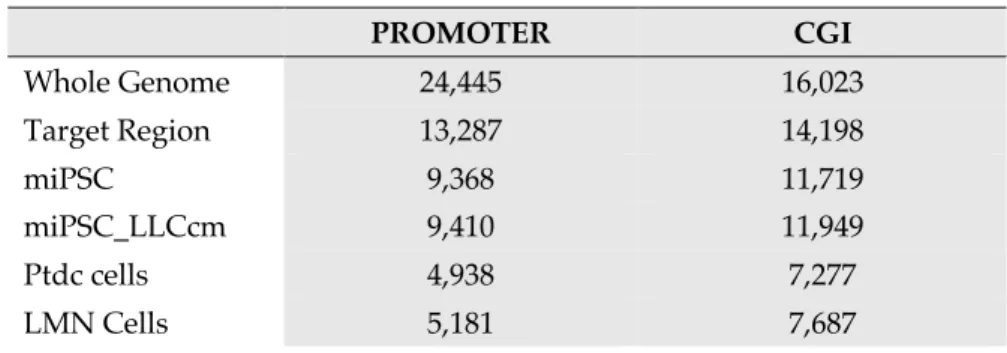
Table 2.1. List of Primers Used in the Experiments 40 Table 3.1. Covered Promoter and CGI number of target regions 53 Table 3.2. Number and length of DMRs in each comparison 56
with distribution in each chromosome
Table 3.3. List of Primers Used in the Experiments 63
Table S1. Data Summary and QC 80
Table S2. Alignment statistics with reference genome 80
Table S3. Proportion of mCG, mCHG and mCHH 80
Table.S4. Average methylation level of mC 80
Table S5. KEGG pathway analysis on hypomethylated 81 DMR-associated genes (miPSCs Vs miPS-LLCcm cells)
Table S6. KEGG pathway analysis on hypomethylated 81 DMR-associated genes (miPSCs Vs Ptdc cells)
Table S7. KEGG pathway analysis on hypomethylated 82 DMR-associated genes (miPSCs Vs LMN cells)
We have been investigating the new insight in the cancer stem cells (CSCs) by developing a CSC model that is derived from induced pluripotent stem cells (iPSCs). The evidence of CSCs was widely accepted as small percentage of cell population in tumor that have a self-renewal capability and are malignant. Microenvironment is crucial to regulate the proliferation, self-renewal ability and differentiation of normal stem cells. By extending this concept, microenvironment distorted by cancer cells could affect the diverse directions of stem cells and leading to the characteristics of CSCs. Even though CSC shared with normal stem cells in the characteristics of maintaining the stemness and differentiation potential, multiple genetic and epigenetic regulations are different to acquire the features of CSCs. Epigenetic mechanisms, such as DNA methylation, histone modification and non- coding RNA elements, are involved in stem-cell maintenance and in the regulation of differentiation of stem cells. On the other hand, the epigenetic alterations are relating to the tumorigenesis with the activation of oncogenes and silencing of tumor suppressor genes.
We have succeeded in converting mouse iPSCs (miPSCs) into CSC-Like cells (miPS- LLCcm) by treating the miPSCs with conditioned medium (CM) of Lewis Lung Carcinoma (LLC) cells. miPS-LLCcm cells developed highly angiogenic and malignant adenocarcinoma as well as lung metastasis when subcutaneously transplanted into nude mice. By the treatment, miPSCs obtained the ability of unlimited growth and the capacity to maintain their stemness, while they were allowed to differentiate without LIF. The subcutaneous transplantation of the survived cells into the mouse formed malignant tumors and metastasized into lung tissues. Thus, we concluded that miPSCs should be converted into CSCs without any intended genetic manipulation. Immunohistochemical
analysis revealed the heterogeneity of the tumors, in which miPS-LLCcm cells should on one hand maintain the undifferentiated population expressing GFP and differentiate on the other into adenocarcinoma phenotype expressing MUC1. The cells from tumors at primary site and metastatic nodules can be maintained in vitro.
In order to further confirm the acquisition of CSC-like phenotype, we characterized the miPS-LLCcm cells and its derived cells form tumor (Ptdc) and from lung metastatic nodules (LMN) with commonly known CSC markers. We evaluated the significantly higher expression of ALHD1 and CD44 in the Ptdc and LMN cells. Epithelial to mesenchymal transition (EMT) is believed to be the primary mechanisms in the transition of cellular stages during development, wound healing and cancer metastasis. The upregulated expression of EMT markers in Ptdc cells was suggesting the potential of partial and metastable epithelial-to-mesenchymal transition (EMT) phenotype in Ptdc cells.
Since this conversion is triggered only by the factor(s) contained in the conditioned medium, we have hypothesized that epigenetic alterations can induce CSCs from normal stem cells in the cancer microenvironment. Methylation and demethylation is generally considered to silence and activate gene expression, respectively. We tried to evaluate the epigenetic changes in early stages of cancer development in this research that has not yet been assessed. We traced the development of CSCs by the change of DNA methylation levels, which would provide the difference between the miPSCs and derived CSCs. Then, we compared the methylation in miPS-LLCcm, LMN and Ptdc cells with that in miPSC by three sets; (1) miPSCs vs miPS-LLCcm cells, (2) miPSCs vs Ptdc cells and (3) miPSCs vs LMN cells. All comparisons between the different cell populations were found to exhibit hypomethylation as compared to miPSCs and 926, 583 and 1105 differentially
methylated regions (DMRs) were identified respectively from the comparisons. DMRs- associated genes were further identified and segregated into hypo- and hyper-methylated genes categories. As the results, hypomethylation was found superior to hypermethylation in the CSCs.
The analysis of KEGG pathways relating to hypomethylated genes revealed the several notable pathways important in cancers. Validating certain pathways in the CSCs, that are corresponding to DMR-related genes showed the pathways relevant for carcinogenesis including Focal adhesion, PI3K-Akt signaling pathway, Calcium signaling pathway, Pathway in cancer and Transcriptional misregulation in cancer. Checking the upregulated expression of the genes included in the enriched pathways that are concordant with hypomethylation showed the trace of candidates relating to oncogenic potential of CSCs. The expression of hypomethylated genes relating to PI3K-Akt pathway was found significantly high among those of the other genes. We found Pik3r5, which is a regulatory subunit of Pik3cg enzyme, as a hypomethylated and highly up-regulated gene relating to PI3K-Akt pathway. In the recent reports, the PIK3CG and PIK3R5 were evaluated as a potential oncogene that are overexpressed in human cancers leading to oncogenic cellular transformation and malignancy. Overexpression of PI3K-Gamma candidates were relating to oncogenic potential of the CSC model. Together with the findings, the constitutive expression of Pik3r5 was detected by immunoblotting with anti-p101 antibody in miPS- LLCcm and its derivatives.
Activation of PI3K-Akt signaling pathway has been commonly reported as key driver of carcinogenesis. The upregulated expression of Pik3r5 should induce the activation of Akt resulting in the onset of tumorigenic and metastatic potential of miPS- LLCcm and Ptdc cells. We assessed the Akt activation with anti-phosphorylated Akt (p-
Akt) antibody and we found Akt was constitutively activated in miPS-LLCcm, Ptdc and LMN cells. Therefore, the hypomethylation of Pik3r5 gene was leading to the up- regulation and is closely related to the activation/phosphorylation of AKT that is the downstream target molecule.
Significant overall DNA hypomethylation during the conversion activated the certain proto-oncogene, with the activation of PI3K-Akt signaling pathway, which represent the malignant conversion even without mutations. In our study, we have successfully demonstrated the CSCs generated from iPSCs by the treatment with CM from cancer derived cells acquired the DNA hypomethylation that might be considered to be the new aspect in the early stage of CSCs.
CHAPTER (1)
GENERAL INTRODUCTION
1.1. Cancer Stem Cells
Cancer has been defined as an abnormal growth of cells with potential character of invasion or spreading into other parts of body [1].
These abnormal cells have the ability to proliferate rapidly, make the malignant tumors and invade the native tissue by producing enzymes. By contrast, benign tumors don't generally invade and usually push the normal tissue to the side [2,3]. There are over 100 types of cancer that can affect in human and their symptoms vary depending on the types of tissue, such as breast, skin, lung, colon, prostate, and lymphoma [1,4].
Research into cancers, nowadays, has been changed enormously with new and advance technologies. In the area of cancer research, there is still questioning how the research projects have been done and how the effectiveness of therapies come from the cancer research in the past. To get the fundamental progress in treating cancers, the cancer researchers are currently focusing into the advanced and more understanding of two areas; (a) the genetic underpinning of cancers and (b) the biology of cancers.
Progressive research outcomes developed many types of cancer treatments which depend on the type of cancer and how advanced it is. Conventional therapy is widely accepted in cancer treatment and is different from alternative or complementary therapies [5,6]. Examples of conventional treatment for cancers include chemotherapy, radiation therapy, and surgery. With the new findings in the area of cancer research, the cancer treatment has also been improved dramatically, such as the adjuvant chemotherapy, hormonal therapy, immunotherapy, and target therapy.
Recent chemotherapeutic treatment can give the promising results in treating the different types of malignancies by combining with surgical removal or radiotherapy. After decades of making the drugs to kill proliferating tumor cells, the notable progresses in cancer treatment was resulted [7,8]. However, cancer researchers are facing the divergent nature of cancers such as, chemo-resistance, turning into more tumorigenic form, metastasis and cancer relapse. One of the reason is the heterogeneity of cancer where certain population of cancer cells have ability to survive against several cancer treatments. The chemotherapeutic agents can remove and kill the rapidly dividing cells of the bulk tumor but often miss the certain population of cells with distinct morphological and functional profile [9]. These issues could be explained by the presence of stem cell population in the tumor as subgroup of cancer cells. Previous findings strongly suggested that these stem cells are responsible for chemoresistance and caner relapse [10]. The remaining stem cells are able to comprise the whole tumor even in a few number left. These population was broadly named as cancer stem cells (CSCs) and thought to be involved in driving the cancer [11].
Cancer stem cell is normally identified as a cell within a tumor that possess the ability to self-renew and to cause the heterogenous lineages of tumor cells. There are many hypothesizes of CSC with their different points of understanding. The hypothesis has to be tested whether what evidence is consistence with it or not and cancer researchers refine the way of treating the cancers according their assessment.
The cancer research over several decades has been led by the mutation theory in that any cell in body could become cancer when it acquires a package of mutation [12,13]. Because of single and/or series of mutation, single gene or some group of genes, called oncogenes, are activated and some genes, called suppressor genes, are inactivated in cancer. In some cases, the combination of these two events was also involved in the carcinogenesis. Therefore, oncologists defined cancers as some kind of genetic mistake. Form this concern, all types of cells in the multicellular organisms could become cancer with certain reasons [14].
Another concern of cancers was evaluated with the evidence of cancer stem cells in the tumor. In this case, only a certain population of cells that have the stem cell properties are really prone to becoming malignant [8]. The hypothesis about the cancer stem cells is in an ongoing debate. There is still needed to answer whether CSCs represent a mature tissue stem cell which has undergone malignant change or whether differentiated cells dedifferentiated into stem cell program together with the malignancy [15]. Until now, cancer stem cells are purely defined only with their capacities of self-renewing and differentiation, considering independently from the concept of origin of cancers [7,16].
Cancers themselves organized through in very similar way to a normal organ, but the cells are proliferating abnormally [17]. Normal organ is organized in a hierarchy where at the apex of the hierarchy it is the stem cell and give rise to the other cells which form the bulk of an organ. In the
tumor, it is generally thought that cancer stem is the master cell that gives rise to heterogeneity of cancers [7].
Tumor initiating capacity of CSCs in tumor was shown by serial transplantation of limited tumor cells into immunocompromised animal models where undifferentiated cancer cells show more than the differentiated cancer cells [8,18]. Researchers had been trying to evaluate the markers to identify and isolate the population of stem cells from tumor cells. Even though the validity of CSC markers depends on different types of tissues, and/or modes of tumorigenesis, it is possible to address the population of CSC in the tumors with CSC markers detected in specific malignancies. Currently, CSCs were identified in human brain, breast, prostate, head and neck, pancreas, liver, ovary, and colon cancers, by using different markers, such as CD133, EpCAM, CD44, CD24, Lgr5 and ALDH1 [19]. With many questions about CSCs, the nature and characteristics of CSCs become interested and therapeutic CSC-targeting approaches popular in past decade of cancer research. Experimental limitation for the researchers is that CSCs are very small percentage of tumor cells, and the culturing and maintenance of these cells are still difficult [9]. Many researchers evaluated the properties and characteristics of CSCs by using the model of CSCs that are selected by the help of CSC-markers. The evaluations about CSCs are still depending on the validity of markers on these cancer types [19,20].
In conclusion, cancer stem cell is defined as a certain type oncogenic cell that have self-renewing and differentiation ability. Their self-renewing
properties drives the generation of more CSCs, and differentiation capacity generate the bulk of tumors. The metastasis and tumor relapse are also related with the properties of CSCs. Due to the properties of CSCs, it is very important to provide the therapy that eradicated CSCs completely.
1.2. iPSC-CSC
The tumor microenvironment (TME) is the cellular environment surrounding the tumor in which stromal fibroblasts, immune cells, macrophages, endothelial cells, leucocytes, and extracellular matrix exist and interact by releasing signaling molecules to promote tumor growth and metastasis. It has been reported the significant role of TME in disease progression like as cancer, but the clear-cut role has not been understood.
A major concept of TME is that cancer cells interact closely with the surrounding cells, which together form the major construct of the TME leading into more complexity of cancer biology. In the case immune cells, the factors secreted in TME drive a chronic inflammatory, immunosuppressive, and pro-angiogenic environment. By getting adaptation in such environment, cancer cells are able to avoid the host immunosuppressive action and to get the new properties of metastasis and angiogenesis. The aberrant pathological process in tumor microenvironment can be related with divergent nature of cancers by changing the genetic or epigenetic regulation. Although it is a transient effect, it could activate the certain signaling pathways regulating cellular
proliferation and migration or in the case of stem cells it could be related with cell fate determination and differentiation.
Stem cells are found in certain population in multicellular organisms with their special characteristics different from others in the body. Stem cells are thought to be in blank state that can develop into cells with different specialized functions in different parts of the body during early life and growth. Stem cells have been termed as undifferentiated and self- renewing cells that can divide and make the unrestricted numbers of copies of themselves. Difference between stem cells and any other cells in the tissues is that when a stem cell divides, one remains by self-renewing as stem cell at exactly the same stage of differentiation and the other turns into a next stage of the differentiation down to a differentiated cell, such as a muscle cell or red blood cell, etc. [16,21]. Generally, type of stem cells can be divided into three types; (a) Embryonic stem cells (ESC) (b) Adult stem cells (c) Cancer stem cells. Embryonic stem cells are pluripotent, meaning they can develop into more than 200 cell types of the adult body and also have the ability to replicate indefinitely [22]. Adult stem cells found in adults can produce only a limited number of cell types. Cancer stem cells (CSCs) are tumor cells that have the principal properties of self-renewal, clonal tumor initiation capacity and clonal long-term repopulation potential [23].
Like the normal stem cells, CSCs are believed to reside in their own niches [24]. The niches for normal stem cells are crucial for proper differentiation of stem cells into certain progenitor cells and differentiated
cells. In contrast, the TME could affect on the differentiation of stem cells.
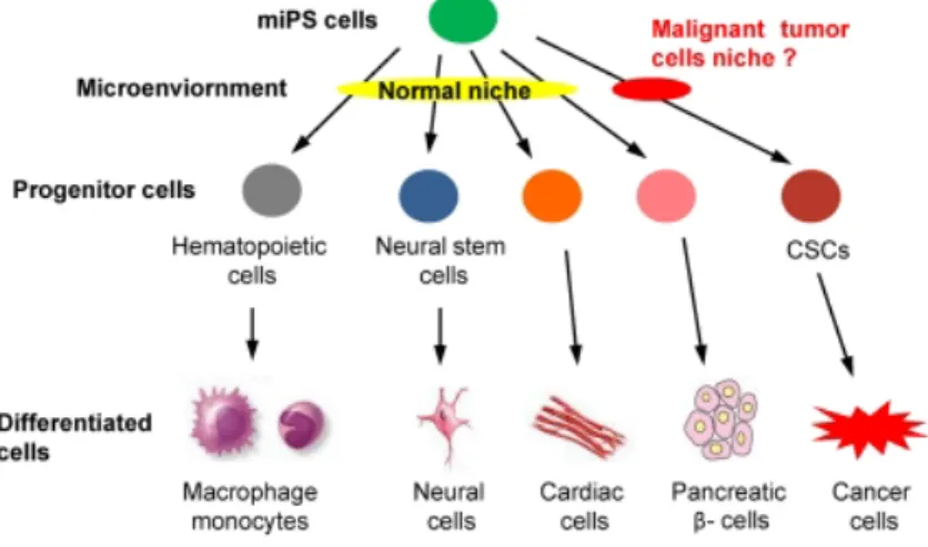
Seno et.al explained the fate of stem cells distorted by TME using the conditioned medium of Lewis Lung Carcinoma (LLC) [24,25]. In their hypothesis, mouse induced pluripotent stem cells (miPSCs) should be induced to some kinds of progenitor cells, such as hematopoietic cells and neural stem cells, differentiating into various phenotypes, such as macrophage, monocytes, neural cells, cardiac cells and pancreatic b-cells, when they were exposed to the normal niche. On the other hand, they hypothesized that CSCs may also be derived from miPS cells only when exposure to a malignant niche [Figure 1.1].
Figure 1.1 The hypothesis of miPSCs conversion into iPS-CSCs. CSCs
are considered derived from normal stem cells affected by the microenvironment being influenced by cancer cells. [25]
They evaluated the new insight in the CSCs developed in cancer microenvironment, by extending the concept that the niche of the normal
cells is involved in the differentiation of stem cells into normal tissues.
While the mouse induced pluripotent stem cells (miPSCs) were allowed to differentiate in the presence of cancer conditioned medium, they acquired the ability to maintain their stemness and differentiation potential, as well as formed malignant tumors with the features of adenocarcinoma and metastasized into lung tissues in vivo, considering the sign of CSCs. These models of cancer stem cell will provide the great advantages in cancer research and its applications in the future.
In iPS-CSC model, miPSCs were converted into CSCs without any intended genetic manipulation. The conversion could be considered through the transcriptional or translational changes determined by genetic or epigenetic alterations that are promoting tumorigenesis. Furthermore, they successfully generated various types CSC models converted from iPSCs with the aid of CM of various cancer cell lines such as human pancreatic carcinoma cell lines PK-8 cells and KLM-1 cells, human breast cancer cell lines T47D cells and BT549 cells, mouse carcinoma cell lines LLC cells, P19 cells, B16 cells and MC.E12 cells [14–16]. Anna et.al evaluated organ specific feature of xenografts tumors developed from iPS-CSC that were converted with conditioned medium of PDAC cells [26]. Furthermore, the story and origin of cancer associated fibroblast was explained with iPS- CSC model developed in the conditioned medium of breast cancer cells [27]. The iPS-CSC have differentiated into the fibroblast that support the tumorigenesis and metastasis of iPS-CSC. Interestingly, iPS-CSC model can also differentiate into the progenies of CSCs containing vascular endothelium [24,28].
In conclusion, the iPS-CSC is a model of CSCs that can explain biology and characteristics of CSC in nature. In addition, CSC generation from various kinds of cancer cells could be a source that provide a library of CSCs for customized cancer treatment. iPS-CSC model could be used in the evaluation of bona fide CSC markers and also the screening of chemotherapeutic drugs that are targeting the CSC population, to get better therapeutic approach of cancer.
1.3. CSC and Epigenetics
Carcinogenesis has been explained by the classical cancer initiation theory; evolutional accumulation of one or more mutations in a single or a few cells resulting in uncontrolled growth [29]. Genetic mutation was considered as the major causes of neoplasia [30]. However, it is now accepting the involvement epigenetic regulatory mechanisms in carcinogenesis. Genetic mutation is responsible for activation of tumor driver genes and silencing of tumor suppressors. In the second part, disruption of epigenetic regulation is leading to overexpression of oncogenes and downregulation of the tumor suppressor genes [31,32]. The genetic and epigenetic regulation were viewed as sole reason of abnormal gene expression.
The regulation performed by epigenetic mechanisms includes histone modification, chromatin remodeling factors, DNA methylation, microRNAs and post–translational modifications. The expression of
certain gene was determined by the status of DNA packaging at the regulation regions. These regions are normally the promoters and/or enhancers and insulators in chromatin. Some regulations were performed by the presence of transcription factors and chromatin modifying enzymes [33].
Without changing in DNA sequence, the ability to change the expression of genes is the primary role of epigenetic regulation. The epigenetic abnormalities involved in the tumorigenesis is not simple as the gain or loss of genes expression because the types of regulation will be different and complex depending on the stage and nature of carcinogenesis.
During the tumor initiation and progression, DNA hypomethylation pattern was occurred in the cancer associated genes when comparing with normal tissues. Previously, the cancer-specific DNA methylation patterns was reported by comparing with associated tissues. The development of technologies in sequencing and microarray analysis are very supportive to examine and understand the aberrant epigenetic regulation specific to cancer.
During differentiation, ESCs start in a pluripotent state from which they sequentially develop into unique cell type with a narrower pluripotency. Histone methylation is short-term silencing mechanism by which some set of genes required for development were repressed at the stage of stem cell [34,35]. On the other hand, DNA methylation are long- term silencing mechanisms. Pluripotent stem cells express the set of transcription factors that are important for maintenance of stemness and
after development these genes are repressed by long-term silencing mechanisms. Therefore, in somatic cells the imprinted genes and pluripotency-associated genes were off by DNA hypermethylation [36,37].
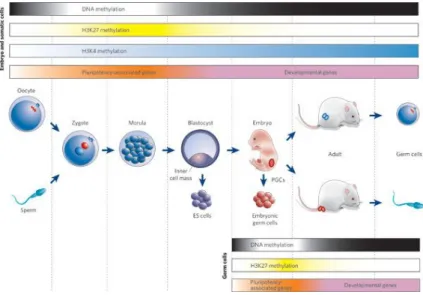
The key developmental events with global epigenetic modifications and gene-expression patterns in mammalian cells were shown in Figure 1.2.
Figure 1.2 Key developmental events with global epigenetic modifications and gene-expression patterns mammalian cells. [37]
Some characteristics of CSC shared with normal stem cells including self-renewal and differentiation. Normal stem cells differentiated into the certain cell type in proper epigenetic regulation.
Altered or aberrant epigenetic regulation was thought to be involved in CSC development required for the features of CSCs, highly proliferative, tumor formation, and metastatic ability to form the new tumor at distant site. The epigenetic regulation pattern for maintaining the stemness was occurred in CSC. The possibility of this evidence can be explained in two ways; one is dedifferentiation mechanisms gained in non-stem cancer cells
and another is the failure of DNA methylation mechanism in normal stem cell during differentiation.
Several key developmental or signaling pathways have been shown to play essential roles to get the CSCs functions, especially tumorigenesis, cancer initiation and maintenance of self-renewing. Epigenetic deregulation may contribute the alteration in such kinds of pathways.
Generally, the Jak-STAT, Wnt, Notch, Hedgehog, PI3K, and NF-kB signaling were shown to be involved in mediating various stem cell properties, such as self-renewal, cell fate decisions, survival, proliferation, and differentiation [38–40]. Recent reports interestingly had reported that the regulation of these signaling pathways are imbalanced in cancers. One of the reasons for these evidences could be the abnormal epigenetic regulations. Recently, the distinct DNA hypomethylation of specific gene sets in MCF7-derived mammosheres caused the activation of Jak-STAT pathway that was considered for the maintenance of CSC properties to be involved in the regulation of stem cells [38,41]. In gastric cancers, activation of Wnt pathways in were more frequently affected by epigenetic alterations than by genetic alterations in the related genes [42]. Moreover, it was reported that the activation of Hedgehog was related with epigenetic changes in which, Shh promoter hypomethylation was suggested as a critical event in breast carcinogenesis [43]. The Notch signaling pathway play important roles in developmental process, and cell-cell communication for regulating the cell proliferation, differentiation and cell lineage progression but it is also dysregulated in many cancers [44]. One of the growing evidences in epigenetic dysregulation of notch signaling
pathway showed that overexpression of Notch ligand has been gained from enhanced histone acetylation at promoter region of this ligand in multiple myeloma.
In conclusion, although the experimental evidences of CSCs relating to their genetic and epigenetic regulation have been growing in number, the hypothesis of CSC still remain needed to understand how they develop, how different in characteristics depending on tissue, and dysregulation of key signaling pathways specific for their functions. It is very important to develop a CSC model that can sufficiently explain about the genetic or epigenetic profiles of CSC. The alteration mechanism in CSCs could be expected to be useful for development of specific therapy for cancer patients.
REFERENCES
1. GBD 2015 Mortality and Causes of Death Collaborators, G. B. D. 2015 M.; of Death, C. Global, regional, and national life expectancy, all- cause mortality, and cause-specific mortality for 249 causes of death, 1980-2015: a systematic analysis for the Global Burden of Disease Study 2015. Lancet (London, England) 2016, 388, 1459–1544, doi:10.1016/S0140-6736(16)31012-1.
2. National Cancer Institute NCI Dictionary of Cancer Terms:
https://www.cancer.gov/publications/dictionaries/cancer- terms/def/benign-tumor.
3. PubMed Health Benign Tumor:
https://www.ncbi.nlm.nih.gov/pubmedhealth/PMHT0025285/.
4. Plummer, M.; de Martel, C.; Vignat, J.; Ferlay, J.; Bray, F.; Franceschi, S. Global burden of cancers attributable to infections in 2012: a synthetic analysis. Lancet Glob. Heal. 2016, 4, e609–e616, doi:10.1016/S2214-109X(16)30143-7.
5. Tannock, I. F. Conventional cancer therapy: Promise broken or promise delayed? Lancet 1998, 351, doi:10.1016/S0140- 6736(98)90327-0.
6. National Cancer Institute NCI Dictionary of Cancer Terms:
https://www.cancer.gov/publications/dictionaries/cancer- terms/def/conventional-treatment.
7. Magee, J. A.; Piskounova, E.; Morrison, S. J. Cancer Stem Cells:
Impact, Heterogeneity, and Uncertainty. Cancer Cell 2012.
8. Reya, T.; Morrison, S. J.; Clarke, M. F.; Weissman, I. L. Stem cells, cancer, and cancer stem cells. Nature 2001, doi:10.1038/35102167.
9. Klonisch, T.; Wiechec, E.; Hombach-Klonisch, S.; Ande, S. R.;
Wesselborg, S.; Schulze-Osthoff, K.; Los, M. Cancer stem cell markers in common cancers - therapeutic implications. Trends Mol.
Med. 2008.
10. Dean, M.; Fojo, T.; Bates, S. Tumour stem cells and drug resistance.
Nat. Rev. Cancer 2005.
11. Dalerba, P.; Cho, R. W.; Clarke, M. F. Cancer Stem Cells: Models and
Concepts. Annu. Rev. Med. 2007,
doi:10.1146/annurev.med.58.062105.204854.
12. Soto, A. M.; Sonnenschein, C. The somatic mutation theory of cancer:
Growing problems with the paradigm? BioEssays 2004.
13. Blagosklonny, M. V. Molecular theory of Cancer. Cancer Biol. Ther.
2005.
14. Brücher, B. L. D. M.; Jamall, I. S. Somatic mutation theory - Why it’s wrong for most cancers. Cell. Physiol. Biochem. 2016.
15. Hermann, P. C.; Huber, S. L.; Herrler, T.; Aicher, A.; Ellwart, J. W.;
Guba, M.; Bruns, C. J.; Heeschen, C. Distinct Populations of Cancer
Stem Cells Determine Tumor Growth and Metastatic Activity in Human Pancreatic Cancer. Cell Stem Cell 2007, 1, 313–323, doi:10.1016/j.stem.2007.06.002.
16. Li, L.; Neaves, W. B. Normal stem cells and cancer stem cells: The niche matters. Cancer Res. 2006, 66, 4553–4557.
17. Egeblad, M.; Nakasone, E. S.; Werb, Z. Tumors as organs: Complex tissues that interface with the entire organism. Dev. Cell 2010.
18. Al-Hajj, M.; Wicha, M. S.; Benito-Hernandez, A.; Morrison, S. J.;
Clarke, M. F. Prospective identification of tumorigenic breast cancer cells. Proc. Natl. Acad. Sci. 2003, doi:10.1073/pnas.0530291100.
19. Karsten, U.; Goletz, S. What makes cancer stem cell markers different? Springerplus 2013, doi:10.1186/2193-1801-2-301.
20. Lobo, N. A.; Shimono, Y.; Qian, D.; Clarke, M. F. The Biology of Cancer Stem Cells. Annu. Rev. Cell Dev. Biol. 2007, doi:10.1146/annurev.cellbio.22.010305.104154.
21. He, S.; Nakada, D.; Morrison, S. J. Mechanisms of stem cell self- renewal. Annu. Rev. Cell Dev. Biol. 2009, doi:10.1146/annurev.cellbio.042308.113248.
22. Keller, G. Embryonic stem cell differentiation: emergence of a new era in biology and medicine. Genes Dev. 2005, doi:10.1101/gad.1303605.
23. Plaks, V.; Kong, N.; Werb, Z. The cancer stem cell niche: How essential is the niche in regulating stemness of tumor cells? Cell Stem Cell 2015.
24. Matsuda, S.; Yan, T.; Mizutani, A.; Sota, T.; Hiramoto, Y.; Prieto-Vila, M.; Chen, L.; Satoh, A.; Kudoh, T.; Kasai, T.; Murakami, H.; Fu, L.;
Salomon, D. S.; Seno, M. Cancer stem cells maintain a hierarchy of differentiation by creating their niche. Int. J. Cancer 2014, doi:10.1002/ijc.28648.
25. Chen, L.; Kasai, T.; Li, Y.; Sugii, Y.; Jin, G.; Okada, M.; Vaidyanath, A.; Mizutani, A.; Satoh, A.; Kudoh, T.; Hendrix, M. J. C.; Salomon, D.
S.; Fu, L.; Seno, M. A model of cancer stem cells derived from mouse induced pluripotent stem cells. PLoS One 2012, 7, doi:10.1371/journal.pone.0033544.
26. Calle, A. S.; Nair, N.; Oo, A. K.; Prieto-Vila, M.; Koga, M.; Khayrani, A. C.; Hussein, M.; Hurley, L.; Vaidyanath, A.; Seno, A.; Iwasaki, Y.;
Calle, M.; Kasai, T.; Seno, M. A new PDAC mouse model originated from iPSCs-converted pancreatic cancer stem cells (CSCcm). Am. J.
Cancer Res. 2016, 6, 2799–2815.
27. Nair, N.; Calle, A. S.; Zahra, M. H.; Prieto-Vila, M.; Oo, A. K. K.;
Hurley, L.; Vaidyanath, A.; Seno, A.; Masuda, J.; Iwasaki, Y.; Tanaka, H.; Kasai, T.; Seno, M. A cancer stem cell model as the point of origin of cancer-associated fibroblasts in tumor microenvironment. Sci. Rep.
2017, 7, 6838, doi:10.1038/s41598-017-07144-5.
28. Prieto-Vila, M.; Yan, T.; Calle, A. S.; Nair, N.; Hurley, L.; Kasai, T.;
Kakuta, H.; Masuda, J.; Murakami, H.; Mizutani, A.; Seno, M. iPSC- derived cancer stem cells provide a model of tumor vasculature. Am.
J. Cancer Res. 2016, 6, 1906–1921.
29. Rozhok, A. I.; Salstrom, J. L.; DeGregori, J. Stochastic modeling indicates that aging and somatic evolution in the hematopoietic system are driven by non-cell-autonomous processes. Aging (Albany.
NY). 2014, 6, 1033–1048, doi:10.18632/aging.100707.
30. Hanahan, D.; Weinberg, R. A. Hallmarks of cancer: The next generation. Cell 2011.
31. Baylin, S. B.; Herman, J. G. S.B. Baylin, J.G. Herman, DNA hypermethylation in tumorigenesis: epigenetics joins genetics, Trends Genet. 16 (2000) 168–174. - Google Search. Trends Genet. 2000, 16, 168–174.
32. Sandoval, J.; Esteller, M. Cancer epigenomics: Beyond genomics.
Curr. Opin. Genet. Dev. 2012.
33. De Carvalho, D.; Sharma, S.; You, J. S.; Su, S. F.; Taberlay, P. C.; Kelly, T. K.; Yang, X.; Liang, G.; Jones, P. A. DNA Methylation Screening Identifies Driver Epigenetic Events of Cancer Cell Survival. Cancer Cell 2012, doi:10.1016/j.ccr.2012.03.045.
34. Boyer, L. A.; Plath, K.; Zeitlinger, J.; Brambrink, T.; Medeiros, L. A.;
Lee, T. I.; Levine, S. S.; Wernig, M.; Tajonar, A.; Ray, M. K.; Bell, G.
W.; Otte, A. P.; Vidal, M.; Gifford, D. K.; Young, R. A.; Jaenisch, R.
Polycomb complexes repress developmental regulators in murine embryonic stem cells. Nature 2006, doi:10.1038/nature04733.
35. Azuara, V.; Perry, P.; Sauer, S.; Spivakov, M.; Jørgensen, H. F.; John, R. M.; Gouti, M.; Casanova, M.; Warnes, G.; Merkenschlager, M.;
Fisher, A. G. Chromatin signatures of pluripotent cell lines. Nat. Cell Biol. 2006, doi:10.1038/ncb1403.
36. Ohm, J. E.; McGarvey, K. M.; Yu, X.; Cheng, L.; Schuebel, K. E.; Cope, L.; Mohammad, H. P.; Chen, W.; Daniel, V. C.; Yu, W.; Berman, D.
M.; Jenuwein, T.; Pruitt, K.; Sharkis, S. J.; Watkins, D. N.; Herman, J.
G.; Baylin, S. B. A stem cell-like chromatin pattern may predispose tumor suppressor genes to DNA hypermethylation and heritable silencing. Nat. Genet. 2007, doi:10.1038/ng1972.
37. Reik, W. Stability and flexibility of epigenetic gene regulation in mammalian development. Nature 2007.
38. Dreesen, O.; Brivanlou, A. H. Signaling pathways in cancer and embryonic stem cells. Stem Cell Rev. 2007, doi:10.1007/s12015-007- 0004-8.
39. Ghoshal, P.; Nganga, A. J.; Moran-Giuati, J.; Szafranek, A.; Johnson, T. R.; Bigelow, A. J.; Houde, C. M.; Avet-Loiseau, H.; Smiraglia, D. J.;
Ersing, N.; Chanan-Khan, A. A.; Coignet, L. J. Loss of the SMRT/NCoR2 corepressor correlates with JAG2 overexpression in multiple myeloma. Cancer Res. 2009, 69, 4380–4387,
doi:10.1158/0008-5472.CAN-08-3467.
40. Toh, T. B.; Lim, J. J.; Chow, E. K.-H. Epigenetics in cancer stem cells.
Mol. Cancer 2017, 16, 29, doi:10.1186/s12943-017-0596-9.
41. Onishi, K.; Zandstra, P. W. LIF signaling in stem cells and development. Development 2015, doi:10.1242/dev.117598.
42. Yoda, Y.; Takeshima, H.; Niwa, T.; Kim, J. G.; Ando, T.; Kushima, R.;
Sugiyama, T.; Katai, H.; Noshiro, H.; Ushijima, T. Integrated analysis of cancer-related pathways affected by genetic and epigenetic alterations in gastric cancer. Gastric Cancer 2015, doi:10.1007/s10120- 014-0348-0.
43. Cui, W.; Wang, L.-H.; Wen, Y.-Y.; Song, M.; Li, B.-L.; Chen, X.-L.; Xu, M.; An, S.-X.; Zhao, J.; Lu, Y.-Y.; Mi, X.-Y.; Wang, E.-H. Expression and regulation mechanisms of Sonic Hedgehog in breast cancer.
Cancer Sci. 2010, doi:10.1111/j.1349-7006.2010.01495.x.
44. Bolós, V.; Grego-Bessa, J.; De La Pompa, J. L. Notch signaling in development and cancer. Endocr. Rev. 2007.
CHAPTER (2)
“iPSC DERIVED CSC MODEL WITH LUNG METASTASIS DEVELOPED IN THE
MICROENVIRONMENT OF LUNG
CARCINOMA”
ABSTRACT
Cancer stem cells (CSCs) are considered to be derived from normal stem cells affected by the tumor microenvironment through the genetic and epigenetic alterations that initiate malignant transformation. We have reported that the conditioned medium of Lewis lung carcinoma (LLC) cells can be used to convert mouse induced pluripotent stem cells (miPSCs) into cancer stem cell phenotype, which named miPS-LLCcm cells. miPS-LLCcm cells developed highly angiogenic and malignant adenocarcinoma after transplanted into nude mice. When miPS- LLCcm cells were subcutaneously injected into a nude mouse, malignant adenocarcinoma-like tumor was formed with lung metastasis.
Immunohistochemical analysis with GFP antibody showed that the nodules formed in lung expressed GFP, of which expression was controlled by Nanog promoter, further proving that these cells were metastatic to lung. This should therefore be a model to study lung metastasis from a tumor formed by subcutaneous injection. In this study, we analyzed the expression of three types of candidate genes for stem cell, tumor driver genes and epithelial–mesenchymal transition (EMT) on four different stages of cells. The CSC-like cells developed from miPSC exhibited self-renewal activity and stem cell marker gene expression.
The expression of EMT markers were analyzed in the primary culture derived from the tumor and nodules metastasized to lung. The primary cells from the tumor highly expressed the EMT markers; snail, slug, Twist and N-cadherin. The results demonstrated in this study indicate that primary cells from tumor are rich in CSCs with high metastatic potential.
2.1. INTRODUCTION
Tumors are composed of heterogeneous cancer cells with distinct morphological and functional profiles. This heterogeneity could be partially explained by the classical cancer initiation theory; evolutional accumulation of one or more mutations in a single or a few cells resulting in uncontrolled growth [1]. Another explanation is based on the presence of stem cells in tumors. Even in a few number the stem cells are considered to comprise the whole tumor in patient [2–4]. The heterogeneous population in cancer tissues are thought to be the progeny of stem cell population resulting from self-renewal and differentiation. Coupled with the malignant tumorigenic potential, the stem cells have been termed as cancer stem cells (CSCs) generating functionally hierarchical structure in a tumor.
There is a considerable evidence that many different cancers have a unique subpopulation of self-renewing cells that can generate the diverse tumor cells.
CSCs were identified in brain, breast, prostate, head and neck, pancreas, liver, ovary, and colon cancers, by using different markers, such as CD133, EpCAM, CD44, CD24, Lgr5 and ALDH1. The question about the origin of CSC is still controversial. The leukemic stem cells were the first identified with the cell- surface markers, CD34+ CD38- differentiating in vivo into leukemic blasts [2]. This particular approach led to emerge new studies which described tissue-specific markers for solid tumors. The phenotype associated with cancers including motility, invasion and chemo-/radio-resistance could be traced to CSCs.
Metastasis through the activation of CXCR4 receptor was previously demonstrated by the migration of invasive CSCs defined with CD133 in
pancreatic cancer [5]. On the other hand, other studies attributed their metastatic potential to the epithelial-mesenchymal transition (EMT) events showing that differentiated cancer cells could turn into CSC-like mesenchymal cells [6].
CSCs is normally characterized as fraction of cancer cells that have ability for self-renewal, pluripotency and sustaining of the bulk of cancer. The best evidence for the existence of CSCs was demonstrated with cancer stem cells model that is derived from mouse induced pluripotent stem cells (miPSC). When miPSC are cultured in the presence of conditioned medium prepared from various cancer cell lines, it acquires the characters of CSCs with high tumorigenicity and stemness. We have evaluated miPS-LLCcm cells that were derived from miPSCs developed in the conditioned medium of Lewis lung carcinoma (LLC) [7]. This cancer stem cell model has the highly tumorigenic and angiogenic ability and also metastatic potential was observed when spheroid cells were injected into the mouse tail vein, multiple metastatic nodules were found in lung after one month.
Adult somatic cells have been successfully reprogrammed to pluripotent stem cells (iPSCs) with the transduction of four transcription factors [8]. The differentiation potential of iPSCs is largely expected to develop multiple potential avenues for the regenerative therapy. Immune rejection of embryonic stem cells can be avoided by replacing these with iPSCs. However, the risks of potential tumor development and other unpredictable biological changes during transplantation are still unresolved. The cellular interaction of transplanted cells in the microenvironment has been reported important to obtain successful results in regeneration therapy [9].
In this evidence, our hypothesis is that miPSCs are affected by the conditioned medium to become the miPS-CSCs through the post-transcriptional and translational or epigenetic regulations. It is a good model to study the behaviors of CSCs in primary tumor induction and metastasis.
2.2. RESULTS
2.2.1. Conversion of miPSCs into CSC-like cells with tumorigenic and metastatic potential
We have reported a model of CSC-like cells converted from miPSC by the exposure to the conditioned medium (CM) from various cancer cell lines [7,10,11].
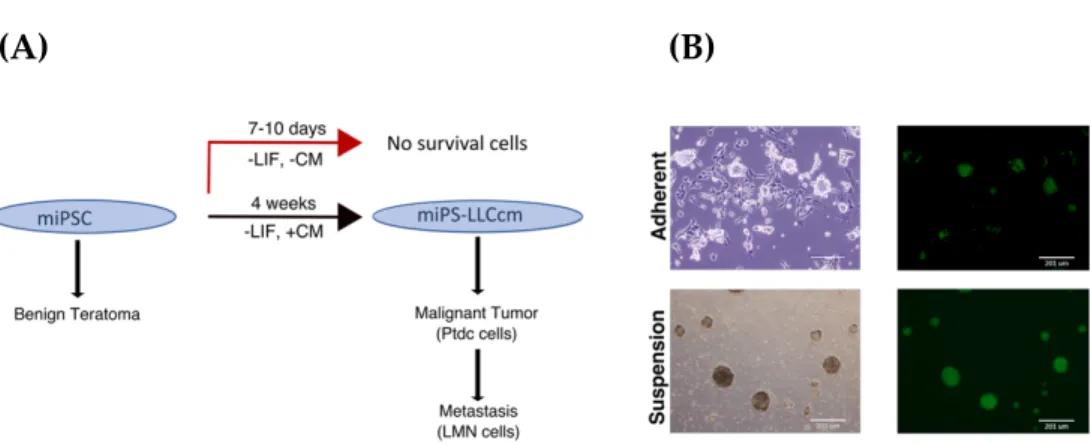
The miPSCs used in the studies had GFP under the control of Nanog promoter wherein undifferentiated stem cells exhibit strong GFP expression and differentiated cells lose green fluorescence. miPSCs were found to be viable in the presence of conditioned medium even when the differentiation was allowed while they started differentiation and failed to survive beyond 10 days without CM. This scheme is briefly summarized in Figure 2.1A. After 4 weeks of treatment with CM, the survived and undifferentiated cells expressing GFP were named as miPS-LLCcm cells. The self-renewal of miPS-LLCcm cells, a specific character of undifferentiated stem cells, was also confirmed by sphere-forming assay as well as its differentiation potential was evident with highly adhesive fibroblast-like phenotype and loss of GFP expression (Figure 2.1B).
(A) (B)
Figure 2.1. Conversion of miPSCs into miPS-LLCcm (A) Summarized scheme of conversion of miPSCs into miPS-LLCcm and its tumorigenic and metastatic activity. (B) Converted miPSCs in adherent culture (top) and in suspension culture (bottom).
The subcutaneous transplantation of miPS-LLCcm cells into immunocompromised Balb/c nude mice generated malignant tumor together with the metastasized nodule-like structures in the lung (Figure 2.2A and 2.2B).
These tumors and lung nodules were subjected to primary cell culturing to isolate GFP expressing cells. The primary cultured cells from tumors was named as Ptdc cells and the cells from lung nodules was named as LMN cells. The Ptdc cells were subsequently transplanted to generate the secondary tumor again. In the subcutaneous injection of 106 cells, the growth of tumors was compared (Figure 2.2C). The Ptdc cells showed the most rapid growth when compared to miPS- LLCcm cells and miPSCs.
Figure 2.2. Tumorigenic and metastatic potential of miPS-LLCcm (A) Tumors and metastatic nodules in lungs generated by subcutaneously transplanted miPSCs, miPS-LLCcm cells and Ptdc cells into the Balb/c-nu mouse. Arrows in lung indicate the positions of nodules. (B) The histogram showed the average number of lung nodules for each of the three cells. (C) The sizes of tumors growing in 4-6 weeks.
Primary cultures derived from the tumors were positive for GFP signal, confirming they were originated from miPS-LLCcm cells and not from host cells, and displayed sphere-forming activity suggesting they preserve stem-like characteristics (Figure 2.3A and 2.3B). miPS-LLCcm cells, Ptdc cells and LMN cells maintained the expression of endogeneous stemness markers, such as
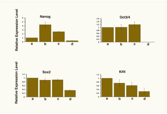
Nanog, Oct3/4, Sox2 and Klf4 without LIF similar with the untreated miPSCs cultured in the presence of LIF (Figure 2.4). Therefore, the stemness can be maintained in the presence of CM and this conversion supports the establishment of CSC-like features.
Figure 2.3. Primary cultured cells (A) Ptdc cells in adherent culture (top) and sphere formation in suspension culture (bottom) with the expression of GFP.
(B) LMN cells in adherent culture (top) and sphere formation in suspension culture (bottom) with the expression of GFP.
Figure 2.4. Comparison of the expression levels of stemness markers by rt- qPCR. a, miPSCs; b, miPS-LLCcm cells; c, Ptdc cells; d, LMN cells.
Histological analysis of the tumor showed poorly differentiated phenotype, high nuclear to cytoplasmic ratio, micrometastasis and some epithelial ductal-like structure, which are the signs of malignancy (Figure 2.5A). Metastatic node-like structures were observed in the lung of the mice (Figure 2.5B). Untreated miPSCs tumor displayed teratoma like phenotype with various germ layers (Figure 2.5C).
These observations confirmed the self-renewing and tumorigenic potential of miPS-LLCcm cells.
Figure 2.5. Histological analyses of allografts of miPS-LLCcm cells. (A) High nuclear to cytoplasmic ratio (a) and with the mass of undifferentiated cells (asterisks in (b)), granular epithelial structure (arrows in (c)) and micrometastasis (arrows in (d)). (B) Metastatic nodules-like tumor in the lung pulmonary tissue (left), in the lung tissue (middle), and in the chest (right). (C) Teratoma from untreated miPSCs showing the three germ layers:
squamous epithelial tissue (asterisks) in ectoderm (left), muscle tissue in mesoderm (middle) and gland-like structures (asterisks) in endoderm (right). (A-C) H&E staining.
2.2.2. Characterization of miPS-LLCcm, Ptdc and LMN cells
In order to further confirm the acquisition of CSC-like phenotype, we assessed the expression of the commonly known CSC markers, CD44 and ALDH1 by rt-qPCR as well as immuno-histochemical analysis. (Figure 2.6A and 2.6B). The LMN and Ptdc cells showed the significantly higher expression of ALHD1 and CD44 than the miPSCs. The miPS-LLCcm derived tumor also showed the expression of these two markers.
Figure 2.6. The localization and expression of CSC markers. (A) IHC analysis showing the expression of CD44 and ALDH1 in the tumor of the miPS- LLCcm cells. (B) The comparison of the expression of CD44 and ALDH1 the Ptdc and LMN cells with miPS-LLCcm by rt-qPCR analysis. ***P < .001, **P
< .01, *P < .05.
2.2.3. In vivo tumorigenic differentiation of miPS-LLCcm
MUC1 plays a crucial role in cancer progression and is considered as a suitable marker for the adenocarcinoma tumor phenotype [12]. Immuno- histochemical analysis showed that tumor associated MUC1 expression was detected in tumor of miPS-LLCcm (Figure 2.7A). Mass of highly proliferating cells showed the expression of GFP as population of cells that are maintaining self- renewal ability.
Ptdc cells showed the upregulation of both E-cadherin, an epithelial marker, and N-cadherin, a mesenchymal marker, as compared to miPS-LLCcm and LMN cells (Figure 2.7B and 2.7C). The expression of Snail, Slug, Twist1 and Twist2 were also upregulated in Ptdc cells suggesting the potential of epithelial-to- mesenchymal transition (EMT) in Ptdc cells.
Figure 2.7. The localization and expression of EMT markers in miPS-LLCcm derived tumors. (A) Immuno-histochemical (IHC) analysis showed that ductal epithelial structure expressed MUC1 (left) and the undifferentiated mass of cells expressed the GFP (right). Comparison of the expression of (B) epithelial and mesenchymal markers (E-cadherin and N-cadherin) and (C) EMT markers in a, miPS-LLCcm cells; b, Ptdc cells and c, LMN cells.
2.3. MATERIALS AND METHODS
2.3.1. Cell Culture
Mouse Lewis Lung Carcinoma cell lines (LLC) were purchased from ATCC (USA) and maintained in DMEM (D5796 Sigma) medium containing 10% fetal bovine serum (FBS,Gibco,NY) and 100 U/ml penicillin/streptomycin (Wako,Japan). Mouse induced pluripotent stem cells (miPS, iPS-MEF-Ng-20D-17;
Lot No.012, Fiken Cell Bank, Japan) were cultured in DMEM containing 15% FBS, 0.1 mM NEAA (100X NEAA, Gibco, NY), 2mM L-Glutamine (Nacalai Tesque, Japan), 50U/ml of penicillin/streptomycin (P/S), 0.1 mM 2-mercaptoethanol (Sigma) and 1000 U/ml of Leukemia Inhibitory Factor (LIF, Millipore, MA) on feeder layers of mitomycin treated mouse embryonic fibroblast (MEF) cells (Reprocell, Japan). In the case of feeder-less, the miPS cells were cultured on gelation (0.1%) coated dishes. The expression of GFP and cell morphology was observed and photographed using Olympus IX81 microscope equipped with a light fluorescence device (Olympus, Japan).
2.3.2. Conversion of miPSCs into the CSC-like cells
According to the methods reported by Chen L and Kasai T et al., we cultured the miPSCs in the presence of conditioned medium of LLC cells [7]. Nanog-GFP reporter expression was used in miPSC cells and the expression of GFP reflects the maintenance of stemness [13]. LLC cells were cultured and prior to collecting conditioned medium, the cells were changed into 5% serum medium at 70-80%
confluency. After 48hrs incubation, the conditioned medium (CM) from LLC cells was collected and filtered through a 0.22 µm filter (Millipore, Ireland). The
miPSCs were treated with the CM for 4 weeks and miPSCs were cultured in the presence or absence of LIF as controls. The miPSCs cultured under feeder-less conditions were treated with the CM in 1:1 ratio of miPS medium and CM for four weeks.
2.3.3. Sphere Formation Assay
To generate the spheroids, serum free medium (DMEM 97.5%, NEAA 1%, L-Glutamine 1%, 100X Pen/Strep 0.5%, 0.1 mM 2-mercaptoethanol, and Insulin- transferrin-selenium-X 1/100 v/v (ITS-X, life technologies, CA) was used and single cells were plated on ultra-low attachment dishes (Corning incorporated, NY) at cell density of 1x10⁴ cells/ml [14].
2.3.4. Animal experiments
The plan of animal experiments was reviewed and approved by the ethics committee for animal experiments of Okayama University under the IDs OKU- 2013252, OKU-2014157, OKU-2014429 and OKU-2016078. All experiments were performed according to the Policy on the Care and Use of the Laboratory Animals, Okayama University. Nude mice (Balb/c-nu/nu, female, 4 weeks) were purchased CharlesRiver, Japan. Cells at 1x10⁶ were suspended in 200 !l of HBSS (Hanks Balanced salt solution, Gibco, NY) and were subcutaneously transplanted into nude mice.
2.3.5. Preparation of primary cell culture
To prepare the primary culture from a mouse allograft, the tumor was excised and cut into small pieces (approximately 1 mm3) and washed in the
Hank’s buffered salt solution (HBSS) for three times. These pieces were transferred into a 15-ml tube with 4 ml of dissociation buffer prepared in PBS containing 0.25% trypsin, 0.1% collagenase, 20% KnockOut™ Serum Replacement (Gibco, NY), 1 mM of CaCl2 and incubated at 37°C for 40 mins. To terminate the digestion, 5 ml of DMEM containing 10% FBS was then added. The cellular suspension transferred into the new tubes and centrifuged at 1000 rpm for 5 mins.
The cell pellet was suspended in 5 ml of HBSS, and centrifuged at 1000 rpm for 5 min. The cell pellet was placed into an appropriate volume of miPS medium without LIF and the cell number was counted with hemocytometer. Then the cells at 5×10⁵ were seeded per 60-mm dish. After a passage, the cells derived from mouse allografts were cultured in the presence of 1 !g/mL of puromycin for 24 hours to remove the host derived cells.
To prepare the primary culture from metastatic nodules in a lung, the lung tissue was excised and cut into small pieces (approximately 1 mm3) and washed in the HBSS for three times. And the same procedures with those for the cells from a tumor allograft were employed to prepare the cells. Finally, the expression of GFP and cell morphology was observed and photographed using Olympus IX81 microscope equipped with a light fluorescence device (Olympus, Japan).
2.3.6. RNA extraction, cDNA synthesis and qPCR mRNA expression analysis
Total RNA was extracted using RNAeasy Mini kit (QUIAGEN, Germany) according to the manufacturer's instructions and 1 !g of RNA was reverse transcribed using Superscript First strand kit (Invitrogen, CA). Quantitative real time PCR was performed with cycler 480 SYBR green I Master Mix (Roche,
Switzerland) according to manufacturer’s instructions. Primers used for qPCR are listed in Table 2.1.
Table 2.1. List of Primers Used in the Experiments
No Names Forward Primer Sequence Reverse Primer Sequence 1 Nanog AGGGTCTGCTACTGAGATGCTCTG AACCCAAGCACGTATCAGGG
2 Oct3/4 TCTTTCCACCAGGCCCCCGGCTC TGCGGGCGGACATGGGGAGATCC
3 Sox2 TAGAGCTAGACTCCGGGCGATGA TTGCCTTAAACAAGACCACGAAA
4 Klf4 GGACTTACAAAATGCCAAGGGGTG TCGCTTCCTCTTCCTCCGACACA
5 CD44 AGAAAAATGGCCGCTACAGTATC TGCATGTTTCAAAACCCTTGC
6 ALDH1 AACACAGGTTGGCAAGTTAATCA TGCGACACAACATTGGCCTT
7 E-cadherin AACCCAAGCACGTATCAGGG GGGGTCTGTGACAACAACGA
8 N-cadherin CCTTGCTTCAGGCGTCTGTG CTTGAAATCTGCTGGCTCGC
9 Snail GGAGTTGACTACCGACCTTGC TGGAAGGTGAACTCCACACAC
10 Slug GCCCTTAAAGGCACTAACGAG ATTCACGAAGGTGACGAGCC
11 Twist1 GCCGGAGACCTAGATGTCATTGT TTAAAAGTGTGCCCCACGCC
12 Twist2 CTCACGAGCGTCTCAGCTAC TTGTCCAGGTGCCGAAAGTC
13 GADPH AACGGCACAGTCAAGGCCGA ACCCTTTTGGCTCCACCCTT
2.3.7. Histological analysis and immunohistochemistry (IHC)
Paraffin embedded tumor sections (5!m) were stained with Hematoxylin (Sigma Aldrich,USA ;0.5%) and Eosin Y (Sigma Aldrich, USA) (HE) for histological analysis. Primary antibodies and dilutions used for IHC were used as follows; anti-GFP antibody 1:200 (#2956, Cell Signaling, USA), anti-MUC1
antibody 1:100 (Abcam/ab15481, UK), anti-ALDH1 antibody 1:200 (Abcam/
ab52492, UK) and Anti-CD44 antibody 1:200 (Abcam/ ab24504, UK).
2.3.8. Statistical analysis
The data were analyzed using two-tailed student’s t-test and are presented as the mean ± standard deviation (SD) at least three-time determinations. A P- values less than 0.05 was considered to be statically significant, while less than 0.01 was highly significant.
2.4. DISCUSSION
The current study focused on the DNA methylation in the CSC model converted from iPSCs by the treatment with conditioned medium of cancer cells.
By the treatment, miPSCs obtained the ability of unlimited growth and the capacity to maintain their stemness, while they were allowed to differentiate without LIF. The subcutaneous transplantation of the survived cells into the mouse formed malignant tumors and metastasized into lung tissues. Thus, we concluded that miPSCs should be converted into CSCs without any intended genetic manipulation. The immunohistochemical analysis revealed the heterogeneity of the tumors, in which miPS-LLCcm cells should on one hand maintain the undifferentiated population expressing GFP and differentiate on the other into adenocarcinoma phenotype expressing MUC1 while miPSCs developed benign teratoma showing the three germ-layers differentiation and the loss of undifferentiated phenotype. Furthermore, the cells from benign teratoma
cannot be maintained but the cells from tumors at primary site and metastatic nodules can be maintained in vitro.
The heterogeneous phenotypes characterized by the expression of E- cadherin and N-cadherin in the Ptdc cells should be implying the progression of cancer and metastatic potential of the tumor maintaining the plasticity of the transition between epithelial and mesenchymal states. Further study is required to confirm the cells are undergoing the EMT being involved in maintenance of stemness, invasiveness and metastasis of tumor cells.
Recently our group generated CSC models converted from iPSCs with the aid of CM of various cancer cell lines such as human pancreatic carcinoma cell lines PK-8 cells and KLM-1 cells, human breast cancer cell lines T47D cells and BT549 cells, mouse carcinoma cell lines LLC cells, P19 cells, B16 cells and MC.E12 cells [7,10,11]. In the conversion of pancreatic duct like adenocarcinoma (PDAC) like CSC model, there was no evidence relating to single point mutations even in Kras oncogene and its xenografts tumors showed the features of acinoductal metaplasia, pancreatic intraepithelial neoplasia and PDAC lesions[10]. We postulated that CSCs may be induced epigenetic changes without any known mutations.
Premature termination of reprogramming were reported to result in tumor development in various tissues with undifferentiated dysplastic cells exhibiting global changes in DNA methylation at H19 DMRs identifying IGF-2 expression up-regulated in the tumor initiating cells [31]. Since the changes in DNA methylation was considered responsible for the conversion of iPSCs into CSCs, the patterns of DNA methylation were compared between the converted cells
(miPS-LLCcm cells), tumor derived cells (Ptdc cells and LMN cells) and miPSCs.
As the results of bisulfite sequencing, we evaluated the list of epigenetically affected genes regarding to the DMRs in the miPS-LLCcm cells and Ptdc cells and LMN cells. Hypo- and hypermethylated genes were identified and hypomethylation was found overall superior to hypermethylation in all CSCs when compared to the parental cell line miPSCs.
The analysis of KEGG pathways relating to hypomethylated genes revealed the several notable pathways important in cancers. Checking the expression of genes associated with these pathways, the expression of hypomethylated genes relating to PI3K-Akt pathway was found significantly high among those of the other genes. PI3K-Akt-mTOR signaling pathway has previously been reported as a key driver of carcinogenesis in several cancer types [32,33]. In this study, we found Pik3r5 (p101), which is a regulatory subunit of Pik3cg enzyme, as a hypomethylated and highly up-regulated gene relating to PI3K-Akt pathway. In the recent report, the evidence of PIK3CG as a potential oncogene were evaluated by analyzing the differential role each unit of PIK3CG, of which overexpression of the catalytic subunit PIK3CG (p110γ) or the regulatory subunit PIK3R5 (p101) leads to oncogenic cellular transformation and malignancy [34]. Therefore, the hypomethylation of Pik3r5 gene leading to the up-regulation is closely related to the activation/phosphorylation of AKT that is the downstream target molecule and Pik3cg should play a key role in carcinogenesis. In fact, the multiple myeloma cells derived from patients, the upregulation of PI3K components, in which PIK3CG has been proved to be a main regulator of cells adhesion and migration [35]. The PIK3CA gene has been reported to be hypomethylated in esophageal cancer cases when compared to the adjacent normal tissues [36]. On the other
hand, both Pik3r5 and Pik3cg were overexpressed resulting in the up-regulation of PI3K-gamma in the class IB PI3Ks, but not the PI3K-alpha in the class IA in our CSCs. Collectively, the activation of PI3K-Akt signaling pathway should significantly be relating with the conversion of miPSC into miPS-LLCcm cells resulting in the constitutive activation of Akt in Ptdc and LMN cells.
According to the recent reports, the tumor cells produced a variety of molecules such as growth factors, cytokines and chemokines, which exhibited various effects such as on tumor growth and angiogenesis, providing them with various microenvironments [37,38]. In our study, we have successfully demonstrated the CSCs generated from iPSCs by the treatment with CM from cancer derived cells acquired the DNA hypomethylation.
2.5. CONCLUSIONS
Significant overall DNA hypomethylation during the conversion should lead to the activation of certain proto-oncogene, which represent the malignant conversion even without mutations. In this context, the hypomethylation might be considered to contribute to the progression and metastasis of the cancer stem cells.