\~:jj(['JIJ;JH11\lilf¥f1 1'f?; 5 i'} : 119~ 122, 1981
EFFECTS OF TEMPERATURE AND ANESTHETICS ON
THE TENSION VALUE IN THE STRETCHED NERVE
FIBRES OF THE CRAYFISH
Takeshi
KAsAcr*
and Osamu
Icr-IIKAvVA **
It has been reported that there is a phase transition in the biological membrane. For example, the transition in the cell membrane Esclze!iclzia coli is exibited by X -ray diffraction techniques (l). It is also mentioned that the phase transition is originated from the phospholipid which is one of the membrane components and that the temperature
of transition is in the equilibrium state between solid and liquid-crystalline phase where the normal growth of the organism is performed (4, 6). Presumably there occur some mechanical or structural changes in the membraneous materials at these transition temperatures. In the present experiment, an attempt was made to evaluate the phase transition of the excitable mambrane by measuring the tension at various temperatures in the crayfish nerve fibres. At the same time, the alteration or the blockade of the action potential of the nerve fibres was also examined by the treatments of various kinds of local anesthetics as well as by temperature changes, for ascertaining that the tension obtained in the present experiment was due to the stretching of the nerve membrane in a fair part.
MATERIALS AND METHODS
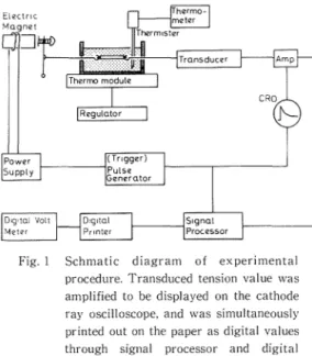
The abdominal nerve cord was used, dissected free from the abdomen of crayfish (Procambarus clarkii weighing about 9-12 g). The outer sheath of the nerve cord was peeled and both ends of the nerve between the third and fourth ganglia, which was 5 mm in length, were tied with threads at the ganglion regions. The schematic diagram of the experiment is shown in Fig.l. The stretching procedure is modified from that of Eyzaguirre and Kuffler (2). One end of the nerve was hooked to a magnet core for an instantaneous stretch of the nerve and the other end to a strain gauge led to an oscilloscope which was designed to measure the tension in the stretched nerve. The nerve was always bathed in Harreveld's solution in order to keep alive, and was stretched at a rate of 20 or 30 per cent of its natural length by the electrical magnet during a period of 4 sec. The temperature in solution was controlled by a thermomodule applying the Peltier's effect. Before beginning the experiment, the preliminary stretch was repeated about twenty times at 30°C until the magnitude of the tension was settled to a constant value. Subsequently, the
*Department of Nursing, Tottori University College of Medical Care Technology **Department of Physiology. Tottori University School of Medicine
120 Takeshi KASAGI • Osamu ICHIKAWA
I
Fig. I Schmatic diagram of experimental procedure. Transduced tension value was amplified to be displayed on the cathode ray oscilloscope. and was simultaneously printed out on the paper as digital values through signal processor and digital printer for the purpose of after analyses.
tensions at the control temperature, i.e. 30 'C, and at each one of the varying test temperatures were alternatin·ly examined. The survival of the nerve through the experiment was ascertained by recording its action potential evoked by an electrical stimulator with Ag-AgCI electrodes.
In order to check whether or not tension obtained in the present experiment was due to the biological membrane, the alteration of action potential was recorded by using a conventional moist chamber (5) after the application of various kinds of local anesthetics to the nerve. The nerve was stretched under the anesthtized condition by the same way as the controlled one.
RESULTS AND DISCUSSION
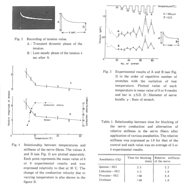
A pattern of the tension produced by the nerve stretch was such that a transient dynamic phase was follo\1·ed by a late steady phase. A typical recording of tbe tension is shown in Fig. 2. The initial maximum value (A) and that after 4 sec (B) are considered as requisite values for following references of data. In Fig. 3, values A and B are displayed in the order of the repetitive number of nerve stretches, varying the test temperature after every other 30
C. These values at test temperatures are plotted in Fig. 4 with relative magnitude to those at 30 "C, respcctiwly. They increased gradully with a decrease of the temperature until 10
·c,
while this tendency become stronger below 10·c.
The slope of the straight line, for example, are 1/70 and 1/9 (deg-1) above and below 10
·c,
respectin:ly. Thismay indicate that some kind of alteration occured in the membrane structural component at 10
·c.
The conduction velosity of the action potential was also measured in the same kind of nerve cord and its results are plotted in this same figure. Above 10 'C the velosity was constant of about 7 m/sec, while it was diminished abruptly bel(lw 10 C, resulting later than 3.1 m/sec or sometimes blocked.The results of the treatments of local anesthetics are shown in the table. The anesthetics such as Quinine-HCI and Lidocaine-HCI easily blocked the nerve conduction within 2.5-4.5 min after their application. On the contrary. Procain-HCI and Urethane could not block the conduction in so far as an hour of their treatments. Former two anesthetics showed the increased magnitude of the tension (both A and B) as well as the high efficacy for anesthesia, while the latter ones produce not only fewer change of tension but also less anesthetic efficacy. It can be said that
Stiffness and excitation of the crayfish nerve 121
l'~-~
~ 4 sec ---?>
Fig. 2 Recording of tension value.
A : Transient dynamic phase of the tension.
B : Late steady phase of the tension 4 sec after A.
'I\
.
L--?-?-~-0
.g 2 2\f
!11·-···•--d···,!On ve!ooly , Con uc \ .• / B7I
.c 6 g ~ ·c g' E ~ :Sl .g a:l\___"-?-r-0
TE'nSIOn.
10 20 30 Temperature (•C) 5 c .Q 4 ~3§
Fig. 4 Relationship between temperatures and stiffness of the nerve fibres. The values A and B (see Fig. 2) are plotted separately. Each point represents the mean value of 6 or 8 experimental results and was expressed relatively to that at 30
·c.
The change of the conduction velocity due to varying temperature is also shown in the figure B.~Jli
Temperature(°C)l
10 l.Of 6 8 D = 550um f = 0.23 §: 9\ .. r"-/'1;!\!YIY\
Jjh
c ·~ 0.5 \ / \ ; \+-4sec~
~ f'\. • t '·--c·'·.. B _...f..._I"...-•'t/ i ···-1-l-! 0 20 40 60 80 No. of stretchFig. 3 Experimental results of A and B (see Fig. 2) in the order of repetitive number of stretches with the variation of test temperature. Plotted value of each temperature is mean value of 6 or 8 results and bar is ±S.D. D: Diameter of nerve bundle. y : Rate of stretch.
Table 1. Relationship between time for blocking of the nerve conduction and alternation of relative stiffness in the nerve fibers after application of various anesthetics. The relative stiffness was expressed as 1.0 for that of the control and each value was an average of 3 or 4 experimental results.
Anesthetics (! %) Time for blocking Relative stiffness
(min) of the nerve
Quinine· HCI 2. 5 2.2
Lidocaine· HCI 4. 5 1.8
Procaine· HCI >60 0.8
Urethane >60 1.0
the tension observed in this experiment involves nerve membrane components because of the correlation between the decrement of nerve excitation and the increase of the tension value, in other words, stiffness in the nerve membrane. This fact seems to be confirmed by the result that large tension values were found at a low temperature below 10 ' In this range of the temperature the conduction velocity was remarkably delayed and sometimes the conduction was found to be blocked, that is, the excitability was declined. It comes to the conclusion that the increase of stiffness either by the anesthetics or by the cooling induces the decrement of the excitability in the nerve membrane. Besides, the critical change of tension and the excitability in the biological membrane suggest the phase transition between the liquid-crystalline phase and solid phases as mentioned by Lee et a!. (3). The interpretation of the results is difficult to distinguish between the contribution of phospholipids and proteins and further of their interaction or coaction in the membrane.
122 Takeshi KASAGI • Osamu ICHIKAWA
ABSTRACT
Alteration of the stiffness in the nerves due to temperature changes or application of various local anesthetics (1%) was examined by means of stretching the nerves during a period of 4 sec. Large magnitude of the tension 1ras obtained below 10'C, at which the conduction velocity was markedly delayed or was blocked. The conduction was also blocked by application of Quinine-HCl and of Lidocaine-HCl, while the blockade was hardly observed by Procaine-HCl or Urethane. Former two drugs produced fairly large magnitude of the tension, on the contrary the latter ones differed little from the control. Thus, the present findings of the tension may be of biological significance.
REFERENCES
1. Engelman D. M. X-ray diffraction stadies of phase transitions in the membrane of mycoplasma laicllawii.]. :\Iol. Bioi. 47,115-117,1970
2. Eyzaquirre C. and Kuffler S. W. Processes of excitation in the dendrites and in the soma of single isolated sensory nerve cells of the lobster and crayfish. ]. Gen. Physiol. 39, 87-119, 1955
3. Lee A. G., Birdsall N. ]. M. and Melcalf ]. C. Nuclear magnetic relaxation and the biological membrane. In Methods in Membrane Biology (ed. Korn E. D.) Plenum Press, New York and London, Vol. 2. 2-142. 197-! 4. Linden C., Wright K., McCornnel H. M. and Fox C. F. Lateral phase separations in membrane lipids and the
mechanism of sugar transport in escherichia coli. Proc. Nat!. Acad. Sci. 70, 2271-2275, 1973
5. Kasagi T., Ichikawa 0., Miyoshi !VI., Hiji Y. and Yamada IV!. Effect of nickel ion on the conduction of action potentials in non-myelinated nerve fibre of crayfish. Arch. internat. Physiol. Bioch. 87, 297-310, 1979 6. Wisnieshi B.
J..
Parks ]. G., Huang H. 0. and Fox C. F. Physical and physiological evidence for twotransitions in cytoplasmic membranes of animal cells. Proc. N atl. Acad. Sci. 71. 4381-4385, 197 4