Occurrence and Pathology of a Disease of Foals Caused by Larval Migration of Strongylus vulgaris
Mlheko SUTOH, * •õ Yurlo SAHEKI, * Ru1Zo ISHITANI, * Sumlo INUI, * * Mlnoru NARITA, * * HiroShi HAMAZAKI* * * and Tadashl YOKOTA* * *
In 1969, •gso-called diarrhea of foals•h manifesting fever, colic symptoms, and severe diarrhea and leading animals frequently to death has occurred in the Hidaka district of Hokkaido. Since then, the disease has continued to occur till now, indicating its peak over a period from 1969 to 1971. Forty-four dead animals were examined epizootiologically and pathologically with the following results. The disease occurred in May to November every year. Sixty per cent of the 44 animals died within 3 days. The average interval between the onset of symptoms and death was 6.2 days. Histopathologically, the thread-like elevation of
the intima, thromboendarteritis, panarteritis, and aneurysm were mainly found in the anterior mesenteric artery and its related blood vessels. As the other marked changes, verminous granuloma, hemorrhagic and necrotic enteritis due to embolism, and diffuse
eosinophilic infiltration were observed. All these changes were caused by the larval migration of Strongylus vulgaris, and led the animals to death. Of the occurrence of this equine disease, few reports have been presented in Japan. Discussion was made on the occurrence and
histopathology of the disease.
It was reported by Kikuchi1) in 1926 that the thread-like elevation, thrombo endarteritis, and verminous aneurysm had occurred frequently in the mesenteric ar tery and its branches, and concluded that these changes had been caused by the migration of larvae of Strongylus vulgaris.
Enigk2) verified Kikuchi's conclusion by
the use of a foal infection test. Drudge et
al.,3) Ershov,4) and Duncan and Pirie5,6)
confirmed this verification from their ex
perimental studies on suckling foals free
from equine Strongylus (S. vulgaris, S.
equinus, and S. edentatus). From the results
of these experimental studies, it was clari
fied that aneurysm of the anterior me
senteric artery and its branches, throm
bosis and verminous embolism of the ileo
cecocolic artery, and necrosis and in
flammation of the intestine due to in
farction were caused by the larval mig
ration of S. vulgaris, and that these disor
- Received for publication June 30, 1976.
*Department of veterinary Pathology, Faculty of Agriculture, Tokyo University of Agriculture and Technology, Saiwaicho, Fuchu-shi, Tokyo, 183 Japan.
•õ Tokyo Research Laboratories, Shin Nihon Jitsugyo Co., Ltd. at present.
**Hokkaido Branch Laboratory, National Institute of Animal Health.
* * *Hidaka Livestock Hygiene Service Center , Hokkaido. Address reprint requests to Dr. Ishitani.
This work was supported by a grant-in-aid from the Equine Health Laboratory, Japan Racing Association.
ders frequently led foals to death.
Of the occurrence of this equine di sease, many reports have been published
by Kikuchi,1,7) Ottaway and Bingham, 8,9) Farrely
,10) Poynter,'1 Curtis et al.,12) Gerber et al.,13> Pauli et al.,14) and Ben-net.15) Few papers, however, have been
presented in Japan. In 1969, •gso-called diarrhea of foals•h manifesting fever, colic symptoms, and severe diarrhea and lead-ing animals frequently to death occurred in the Hidaka district of Hokkaido. Since then, the occurrence of the disease has continued till now, indicating its peak over a period from 1969 to 1971. From the results of epizootiological and pathological observations on 44 dead animals, it was strongly suggested that the main cause of death might be the larval migration of S. oulgaris. This paper deals with the oc-currence and pathology of the disease.
Materials and Methods
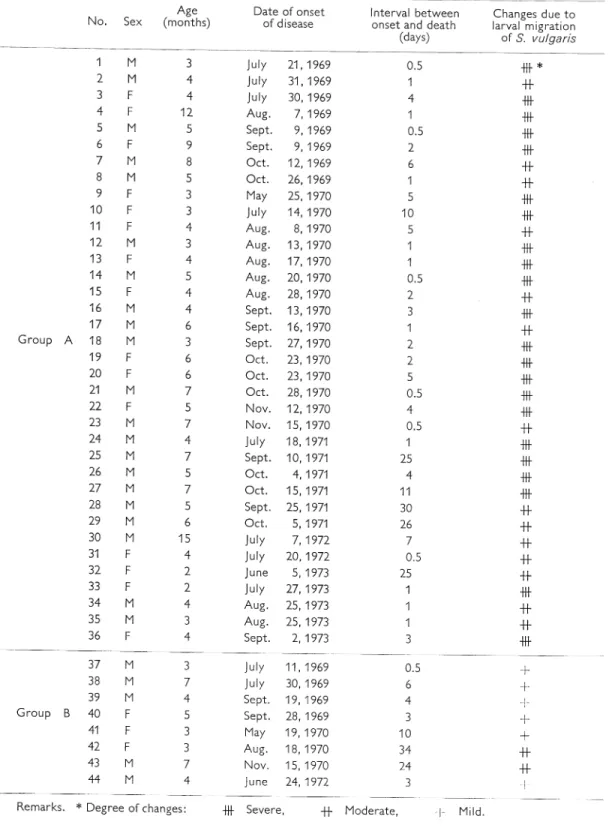
The materials used are shown in Table 1. They consisted of 44 dead foals 2 to 15 months old. Of them, 36 were Thorough breds, 7 Anglo-Arabians, and 1 was Ara bian obtained from the Hidaka district of Hokkaido over a period from 1969 to 1973. They were divided into 2 groups, A and B, according to histopathological changes. The cases of Group A revealed severe al terations caused by the larval migration of S. vulgaris. The cases of Group B showed an association of the changes caused by the larval migration of S. vulgaris with those due to some other causes.
After the postmortem examination,
tissue specimens were collected from the
altered portions of the anterior mesenteric
artery and its branches, jejunum, ileum,
cecum, colon, heart, lung, liver, spleen,
kidney, brain, adrenal gland, skeletal
muscle, thymus, and lymph nodes of
several regions. They were fixed in 10/0
formalin solution, embedded in paraffin,
cut and stained with hematoxylin and
eosin (HE), alcian blue, periodic acid
Schiff (PAS), aldehyde fuchsin, one-step
trichrome, Prussian blue, Sudan black B,
Congo red, thioflavine T, toluidine blue,
and by the Kossa reaction.
Results
Occurrence and clinical signs
About 70 per cent of the 44 cases were
collected in 1969 and 1970. The animah
were taken ill over a period from May to
November, with a peak of occurrence in
August and September, every year. Most
of them were 3 to 7 months of age (Table
1).
In general, the animals were well in growth and fleshy before the onset of symptoms. As clinical signs, a fever of 38.8-39.0•Ž appeared at the beginning, and was followed by pyrexia of over 40.0•Ž, colic symptoms, hemorrhagic diar rhea, tachycardia, and dyspnea. Diarrheal feces were evil in smell, and watery and bloody or tar-like. The visible mucous membranes were cyanotic or sometimes anemic. Peristalsis generally stopped, but was rarely accelerated. Sixty per cent of the 44 animals died within 3 days. The average interval between the onset of symptoms and death was 6.2 days.
Vascular lesions and related changes of organs
(Table 2)
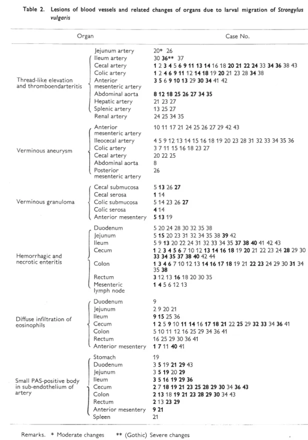
1. Thread-like elevation and thrombo endarteritis. The thread-like elevation of the intima caused by the migration of S. vulgaris larvae was observed in the an terior mesenteric artery and its related
Table 1. Experimental materials
Table 2. Lesions of blood vessels and related changes of organs due to larval migration of Strongylus vulgaris
blood vessels in all the cases. It was a mural
thrombus of fibrin deposit with small tun
nels formed by the larval migration (Fig .
1). It was affected with a slight infiltration
of neutrophils,
eosinophils, and lympho
cytes. Most parts of the intima to which a
fibrin thrombus was attached lacked en
dothelial cells. The surface of the thrombus
was covered with proliferating endothelial
cells. Two cases, Nos. 10 and 11, revealed
young fourth-stage larvae of S. vulgaris in
small tunnels of fibrin thrombus in some
branches of the anterior mesenteric artery
(Fig. 2). Two cases, Nos. 6 and 14,
re-Fig. 1. Thread-like elevation showing mural throm bus of fibrin deposit with a small tunnel (arrow) formed by larval migration of Strongy lus vulgaris
Case No. 10. Artery of adrenal gland. Alcian blue (AB) and periodic acid-Schiff (PAS) stain,
x 100.
Fig. 2. Multiple thread-like elevation
Young fourth-stage larvae (arrows) and eosinophils in fibrin tunnel of arteries of medium caliber. Case No. 11. Branch of anterior mesenteric artery. Hematoxylin and eosin stain (HE), x100.
Fig. 3. Young fourth-stage larvae (arrows) attached to extensive fibrin throbmus of thread-like
elevation
Fig. 4. Verminous nodule formed by emboli of young fifth-stage adults and thrombus (arrow) Circulatory disturbance and necrosis are observed in surrounding tissues. Case No. 4. Cecocolic
artery. Aldehyde fuchsin (AF) and one-step trichrome stain, x 3.5. Fig. 5. High-power magnification of part of the lesion shown in Fig. 4
Young adult (arrow), necrotic arterial wall, and proliferating granulation tissue. AF and one-step trichrome stain, x100.
Fig. 6. Necrotic and granulation tissues and newly formed blood vessels (arrow) in lumen of artery of small caliber
Case No. 15. Anterior mesentery. HE, x50.
Fig. 7. Granulation tissue and newly formed blood vessels having muscle layer (arrows) in lumen of artery of small caliber
Case No. 33. Submucosa of cecum. AF and one-step trichrome stain, x 250.
vealed larvae of the same stage in ex
tensively accumulated fibrin thrombi at
tached to the thread-like lesion in the
cecocolic and splenic arteries (Fig. 3).
Various degrees of nodular thickening of
the intima were present in the other parts
of arteries of the mesentery, cecum, colon ,
liver, and kidney. Stale or fresh thrombi of
various sizes were attached frequently to
this portion. Eosinophils infiltrated focally
or diffusely in the intima and sometimes in
the media (Fig. 2). Lesions of edematous
looseness of connective tissue were also
found in the media and adventitia.
Furthermore, thrombi were present, oc cluding completely arteries of small and medium caliber in the cecum and colon in many cases. There were emboli with fifth-stage larvae in arteries of medium caliber in the serous membrane of the cecum and colon of two cases, Nos. 1 and 4, and those with fourth-stage larvae in arteries of small and medium caliber in the submucosa of the cecum and colon in Case No. 27 and of the ileum in Case No. 39. The arterial walls became necrotic.
Fig. 8. Verminous arteritis accompanied with throm bus and S. vulgaris larvae (arrows)
It shows complete occulusion of arteries with thrombus. Case No. 16. I1eocetal artery. HE, x 5.
Fig. 9. Verminous arteritis with thickened, folded intima containing thrombus and S. vulgaris larvae (arrows)
Case No. 11. Root of the anterior mesenteric artery. HE, x 2.5.
Fig. 10. Verminous aneurysm with marked thinning of vascular wall and dilatation of lumen con taining thrombus
Case No. 22. Anterior mesenteric artery. AF and onestep trichrome stain, X4.
Circulatory disturbances and inflamma
tory changes were observed in the
sur-rounding tissues (Figs. 4 and 5). In some
instances, the lumina of arteries were oc
culuded almost completely with granula
tion tissue which involved newly formed
blood vessels having a muscle layer and
containing erythrocytes in the lumen
(Figs. 6 and 7). Some arteries showed loss
of structure by the replacement of granu
lation tissue, and aggregation of macro
phages containing hemosiderin.
In only one case (No. 16), fourth-stage
larvae were in the media of vasa vasorum
in the large mesenteric artery with their
lumina filled with granulation
tissue.
No particular relationship could be
ob-served between the changes of arteries
stated above and fatty substances as de
monstrated
by Sudan black B staining.
2.
Verminous aneurysm. Aneurysms
caused by S. vulgaris larvae were
ob-served in 32 cases of Group A and 2 cases
of Group B. Most of them were found in
the ileocecal artery. Macroscopically, they
were about 1.5 to 10 cm in diameter,
found singly or in a clump of a few, and
occluded with thrombi containing nume
rous larvae of Strongylus (Figs. 8 and 9).
There were few genuine aneurysms mani
festing a marked dilatation of the lumen
and thinning of the vascular wall (Fig. 10) .
In mild lesions, the intima had large mural thrombi attached and containing fourth- and fifth-stage larvae, and was
thickened, elevated, and replaced en tirely with fresh granulation tissue con sisting of mononuclear cells, lymphocytes,
eosinophils, fibroblasts, and new capil laries. Macrophages containing hemosi
derin, a few giant cells, and epithelioid cells were found in some parts of the gra
nulation tissue. The tunica elastica interna was destroyed and disappeared in some places. Degeneration of elastic fibers and infiltration of large mononuclear cells and lymphocytes, however, were slight in the tunica media. No changes were ob-served in the adventitia.
In moderate lesions, there were changes
spreading from the intima to the media
and adventitia. Vascular lumina were
almost completely occluded with thrombi
containing many larvae, numerous eosi
nophils and lymphocytes, and much fibrin
(Fig. 11). There were severe infiltration of
large mononuclear cells, eosinophils, lym
phocytes, and plasma cells, destruction
and disappearance of the tunica elastica,
dissociation of elastic fibers, and hyaline
swelling of smooth muscle fibers. Part of
the smooth muscle was replaced with con
nective tissue in the media (Fig. 12).
In arteries affected with severe aneurys mal changes, most of the media and part of the adventitia were replaced with con nective tissue, and necrotic muscles re mained like islets. So that the normal structures of the intima and media were lost completely. Deposition of calcium was present in the necrotic parts of the tunica intima.
In Case No. 14, masses of bacteria were
found in thrombi
and large abscesses
spread in the intima and media. Severe
necrosis was seen in the arterial wall, and
congestion and hemorrhage were noticed
in the adjacent mesentery in three cases,
Nos. 4, 5, and 30.
3. Verminous granuloma. Verminous granulomas consisted fundamentally of three areas. The central area contained necrotic masses, necrotic tissue of the arte rial wall, and sometimes larvae. The
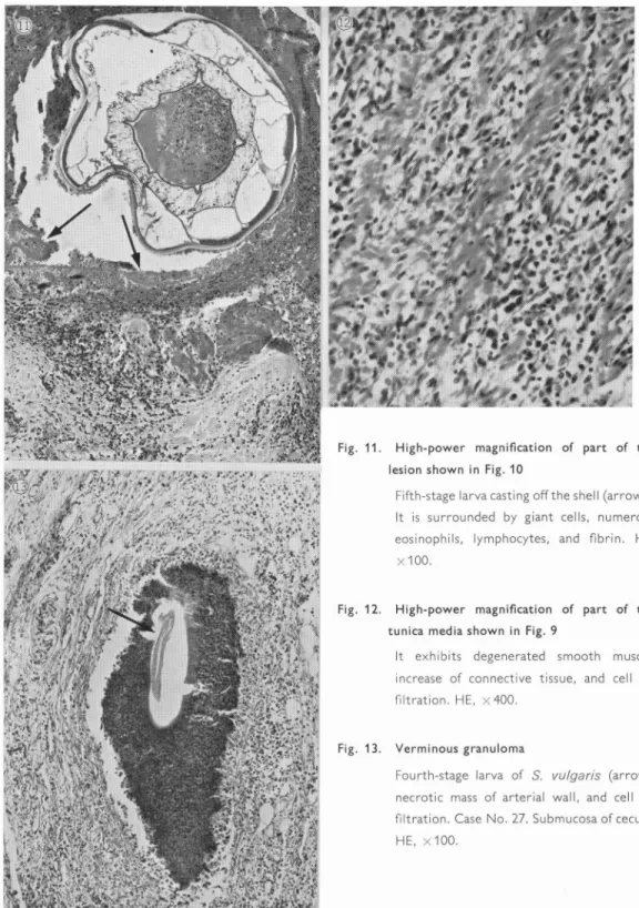
inter-Fig. 11. High-power magnification of part of the lesion shown in Fig. 10
Fifth-stage larva casting off the shell (arrows). It is surrounded by giant cells, numerous eosinophils, lymphocytes, and fibrin. HE,
x 100.
Fig. 12. High-power magnification of part of the tunica media shown in Fig. 9
It exhibits degenerated smooth muscle, increase of connective tissue, and cell in filtration. HE, x400.
Fig. 13. Verminous granuloma
Fourth-stage larva of S. vuigaris (arrow), necrotic mass of arterial wall, and cell in filtration. Case No. 27. Submucosa of cecum. HE, x100.
mediate area was composed of epithelioid
cells with a few giant cells. The outer area
surrounded it and consisting of fibroblasts,
fibrous connective tissue, and numerous
inflammatory cells, including lymphocytes
and large mononuclear cells (Fig. 13).
In small granulomas, giant cells, epithe
lioid cells, and lymphocytes accumulated
around the necrotic tissue, and were
sur-rounded by a thin layer of connective
tis-sue and a few eosinophils. There were
some granulomas containing a few giant
cells and a dense accumulation of eosino
phils. In arteries of medium caliber in
the serous membrane of the cecum and
colon in two cases, Nos. 1 and 4, and in
the arteries of small and medium caliber in
the submucosa of the ileum in Case No.
39, larval emboli were seen,
accompany-ing a complete necrosis of the arterial wall.
Such lesions were surrounded by epithe
lioid cells and a few giant cells. The outer
area exhibited diffuse hyperplasia of
granulation tissue and severe infiltration
of large mononuclear cells, lymphocytes,
and eosinophils. Regressive changes were
found spreading all over the affected
tis-sues, where there were many macrophages
containing eosinophilic granules and
foam-like macrophages characterized by a large
nucleus and wide cytoplasm of foamy
structure.
In verminous granuloma containing larvae outside of blood vessels, it was difficult to distinguish the larva of S. vulgaris from that of any other parasite. So that other parasite larvae might be contained in the lesion.
4. Hemorrhagic and necrotic enteritis. Caused by the severe thrombo-embolic changes of blood vessels in the intestine and mesentery, marked hemorrhage and
necrosis occurred mainly in the cecum
and colon (Fig. 14). Macroscopically,
severe hyperemia, petechial, spot-like, and
diffuse hemorrhages, severe necrosis, and
ulcer were observed. Microscopically,
edema was marked in the submucosa
and spread into the muscle layer and
subserosa (Fig. 15). There were also
necrosis of the mucous membrane, ede
matous swelling and vacuolization of
the arterial wall (Fig. 16), and decrease
of stainability, and hyaline swelling and
fragmentation of smooth muscle fibers
(Fig. 17).
In areas showing severe circulatory
disturbances, arteries of small and medium
caliber were markedly extended with
blood and thrombi, and the tunica
elastica and elastic fibers were stretched
to form a thin memberane (Fig. 14). In
some other cases, infiltration of eosino
phils, large mononuclear cells, lymphocy
tes, and macrophages containing hemo
esiderin, as well as hyperplasia of con
nective tissue, was present in lesions of
the intestines.
In the lymph follicles of the ileum, cecum, colon, rectum, and lymph nodes of the mesentery, karyorrhexis and kary opyknosis of lymphocytes and deposition of hyaline substance were found in many cases. In the periphery of the degenerated lymph follicles, macrophages containing light yellowish-brown fatty pigment gra nules were present here and there. Edema and necrosis of the adipose tissue and nerve cells were observed in the lesion.
5. Diffuse infiltration of eosinophils. A
diffuse infiltration of eosinophils was
observed in the alimentary tract, especially
in the cecum and colon, and the mesen
terv in many cases (Fig. 18). It was mainly
Fig. 14. Anemic infarction of intestine and markedly distended arteries with thrombi (arrow)
Mucous membrane shows complete necrosis. Case No. 18. Cecum. HE, x2.4.
Fig. 15. High-power magnification of part of the lesion shown in Fig. 14
Necrosis of mucous membrane and lymph follicles. Edema is marked in the submucosa. HE, x40.
Fig. 16. Vacuolization of wall of artery of small caliver and small PAS-positive bodies (arrows) in the subendothelial tissue
Case No. 19. AF and one-step trichrome stain, x 250.
Fig. 17. Hyaline swelling and fragmentation of smooth muscle fibers and intermuscular edema
Case No. 15. Muscle layer of ileum. AF and one-step trichrome stain, x400.
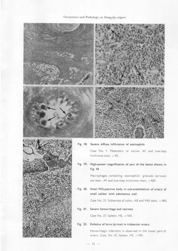
Fig. 18. Severe diffuse infiltration of eosinophils
Case No. 1. Mesentery of cecum. AF and one-step trichromestain, x40.
Fig. 19. High-power magnification of part of the lesion shown in Fig. 18
Macrophages containing eosinophilic granules (arrows) are seen. AF and one-step trichrome stain, x 400.
Fig. 20. Small PAS-positive body in sub-endothelium of artery of small caliber with edematous wall
Case No. 21. Subserosa of colon. AB and PAS stain, x400.
Fig. 21. Severe hemorrhage and necrosis Case No. 27. Spleen. HE, x 100.
Fig. 22. Embolus of larva (arrow) in trabecular artery
Hemorrhagic infarction is observed in the lower part of artery. Case. No, 42. Spleen. HE, x100.
found in the submucosa showing ede
matous swelling and thickening. In severe
cases infiltrating eosinophils were spread
all over the intestinal wall. They fre
quently formed a dense accumulation,
especially in the periphery of lymph
follicles and regional lymph nodes. There
was not always a diffuse infiltration of
eosinophils in the lesion of verminous
granuloma. A large number of
macro-phages containing eosinophilic granules
appeared in areas where an accumulation
of eosinophils was present (Fig. 19).
6. Small PAS-positive bodies in the sub endothelial tissue of arteries. Small bodies stained deeply with PAS were seen in the intima of arteries of small and medium
caliber in the alimentary tract and mesen tery (Figs. 16 and 20). They were round in shape and had jagged protuberances. In some cases many bodies were con tained in almost all the cross sections of blood vessels. They were 1 to 10 in num ber, 5 to 20 ƒÊm in diameter, and covered with or entirely surrounded by endothelial cells. In hematoxylin and eosin staining they showed a concentric configuration with basophilic outer rims and deeply eosinophilic inner portions. Their struc tures could be distinguished quite clearly by one-step trichrome staining. They were also positive for aldehyde fuchsin staining, and pale positive for the Kossa reaction and Prussian blue staining.
•@ The intima of blood vessels with the bodies was often vacuolated and bulged into the lumen. In arteries with many bodies stenosis of the lumen was present. All the intima and endothelial cells of arteries were deeply positive for alcian blue staining. There was no relationship between the appearance of bodies and the
presence of thrombosis or inflammatory changes.
Changes of the other organs
In 15 cases the heart was affected with severe or moderate congestion and sub
epicardial and endocardial hemorrhage. In three cases, Nos. 4, 6, and 35, were revealed such changes as loss of striation, sarcolysis, and hyalinization of myocar
dial fibers, but cellular responses were scarcely observed. There were conges
tion, edema, and hemorrhage in the lungs of 24 cases. In these cases, Nos. 36 and 44 showed purulent brochopneu monia and No. 43 had multiple abscesses in the lung. In the liver, congestion and
edema, focal necrosis, and fatty degenera tion were observed in 10, 6, and 4 cases, respectively. Reticuloendothelial cells were activated in 3 cases, of which Nos. 14 and 42 were diagnosed as the chronic type of equine infectious anemia and strepto coccal infection, respectively, and Nos. 14 and 43 had multiple abscesses.
Thirty-one of the 44 cases revealed
hemorrhage in the spleen. In 14 cases
the hemorrhage was so severe that the
splenic parenchyma looked like a sea of
blood and severe degeneration and nec
rosis were observed (Fig. 21). Case No. 42
exhibited hemorrhagic infarction due to
an embolus of larvae in the trabecular
artery (Fig. 22). Three cases, Nos. 25,
28, and 44, showed hemorrhagic infarc
tion of unknown cause. Severe activation
of reticuloendothelial cells was detected
in Case No. 42 diagnosed as equine infec
tious anemia.
In the kidneys, congestion and hemor
rhage (15 cases), anemic infarction (10
cases), and thrombus formation (12 cases)
were found. In addition, perirenal
toma accompanied with parenchymal necrosis, hyperplasia of granulation tissue , and deposition of hemosiderin were seen in three cases, Nos. 25, 26, and 27, two of which showed hemorrhage in the pelvis . In Case No. 21, an embolus with fourth-stage larvae was found in the arcuate artery, and necrosis and round cell infilt ration were present in the surrounding tissues. Multiple bacterial emboli were observed in glomerular capillaries of Case No. 14. In the adrenal gland, hemor rhage of the cortex was induced in 5 of 13 cases examined, and moderate fatty changes of the zona fasciculata and reticularis in 2 cases. A slight interstitial infiltration of lymphocytes and lympho cytic nodular formation in acini were found in Case No. 42.
The central nervous system was ex
amined in 22 cases. Changes were absent
in all the cases, except one diagnosed as
equine infectious anemia.
This case re
vealed a marked perivascular infiltration
of round cells in the meninges and paren
chyma of the brain. Congestion and small
hemorrhagic foci were seen in the thymus
of 6 cases. Furthermore,
3 of 13 cases
examined revealed hyaline swelling and
hemorrhage in skeletal muscles.
Findings of bacterial cultivation
In 13 of 41 cases in which bacterial cultivation had been performed, Escheri chia coli was isolated from every organ . Streptococci were detected from an abs cess of the liver in Case No. 14 and abs cesses of the liver, lung, and kidney in Case No. 43.
Discussion
Occurrence of the disease
It has been reported by Duncan16) in
England that a count of 5,000 third-stage larvae of Strongylus contained in one kg of herbage was never unusual in the pasture. According to Poynter,11) l to 5 per cent of Strongylus eggs contained in the feces belonged to S. vulgaris. The number of third-stage larvae used by Enigk2) and other research workers3,5,6) in their experiments ranged from 800 to 8,000. In England also, Russel,17) Poyn ter,11) Ogbourne, 18,19) and Duncan16) reported that the number of eggs con tained in the feces of a mare increased in spring and summer, causing a severe contamination of the pasture with third-stage larvae of S. vulgaris in summer and autumn.
In the present occurrence, the foals having marked aneurysms in the mesen teric arteries were very young. Of them, five were 3 months old and two 2 months old. From this fact it was assumed that the environment of raising the foals might have remarkably been contaminated with third-stage larvae of S. vulgaris. "So-called diarrhea of foals" in the present report began to occur in April, showing its peak in October, and finished in December. The mode of this occurrence seemed to resemble the seasonal curve showing a rise and fall of the number of S. vulgaris third-stage larvae in the pasture in England.
As to the pathogenesis of the colic syn-drome of horses due to embolism, Dobber stein20,21) emphasized the changes of the nervous tissues caused by aneurysms in the region of the anterior mesenteric artery. Since then, the changes of the artery itself induced by the larval migration of S. vulgaris have been neglected for a long time. The horses used by Dobberstein in
his experiments, however, seemed to be adult or older in age, and were not likely to be susceptible for S. vulgaris larvae. As stated already, a severe pathomorphism which had completely changed the tradi tional conception of Dobberstein was clarified experimentally by some research workers 26)
There are some reports8,9,22) showing
that verminous aneurysms caused by the
larvae of S. vulgaris were observed at high
percentage in horses. According to Poyn
ter, the incidence of aneurysm was 90%
in yearlings and 47% in foals. Duncan23)
described that the foal was so high in
susceptibility that it showed severe sym
ptoms
in
an
infection
experiment.
Bennet15) and
Velichkin24) stated
that
the elimination
of parasites,
especially
S. vulgaris, would be very important
in
horses manifesting
colic symptoms.
In
the recent years, Gerber et a1.,13) Pauli
et al.,14) and others reported
natural
cases in which death had occurred with
intestinal alterations due to the embolism
of S. vulgaris larvae. The present occur
rence of the disease in the Hidaka district
seemed to be a big one without precedent.
Lesions due to larval migration o f S. vul uaris
Concerning changes in the arteries of horses infected with S. vulgaris larvae, the thread-like elevation, thromboend arteritis, and panarteritis were described by Kikuchi,l,7) Hobmaier,25) O1t,26) Coffman and Carlson,27) and Grea torex.28) Furthermore, Enigk2,29) and other research workers3,4,6,23) clarified in their infection experiments that such severe alterations of the intestines as circulatory disturbance, necrosis, and in flammation were caused by the throm bosis and embolism of branches of the
ileocecal artery. Other lesions apparently caused by the larval migration of S. vulgaris were noticed in a case of sudden death due to the thrombosis of the right coronary artery reported by Cronin and Leader,30) in cases showing changes of the brain due to infarction by Fraser,31) and in a case with infarction in the kidneys by Mahaffey and Adam.32) In the present cases, a large number of thread-like eleva tions were observed in the splenic artery and a small number of them in the arteries of the liver, kidney, and adrenal gland. There were also a hemorrhagic infarction due to the embolus of larvae in the
trabecular artery of the spleen and the embolus of larvae in the arcuate artery of the kidney.
As pointed out by Mathieson,33) Duncan and Pirie5) and Bennet,15) and shown in the present cases, the vascular change named as verminous aneurysm was the inflammation of the anterior mesenteric and ileocecocolic arteries. In the present cases, it was rare to find genuine aneury sms with the dilatation of the lumen and thinning of the arterial wall caused by the degeneration and rupture of the tunica elastica interna and the muscle layer, and it was common to observe verminous arteritis accompanied with marked thick ening of the arterial wall and formation of thrombus.
Bacterial masses were found within the aneurysms of Case No. 14. In this case, abscesses of aneurysms, multiple abscesses of the liver, and embolic glomerulo nephritis were present and streptococci isolated from the liver by cultivation. Enigk29) proved that no pathogenic bac teria were carried into blood vessels of the host animal with S. vulgaris, and
presumed that the abscess of aneurysm seen in literature might have been induced by metastasis from some other purulent lesion caused by bacteria before the parasitic invasion.
Many previous authors mentioned that eosinophilic infiltration had been con spicuous only in vascular lesions and verminous granulomas. However , a severe diffuse and dense accumulation of eosino phils was observed and macrophages containing eosinophilic granules appeared especially in the submucosa of the intestines in the present cases.
The small PAS-positive bodies in the sub-endothelium of arteries of small and medium caliber were identical with the initial bodies observed previously in the horse.34-37) According to Montail et a1 .35) they arose from some of the sub-endo thelial cells, underwent degeneration , and showed a structure consisting of masses of calcium and iron surrounded by degen erated cellular materials . What significance they had was unknown. They were , however, common in healthy horses17)
and seemed to have been formed in the very early stage of arterial changes .35)
In the present cases, it was assumed that the formation of these bodies might be associated with edematous changes of arteries.
Lesions of other organs
Very few previous papers
deal with
histopathological
changes of organs , ex
cept the artery and intestine. A marked
activation of the reticuloendothelial
cells
was described by Ershov.38) It was ,
how-ever, slight or absent in the present cases ,
except two which
were
diagnosed
as
equine
infectious
anemia
and
strepto
coccosis, respectively. Marked circulatory
disturbances, such as congestion, edema, and hemorrhage of the heart, lung, liver, spleen, and regional lymph nodes, and anemic infarction of the kidney were present in many cases. Hemorrhage was seen in the adrenal cortex in 5 of 12 cases examined. Degeneration and necrosis of the lymph follicle were observed in the spleens of almost all the cases. These findings were similar to those of the intestine and mesenteric lymph nodes.
Perirenal
hematoma
was found in 3
cases, two of which had hematoma in the
pelvis. Ohshima
et al.39) reported
an
equine case of perirenal hematoma
en-countered
in a slaughterhouse.
Zschie
sche40) described that a fatal case mani
festing colic symptoms had been caused
by hemorrhage from perirenal hematoma
into the abdominal
cavity. Doll41) con
sidered aneurysm as one of the causes of
perirenal hematoma and emphasized the
necessity of confirming the site of hemor
rhage. In the present cases, a relationship
was unknown between the site of hemor
rhage and the formation of aneurysm.
Cause of death in the present cases
As stated already, aneurysm of the anterior mesenteric artery caused by the larval migration of S. vulgaris, diffuse hemorrhagic and necrotic enteritis result-ing from thromboembolism in the ileo cecocolic artery and its branches, and a marked circulatory disturbance of main organs were enough to explain the case of death. Besides, Escherichia coli was iso lated from the materials of some organs in 13 of 36 cases. It was presumed, how-ever, that invasion of bacteria into organs might occur in the agonal stage, because of the lack of cellular response to micro-organisms.
In Case No. 42 diagnosed histopatholo gically as the chronic type of equine infectious anemia, the direct cause of death was considered to be acute hemor rhagic and necrotic enteritis induced by larvae of S. vulgaris. In two cases, Nos. 14 and 43, from which streptococci had been isolated by cultivation, the direct cause of death was also considered to be necrotic enteritis induced by larvae of S. vulgaris. Even in the other 6 cases of group B, the changes of the artery and intestine were milder than the cases of group A. These changes, however, were considered to be an important cause of death.
Acknowledgments
The authors wish to thank Mr. M. Ito, of the National Institute of Animal Health, Tokyo, for his
technical assistance in photographing.
Literature Cited
1) Kikuchi, K. 1926. Pathology on Strongylus vul garis of horse. I [transl. from Japanese] Nihon Byori Gakkai Kaishi [Soc. Pathol. Jap.] 15, 581-590
[in Japanese].
2) Enigk, K. 1950. Zur Entwicklung von Strongylus oulgaris (Nematodes) im Wirtstiere. Tropenmed. Parasitol. 2, 287-306.
3) Drudge, J. H., E. T. Lyons and J. Szanto. 1966. Pathogenesis of migrating stages of helminths, with
special reference to Strongylus vulgaris. p. 199-214, In Biology of parasites. Academic Press, Inc., New York.
4) Ershov, V. S. 1969. The biology of horse Strongyles and the allergic reactions caused by them, p. 304-309, In J. T. Bryans and H. Gerber
[ed.], Int. Conf. Equine Infect. Dis., 2nd ed., S. Karger, Inc., Basel.
5) Duncan, J. L. and H. M. Pine. 1972. The life cycle of Strongylus vulgaris in the horse. Res. Vet.
Sci. 13, 374-379.
6) Duncan, J. L. and H. M. Pine. 1975. The patho genesis of single experimental infection with Strong vlus vulgaris in foals. Res. Vet. Sci. 18. 82-93. 7) Kikuchi, K. 1928. Beitrage zur Pathologic der
durch Sclerostomum vulgare verursachten Verander ungen den Pferdes. Z. Infektkr. Haustiere 34, 237.
8) Ottaway, C. W. and M. L. Bingham. 1941. Some
records of parasitic aneurysm in clinically affected horses. Vet. Rec. 53, 275-282.
9) Ottaway, C. W. and M. L. Bingham. 1946. Further observations on the incidence of parasitic anuerysm in the horse. Vet. Rec. 58, 155-159. 10) Farrelly, B. T. 1954. The pathogenesis and signi
ficance of parasitic endarteritis in the ascending aorta of the horse. Vet. Rec. 66, 53-61.
11) Poynter, D. 1954. Seasonal fluctuation in the number of Strongyle eggs passed by horses. Vet. Rec. 66, 74-78.
12) Curtis, R. A. 1964. Mesenteric aneurism in a horse. Can. Vet. J. 5, 36-38.
13) Gerber, H., P. Chuit and B. Pauli. 1971. L'in farctus de l'intestin grele chez le cheval. Schweiz. Arch. Tierheilk.113, 678-697.
14) Pauli, B., H. Gerber and P. Chuit. 1971. Dun ndarminfarkte beim Pferd. Schweiz. Arch. Teir heilk.113, 685-697.
15) Bennet, D. G. 1972. Predisposition to abdominal crisis in the horse. J. Amer. Vet. Med. Ass. 161, 1189-1211.
16) Duncan, J. L. 1974. Field studies on the epide miology of mixed Strongyle infection in the horse.
Vet. Rec. 94, 337-345.
17) Russell, A. F. 1948. The development of helmin thiasis in Thoroughbred foals. J. Comp. Pathol. 58,
107-127.
18) Ogbourne, C. P. 1971. Variations in the fecundity of strongylid worms of the horse. Parasitology 63,
289-298.
19) Ogbourne, C. P. 1973. Survival on herbage plots of infective larvae of strongylid nematodes of
the horse. J. Helminthol. 47, 9-16.
20) Dobberstein, J. and H. Hartmann. 1932. Ueber die Anastomosenbildung im Bereich der Blind
und Grimmdarmarterien des Pferdes and ihre Bedeutung fur die Entstehung der embolischen Kolik. Ben. Munch. Tierarztl. Wschr. 48, 397-402. 21) Dobberstein, J. 1938. Die chronische Kolik des
Pferdes. Ben. Munch. Tierarztl. Wschr. 64, 493-499 u. 509-512.
22) Poynter, D. 1960. The arterial lesions produced by Strongylus vulgaris and their relationship to the
migratory route of the parasite in the horse. Res. Vet. Sci. 1, 205-217.
23) Duncan, J. L. 1974. Strongylus vulgaris infection in the horse. Vet. Rec. 95, 34-37.
24) Velichkin, P.A. 1964. Proc. Int. Congr. Parasitol., Rome Vol. 2, p. 868. [Cited from Duncan23)], 25) Hobmaier, P. A. 1925. Uber die Entstehung des
Aneurysma Verminosum equi. Z. Infektkr. Haustiere 28, 165-177.
26) Olt, A. 1932. Das Aneurysma verminosum des Pferdes and seine unbekannten Beziehungen zu
Kolik. Dtsch. Tierarztl. Wschr. 40, 326-332. 27) Coffman, J. R. and K. L. Carlson. 1971. Ver
158, 1358-1360.
28) Greatorex, J. G. 1975. Diarrhoea in horses associated with ulceration of the colon and cecum resulting from S. vulgaris larval migration. Vet. Rec. 97, 221-225.
29) Enigk, K. 1951. Weitere Untersuchungen zur Biologic von Strongylus vulgaris (Nematodes) im Wirtstiere. Tropenmed. Parasitol. 2, 523-535. 30) Chronin, M. T. I. and G. H. Leader, 1952.
Coronary occlusion in a Thoroughbred colt. Vet. Rec. 64, 6-11.
31) Fraser, H. 1966. Two dissimilar types of cerebel lar disorder in the horse. Vet. Rec. 78, 608-612. 32) Mahaffey L. W. and N. M. Adam. 1963.
Strongylus vulgaris in the urinary tract of a foal and some observations upon the habits of the parasite.
Vet. Rec. 74, 561-566.
33) Matieson, A. 0. 1964. A study into the distribu tion of, and tissue responses associated with, some intestinal parasites of the horse. M. Sc. thesis, Uni
versity of Edinburgh [Cited from Duncan23)]. 34) McCraw, B. M. and 0. D. Slocombe. 1974.
Early development and pathology associated with Strongylus edentatus. Can. J. Comp. Med. 38, 124-138.
35) Montail, J. R., J. D. Strandberg, and R. A. Squire. 1970. A histochemical and ultrastructural study of intimal bodies of horse arterioles. Lab.
Invest. 23, 302-306.
36) Nakamura, T., Y. Saheki, H. Tada and M. Kirisawa. 1965. •gColitis X•h in horse. Jui Chikusan Shinpo (J. Vet. Med.) No. 392, 125-128. [in Japa nese].
37) Rooney, J. R., J. T. Bryans and E. R. Doll. 1963. Colitis X of horses. J. Amer. Vet. Med. Ass. 142, 510-511.
38) Ershov, V. S. and M. Y. Naumycheva. 1971. Allergy and autoallergy in the pathology of helminthiasis in animals. p. 173-176, In S. M.
Gaafar et al. [ed], Pathology of parasitic diseases. Purdue University Studies Lafayette, Indiana. 39) Ohshima, K., H. Satoh, S. Nakagawa and T.
Saito. 1952. Two cases of perirenal hematoma observed in the Sapporo Abattoir. Jui Chikusan
Shinpo (J. Vet. Med.) No. 80, 95-97. [in Japanese]. 40) Zschiesche, M. 1924. T. R. Jg., 2, 18-20
[cited from Ohshima39)].
41) Doll. 1929. Bruns' Beitr. klin. Chirurg, p. 147 [cited from Ohshima39)].
Strongylus vulgaris 子 虫 の体 内移 行 に原 因 す る子 馬 の疾 病 の発 生 お よ び病 理 須 藤 三 重 子*† ・佐 伯 百 合 夫*・ 石 谷 類造*・ 乾 純 夫 **成 田 実**・ 浜 崎 裕***・ 横 田 禎*** 1969年,北 海 道 日高地 方 に お い て,発 熱 ・疝 痛 ・激 しい 下 痢 を 主 徴 と し,し ば しぼ 致死 的 に 経過 した"い わ ゆ る幼 駒 の下 痢 症"と 称 され る疾 病 が 多数 発 生 した 。 そ の 後,本 疾 病 は1969年 よ り1971年 を ピ ー ク と して 現 在 もな お 発 生 が続 い て い る。 そ れ らの うち44例 の 死 亡 した 子 馬 に つ い て,発 生 状 況 の 観 察 と病 理 組 織 学 的 検 索 を 実 施 し,下 記 の 結 果 を 得 た 。 本 病 の発 生 は 毎 年, 3月 に 始 ま り11月 に は 終 わ って い る。 発 症 よ り死 亡 ま で の 日数 は,3日 以 内 の も のが 死 亡 した44例 中 の60% を 占 め,平 均 日数 は6.2日 で あ った 。 病 理 組 織 学 的 に は,前 腸 間膜 動脈 お よびそ れ に 関連 の あ る血管に,内 膜 に お け る 糸状 隆 起線,血 栓 性 動脈 内膜 炎,汎 動 脈 炎 や 動脈 瘤 が見 られ た 。 そ の他 の著 明 な変 化 と しては, 寄 生 虫 性 肉芽 腫,塞 栓 に よる 出血 性 壊 死 性 腸 炎,お よ び 瀰 漫1生好 酸球 浸 潤 が認 め られた 。 これ らの病 変 は す べ て 普 通 円 虫 子 虫 の体 内移 行 に よ り生 じた もの で,死 の 原 因 とな っ た もの で あ る 。 馬 の 本 病 の 発 生 につ い て は,日 本 で は 僅 か な報 告 しか な い 。 この 論文 で は本 病 の 発 生 状 況 と病 理 組 織 に つ い て の若 干 の考 察 を行 な っ た 。 昭和51年6月30日 受付 *東京農工大学農学部家畜病理学教 室 **農林省家畜衛生試 験場北海道支場 ***北海道 日高家畜保健衛生所 †現勤務地 新 日本実業株式会社東京研究所 78